6 Oral Examination of the Conscious Patient
 Call Kai
 Learn
Learn Practice Test
 Spaced Repetition
 Match
Match Flashcards
 Knowt Play
1/60

There's no tags or description
Looks like no tags are added yet.
Name | Mastery | Learn | Test | Matching | Spaced | Call with Kai | Chat |
|---|
No analytics yet
Send a link to your students to track their progress
61 Terms
1. evaluate head and neck thru visual, auditory, and palpation (what is the jaw shape? muscle wasting? swelling or draining tracts?)
2. can they close the mouth? is there an increase in mouth opening?
3. soft tissue contracture?
4. spasms of lower jaw, tongue, lips?
5. palpate head and neck = ventrum, sides of mandibles/incisive/maxillary bones, TMJ, zygomatic arches, orbital rims, temporal lines of frontal bones, external sagittal crest, external occipital protuberance
6. palpate muscles of mastication (temporal, masseter, medial pterygoid, digastricus) = hypertrophy? atrophy?
7. palpate salivary glands
8. position of globes? can you retropulse them? (this is a good thing)
9. skin of head
10. nasal discharge and airflow thru nostrils?
11. facial symmetry?
12. malocclusion?
13. recurrent behaviors (licking, chewing?)
14. puffy eyelids?
15. skin fold lesions near lips?
16. weak maxillofacial bones?
17. clicks/pops?
Talk about doing an external orofacial examination in a conscious patient (its a loooong one)
Dolichocephalic (long nose), mesaticephalic, brachycephalic (short nose)
One part of the orofacial exam is to note the skull and jaw type. What are you looking for?
May indicate muscle wasting, swelling, or masses
One part of the orofacial exam is to note increase or decrease of prominent skeletal landmarks. Why do we care about this?
Nerve dysfunction, fractures, intraoral swelling or masses, TMJ luxation
One part of the orofacial exam is to see if the dog is opening its mouth more or wider than normal. Why might this happen?
Tetanus
One part of the orofacial exam is to see if there is soft tissue contracture in the face. What might cause this?
External saggital crest
What is 20?
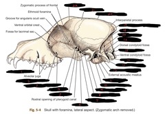
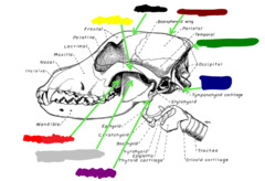
External occipital protuberance
What is hilighted in green?
Hypertrophy, atrophy, pain, swelling
Why would you palpate the muscles of mastication in a dog? Or even the neck muscles?
Trauma or sialoliths (concretions in salivary gland)
One part of the orofacial exam is to palpate the salivary glands. Why would you do this?
Abscess in retrobulbar area (esp. cats), orbital cellulitis from periodontitis or endodontic disease (bacterial infection of soft tissue and fat surrounding eye socket), oral foreign body, extension of oral neoplasia
One part of the orofacial exam is to retropulse the globes and assess their position. Why would you do this? (note it is good if the eyes retropulse when you push on them)
Look for erythema, alopecia, or masses
One part of the orofacial exam is to evaluate the skin of the head. Why would you do this?
Sinusitis, nasal foreign body, neoplasia, nasal fungal infection, oronasal fistula
One part of the orofacial exam is to look for nasal discharge. Why would they have nasal discharge?
Dislocations, fractures, nerve damage, allergies, inflammatory disorders, neoplasia
One part of the orofacial exam is to look for facial symmetry. What may cause abnormal facial symmetry?
Genetics, fractures, TMJ luxation
One part of the orofacial exam is to look for malocclusion. What might cause this?
Dental related or primary skin disease: intertrigo or skin fold dermatitis
One part of the orofacial exam is to look for skin fold lesions near the lips. What may cause these?
Hypothyroidism (bc of slower metabolism and lymphatic drainage), allergies
One part of the orofacial exam is looking for puffiness of eyelids, lips, jaws, or tongue. What might cause this?
Look for "rubber jaw". Must rule out hyperparathyroidism aka fibrous osteodystrophy
One part of the orofacial exam is to evaluate the bones of the maxilla. Why would you do this?
Click/pop: TMJ disorder, malocclusion
Yelp upon yawn: TMJ disorder, stomatitis, periodontal disease, oropharyngeal problem
Honking/reverse sneeze/stridor/stertor: upper resp. issue, oronasal fistulae, tumor
One part of the orofacial exam is to listen for abnormal noises. Name some and why they might occur
1. lips and oral mucosa evaluation
2. dental evaluation (teeth)
3. periodontal exam
4. oral cavity evaluation
What are the 4 stages of an intraoral exam?
1. depigmentation/hyperpigmentation
2. erythema/excoriation
3. puffiness (hypothyroid, allergies)
4. scaling (vit. B deficiency, fungal infection)
5. malodor
6. lip avulsion/trauma
Name the things you evaluate when you are doing a lip intraoral exam
1. lack of moisture
2. color change
3. stomatitis
4. fistulaes
5. ulcers (CUPS, chronic ulcerative paradental stomatitis)
6. lacerations
7. foreign bodies
Name the things you evaluate when you are doing an oral mucosa intraoral exam
1. color variation
2. enlargement (hyperplasia)
3. bulbous appearance
4. hyperemia
5. recession
6. fistulation
7. gingival sulcal exudates
Name the things you evaluate when doing an attached gingiva intraoral exam
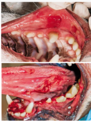
Chronic alveolar osteitis = bulbous appearance
What is this?
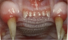
CUPS: chronic ulcerative paradental stomatitis
What is this?
1. tight lip attachment
2. debris collection
3. discoloration
Name the things you evaluate when doing a frenula intraoral exam
1. fractured teeth
2. note permanent/deciduous/mixed dentition
3. missing teeth
4. surface abnormalities
5. tooth staining
6. loss of tooth substance: enamel, caries
7. enamel infractions
Name the things you evaluate when you are doing a dental (teeth) intraoral exam
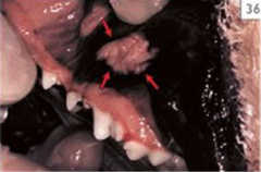
Cheek chewer lesion
What is this?
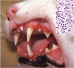
Stomatitis in a cat (inflammation of the mucous membranes in the mouth)
What is this?
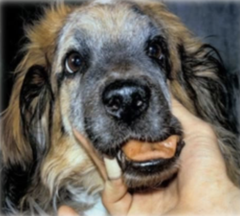
Rubberjaw (hyperparathyroidism)
What is this?
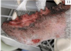
Fistulous tracts from endodontic compromise of mandibular canine teeth
What is this?
Slab fracture of #208
What is this?
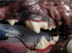
Uncomplicated crown fracture
In this type of dental fracture, there is no pulp exposure
Complicated crown fracture
In this type of dental fracture, there is pulp exposure
Uncomplicated crown root fracture
In this type of dental fracture there is involvement of the crown and root, but no pulp exposure
Complicated crown root fracture
In this type of dental fracture there is involvement of the crown and root, and pulp exposure
Root fracture
In this type of dental fracture, there is fracture involving the root only
Operculum (tough fibrous covering) covering mandibular premolars and molars
What is happening here?
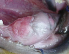
Tetracycline staining
What is happening here?
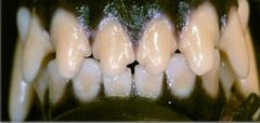
Pulp necrosis
Indicates endodontic compromise and hemoglobin breakdown
What is happening here? Why do we care about this?
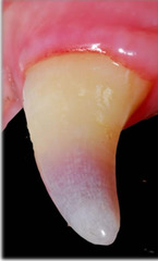
EXTRINSIC staining: plaque, metal, chlorhex, fluoride
INTRINSIC staining: enamel opacity, tetracycline, dental caries, pulp necrosis, tooth wear from abrasion, fluorosis
When you do an intraoral exam and look at the teeth, you look for staining. What kinds of staining are there and name some examples
Enamel attrition with stained tertiary dentin
What is happening here?
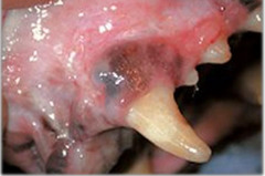
Dullness from endodontic compromise
What is happening here?
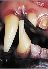
Attrition (wearing away of tooth enamel due to tooth on tooth contact)
What is happening here?
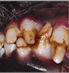
Enamel hypoplasia (enamel is underdeveloped = poor/thin enamel layer)
What is happening here?
Complicated crown fracture
What is happening here?
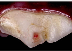
Enamel loss thru enamel hypocalcification/hypoplasia, resorption, or gradual wear, fracture, or caries
Name some things that could cause loss of tooth substance
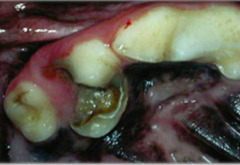
Caries
This may be difficult to discern from tertiary dentin but overall it causes loss of tooth substance because they are areas of damage on the tooth caused by acids produced by bacteria in the mouth (aka tooth decay or cavities)
Dental caries
What is this?
Enamel infraction
What is this?
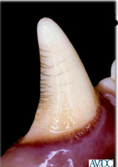
Enamel infraction
This is incomplete fracture (crack) of the enamel without loss of tooth substance
PD0: normal
PD1: gingivitis without attachment loss
PD2: less than 25% attachment loss, maybe stage 1 furcation
PD3: 25-50% attachment loss, stage 2 furcation
PD4: more than 50% attachment loss, stage 3 furcation
How many stages are there of periodontal disease? Explain them
1. overall tongue size
2. lingual molar salivary glands NORMAL in cats
3. tongue mobility, flaccidity, papilla, depressions
4. under tongue for foreign bodies
Name the things you evaluate when you are doing a oral cavity intraoral exam
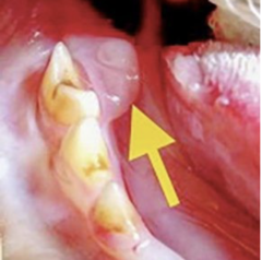
NORMAL = lingual molar salivary gland
What is happening in this cat?
EPIGLOTTIS, make sure you look at it during intubation
What MAIN structure do you want to examine particularly close when you do an oral cavity exam of an animal under anesthesia? (even tho you still examine the structures of the oral cavity as in an awake cavity)
1. periodontal disease
2. foreign body in mouth
3. tumor induced necrosis
4. ketoacidosis
5. uremia
A patient has halitosis (bad breath). Give some differentials?
Maxillary arch: sternal recumbency
Mandibular arch: dorsal recumbency
Which position should you radiograph the maxillary arch of an anesthetized patient? Mandibular arch?
Supragingival scaling (removing plaque and tarter from portion of tooth above gum line) and subgingival calculus removal then probe the sulci in 8 locations after calculi removal. Probe furcations as well. After probing assess gingival index
After you take radiographs and the patient is anesthetized, what can you do for the periodontal exam?
FALSE: when soft tissue inflammation is present in the ABSENCE of inciting (stirring up) plaque and calculi this can indicate tooth resorption, immune mediated disease, tooth resorption, or neoplasia
TRUE OR FALSE: Soft tissue inflammation when stirring up plaque and calculi can indicate tooth resorption, immune mediated disease, tooth retention or neoplasia
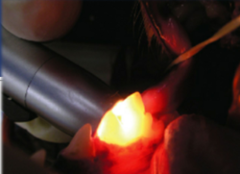
NORMAL = vital tooth. This is transillumination
Is this normal dental light transmission? Or abnormal? What is this process called?
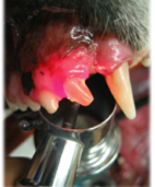
ABNORMAL = nonvital tooth. This is transillumination
Is this normal dental light transmission? Or abnormal? What is this process called?
Treatments = red
Pathology = blue
On a dental chart, what color are treatments in? How about pathologic terms?