CMI 354 Test #1
 Call Kai
 Learn
Learn Practice Test
 Spaced Repetition
 Match
Match Flashcards
 Knowt Play
1/103

There's no tags or description
Looks like no tags are added yet.
Name | Mastery | Learn | Test | Matching | Spaced | Call with Kai | Chat |
|---|
No analytics yet
Send a link to your students to track their progress
104 Terms
Ascending aorta
1

Arch of aorta
2

Descending aorta
3

Left coronary artery
4

Right coronary artery
5

Brachiocephalic artery
6

Right subclavian artery
7

Right common carotid artery
8

Left common carotid artery
9

Left subclavian artery
10

Inferior vena cava
1

Right kidney
2

Right renal artery
3

Right renal vein
4

Abdominal aorta
5

Left renal vein
6

Left renal artery
7

Superior mesenteric artery
9

Left kidney
10

Femoral vein
1
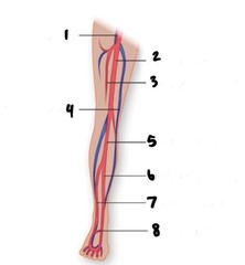
Femoral artery
2

Deep femoral artery
3

Great saphenous vein
4

Popliteal artery
5

Posterior tibial artery
6

Anterior tibial artery
7

Arcuate artery
8

2 cm
Normal aorta diameter
Aneurysm
Vessel dilation >3 cm
Surgical aneurysm
Vessel dilation >6cm
Aneurysmal
Increased diameter greater than 50% qualifies as
infrarenal aorta
What's the most common place of an aneurysm in the abdomen?
Spectral broadening is caused by turbulence in blood flow as the normally homogeneous velocity of reflective red blood cells becomes more diverse, resulting in the apparent broadening of the spectral Doppler waveform
Doppler signal: what is spectral broadening?
triphasic: consists of forward flow in systolic peak, reversal forward flow in late disatole
biphasic: delayed acceleration, decreased systolic peak, increased flow in diastole
monophasic: delayed acceleration, decreased systolic peak, increase of flow in diastole
Doppler signal, what is the flow type: triphasic, biphasic, monophasic

behind the knee
Where is the popliteal artery?

gastrocnemius muscle, medial head
What muscle is by the popliteal artery
Subclavian artery, axillary, brachial (front), deep brachial (back), radial (thumb), ulnar (pinky)
Know the arteries from upper abdomen to the fingers

they are caused by impaired blood flow
- pain
- pallor (pale skin tone)
- paresthesia (numbness feeling)
- pulselessness (faint pulse)
- paralysis (weakness
with movements)
Know the 5 P's for an acute arterial occlusion
- evaluates patency of the wrist arteries and source of arterial supply to the fingers
-Is useful in determining hand viability if the radial artery is to be removed for use as a coronary artery bypass graft, or for various procedures such as dialysis or angiography
- used to assess the hand viability if there is an obstruction of the radial artery
What does the Allen's test for ulnar atery, L test do
-sonographer palpates radial artery at wrist and applies manual compression
-patient balls hand into a fist for one minute and then relaxes
-skin will turn pale when hand clenched and color will return when relaxed
How does a sonographer do an Allen test?
ankle/brachial (ABI) is calculated by dividing the ankle pressure (systolic) by the higher of the two brachial pressures (systolic)
Calculate an Ankle/Brachial Index & what's the equation

Doppler waveform analysis
What test is used to detect the presence and severity of arterial occlusive disease?
a reddening or dark discoloration that occurs when a limb has poor arterial flow
Rubor
bluish discoloration of the skin
Cyanosis
abnormal sensation of numbness and tingling
paresthesia
Condition that exists when symptoms of intermittent ischemia of the fingers or toes occur in response to cold exposure as well as emotional stress
Raynaud's phemomenon
Tibial area, toes, bony prominences
Where do arterial ulcers typically occur?
Severe
What is the level of pain associated with arterial ulceration?
Deep regular shape
What is the appearance of arterial ulcers?
Little
How much bleeding is typically associated with arterial ulcers?
Shiny skin, loss of hair, thickened toenails
What are some trophic changes associated with arterial ulcers?
Near medial and lateral malleolus
Where are venous ulcers typically located?
Mild to severe
What is the pain level associated with venous ulceration?
Shallow, irregular shape
What is the appearance of a venous ulcer?
Venous ooze
What type of bleeding is associated with venous ulcers?
Stasis changes: brawny discoloration, lipodermatosclerosis, varicosities often present
What are some other findings associated with venous ulcers?
-hemodynamically significant stenosis
-a critical narrowing of an artery (stenosis) that results in a major reduction in maximal flow capacity (flow volume and pressure) in a distal vascular bed.
-cross sectional area of arterial lumen is reduced 75% (diameter reduction of 50%)
What constitutes a critical stenosis?
FLOW VOLLUME INCREASES AS PRESSURE INCREASES, BUT ONLY TO A POINT.
► AS FLOW CHANGES FROM STABLE TO DISTURBED, INCREASE IN PRESSURE DOES NOT INCREASE FLOW VOLUME BUT INCREASES FLOW DISTURBANCE CAUSING THE FORMATION OF EDDY CURRENTS.
► RENOLDS NUMBER IS A DIMENSIONLESS NUMBER THAT IS EXPRESSED FOR TURBULANCE.
► TURBULANT FLOW CAUSES VESSELS TO VIBRATE. THIS VIBRATION PRODUCES VASCULAR BRUITS.
Reynold's Number

RELATIONSHIP BETWEEN VOLUME FLOW (Q), PRESSURE (P), AND RESISTANCE (R)
► MAY BE WRITTEN Q = (p1-p2)pie r 4/8 n L
► CHANGE IN THE DIAMETER AFFECTS RESISTANCE
MORE THAN EITHER VISCOSITY OR VESSEL LENGTH
► RADIUS OF A VESSEL IS DIRECTLY PROPORTIONAL TO THE VOLUME OF FLOW
Poiseuille's Law

VELOCITY AND PRESSURE ARE INVERSELY RELATED.
Bernoulli's Principle

-often called a mini-stroke. TIA is a temporary blockage of blood flow to the brain
-most TIA symptoms last from only a few minutes up to 24 hour
Tia(transient ischemic attack)

Is gradual worsening of symptoms of brain ischemia
Stroke in Evolution (SIE)
brief, transient interruption in blood flow to the brain lasting longer than 24 hours but less than a week
RIND reversible ischemic neurologic deficit
completed stroke is usually employed to signify a focal neurological disability that came on abruptly and has become stabilized
Complete Stroke
-coma, absence of brain stem reflexes, and apnea
-the irreversible loss of all functions of the brain, including the brainstem
What constitutes acute brain death
LAMINAR FLOW-->
-PARABOLIC FLOW
-Plug flow
Turbulent flow - a chaotic form of fluid transport in which velocity components randomly fluctuate.
Types of blood flow

resembles the shape of a bullet
velocity highest at lumen
Parabolic flow
rounded; all blood cells and layers travel at the same velocity
Plug flow
Ophthalmic artery
What branch of the internal carotid has clinical significance to us
True: Saccular, Fusiform
Dissecting
Pseudoaneurysm
Types of aneurysms

True - dilation of all three layers of arterial wall
Pseudo - pulsating hematoma; hole in arterial wall allows blood to escape under pressure into a contained area
True VS Pseudo - Aneurysm
Not all layers dilated; high rupture risk.
false aneurysm
determining the changes in volume of an organ part or body
Plethysmography

Caused by swelling within osteofascial compartments of the leg, arm, or abdomen
-results from ischemic injury to capillary bed within subsequent increased fluid leakage out of the circulating volume
-clinical findings include paresthesia, pain, weakness of involved muscle, and tension of the compartment
Compartment syndrome
caused by compression of the popliteal artery by the medial head of the gastrocnemius muscle or fibrous bands
-most commonly found in young athletes
-appears bilaterally in 2/3 cases
Popliteal artery entrapment syndrome
5 P's: pain, pallor, pulselessness, paresthesia, paralysis, polar (cold foot), purplish (cyanosis)
-occlusion may result from thrombus, embolism, or trauma
-consider an emergency
acute arterial occlusion
Claudication
3 areas: buttock, thigh, and calf
chronic arterial occlusion
Aortoiliac disease
What does buttock claudication suggest?
Iliofemoral disease
What does unilateral buttock claudication suggest?
Distal external iliac/common femoral disease
What does thigh claudication suggest?
Femoral/popliteal disease
What does calf claudication suggest?
pain in muscles during exercise but subsides with rest; results from inadequate blood supply to exercising muscle
Claudication
casts/bandages that can't be removed
waveforms may be affected by ambient temperature
congestive heart failure may cause dampened waveforms
continuous wave transducer is unable to differentiate occlusion from stenosis or locate it
sonographer dependent
Limitations of arterial Doppler: upper and lower extremities
can't tell the difference between stenosis and occlusion
can only identify general location of obstruction
can show falsely elevated doppler pressures in patients with calcified vessels
ABI's may appear lower in patients with congestive heart failure
ABI's may appear higher thigh pressures if thigh is too narrow
unable to perform tests on patients with certain conditions (DVT, graft, stent, e.g.)
Limitations of arterial Doppler: segmental pressures
skin must be intact for probe placement
ulcerated skin
patient must rest quietly
can only measure superficial layers of tissue
lack of availability and standardized protocols
Limitations of arterial Doppler: laser Doppler
vasoconstriction
cold
smoking
nervous
ulcerations
gangrene
extensive bandages
dry skin
Limitations of arterial Doppler: digital pressures and plethysmography
presence of dressings/staples/sutures/open wounds
IV site
cold room
obesity
anastomosis sites
Limitations of arterial Doppler: duplex scanning of upper extremities
presence of dressings/staples/sutures/open wounds
incisional tenderness/hematomas
calf vessels of obese patients are difficult to image
calcified arteries can cause artifactual shadowing
Limitations of arterial Doppler: duplex scanning and color flow imaging of the lower extremities
size of the patient, bowel gas, previous abdominal surgery, shortness of breath/rapid respiration, not fasting
Limitations of arterial Doppler: abdominal
body habitus (obesity), lack of fasting, patient can't hold breath, recent surgery and ascites
Limitations of arterial Doppler: renal Doppler
depth, scarring, effects of radiation treatment
Limitations of arterial Doppler: preoperative mapping procedures
-A difference greater than 30 mmHg between 2 consecutive levels is considered significant and would suggest obstruction
-Horizontal difference of 20-30 mmHg or more suggests obstructive disease at or above the level in the leg of lower pressure
Segmental pressures
Externa - external
Media - middle
Intima - inner most
layers of artery wall

first major branch is the ophthalmic artery
Know ICA branches

first major branch is the superior thyroid artery
Know ECA branches

-feeds the stomach, liver, pancreas, duodenum, and spleen
-branches into left gastric, splenic, and common hepatic arteries
Know the celiac trunk and its major branches

-first branch of aortic arch
-arises on right
-divides into right common carotid artery and subclavian arteries
brachiocephalic artery
-second branch of the aortic arch
-terminates at carotid bifurcation
-splits into ICA and ECA
left common carotid artery
-third branch of aortic arch
-terminates at thoracic outlet
left subclavian artery
A condition characterized by abnormal growth of cells in the arterial wall.
What is fibromuscular dysplasia?