Pigment
 Call Kai
 Learn
Learn Practice Test
 Spaced Repetition
 Match
Match Flashcards
 Knowt Play
1/75

There's no tags or description
Looks like no tags are added yet.
Name | Mastery | Learn | Test | Matching | Spaced | Call with Kai | Chat |
|---|
No analytics yet
Send a link to your students to track their progress
76 Terms
What are the 3 exogenous pigment substances?
carbon & dusts
Carotenoid pigments
Tetracycline
What are the 3 endogenous pigments?
melanin
Lipofuscin and Ceroid
Hemtaogenous pigments
Pneumoconiosis
lung disease resulting from inhalation of dusts
ANthracosis
lung disease resulting from inhaling carbon particles
Silicosis
lung disease associated with inhaling silicone particles
True or False: Pneumoconiosis is not associated with clinical disease.
TRUE
What is the gross appearance of carbon and dusts?
black stipping in the lungs and tracheobronchial lymph nodes
What is the miscroscopic appearance of carbon and dust?
fine black granular material and crystalline material in macrophages adjacent to airways and vessel
WHat organs are carbon and dust deposits found?
lungs
Tracheobronchial lymph node

ID Pigment
Carbon/Dust in airway
What are carotenoid pigments abundant in?
leafy green plants
What does carotenoid pigments need to be differentiated from?
icterus
True or False: Carotenoid pigments remain after histological processing.
FALSE
What is the gross appearance of carotenoid pigments?
yellow color in plasma, adipose tissue and other lipid-laden cells
What specific species and breeds of animals can have pronounced carotenoid pigments?
Horses and cattle grazing lush
Especially Jersey cows
Pigs and chickens with confined animal diets
True or False: Carotenoid pigment is only in lipid rich tissue
TRUE
Where does tetracycline bind?
calcium and phosphate in teeth and bones
When would tetracycline cause permanent discoloration?
if its administered when the teeth mineralize
How can bone appear that is stained with tetracycline?
yellow and flurorese under UV light
Melanin
pigment responsible for color of hair, skin and iris this is performed by melanocytes
Where are melanocytes derived from?
neural crest
What is the function of melanin?
shield the nucleus from UV light
Melanosis
localized deposits of melanin
Where can melanin deposit?
leptomeninges
Oral mucosa
Aortica intima
Lungs
What is the microscopic appearance of melanin?
small, brown/black and non refractile
WHat is the gross appearance of melanin?
brown to black coloration

ID pigment
melanin
What diseases could cause diminished melanin?
copper deficiency → fading coat color
Genetic disorders
Immune mediated disease
What is the first step in melanin synthesis?
conversion of tyrosine to DOPA via tyrosinase which uses copper
What diseases could cause hyperpigmentation?
chronic injury
Endocrine diseases
Cancer
Lipofuscin
yellow-brown lipoprotein that accumulates as residual bodies in secondary lysosomes
Where is Lipofuscin typically found?
long lived cells
Cardiac myocytes and neurons
True or False: Lipofuscin has little to no effect on the cell
TRUE
Ceroid
lipofuscin-like pigment that accumulates in disease states
Gove 4 examples of when ceroid would accumulate.
lysosomal storage diseases
Cachexia
Vitamin E deficiency
Oxidative stress

What is this?
Intestinal ceroidosis
How can you differentiate Lipofuscin and Ceroid?
Ultrastructural appearance and history
Lipofuscin: granular appearance; old age
Ceroid: forms whorls; disease state
What are hematogenous pigments derived from?
erthrocytes
What is hemoglobin made up of?
four globular protien subunits with central iron-containing heme group
What does cyanide do?
blocks oxidative phosphorylation
Cells cannot use O2
What is the appearance of cyanide toxicity in blood?
venous blood is as red as arterial blood
What is the appearance of carbon monoxide poisoning?
bright cherry red blood
What does nitrate do to hemoglobin?
oxidizes iron in heme
What appearance does nitrate poisoning have?
brown blood
What effects on pigment of the plasma and kidney does intravascular hemolysis cause?
Plasma: pink
Kidney: dark red to gunmetal blue with hemoglobin casts

ID pigment/structure
hemoglobin casts
Hematin
brown-black iron containing pigment formed by the oxidation of hemoglobin
When does acid hematin form?
forms in tissues fixed in unbuffered acid formalin (formalin pigment)
WHat is the appearance of acid hematin?
dark brown to black granular or crystalline material in vessels or areas of hemorrhage
True or False: Acid Hematin is clinically significant.
FALSE - postmortem change
What parasites cause parasitic hematin?
Liver flukes (fascioloides magna)
Lung mites (pnumonyssus simicola)
Where is hematin found when associated with parasites?
Around the infection or in the migrating tracts
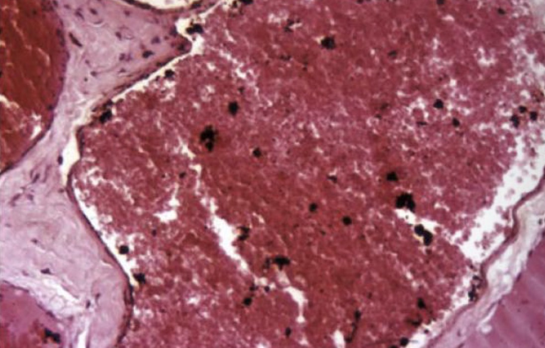
ID pigment
Acid hematin
Hemosiderin
Iron storage complex of partially digested ferritin and lysosomes
What is the appearance microscopically of hemosiderin?
Golden brown intracellular granules in macrophages
can also be in hepatocytes and ends tubular epithelial cells
What stain can make hemosiderin more visible?
prussian blue stain
Prussian blue stain reacts with the ____ in hemosiderin
iron
When can hemosiderosis be seen?
splenic red pulp with increased RBC destruction
Chronic passive congestion in lungs
Hemorrhage response (bruises)
Diet
Iron overload

ID pigment
hemosiderin
Siderotic plaques are considered ______
incidental

ID
Siderotic plaques
Hematoidin
bright yellow crystalline pigment derived from hemosiderin that is deposited in tissues at sites of hemorrhage

ID yellow pigment
Hematoidin
Describe the breakdown of iron to bilirubin.
Iron is removed from heme
Heme is converted by heme oxygenase to biliverdin
Biliverdin converted by biliverdin reductase to bilirubin
How does the liver conjugate bilirubin?
with glucuronic acid
Hyperbilirubinemia will stain tissues _____
yellow
Prehepatic Icterus
hemolysis or increases RBC turnover
Hepatic Icterus
hepatocellular injury that decreases uptake, conjugation, or secretion of bilirubin
Posthepatic Icterus
outflow of bile from liver is reduced or obstructed
Where is icterus easiest to see?
plasma
Sclera
Non-haired skin
Intima of great vessels
fat
Liver
True or False: You cannot see bilirubin histologically.
TRUE
What can you see with cholestasis in the liver?
orange to brown bile casts with bile caniculi
Porphyria
heme synthesis disorder that results in deposition of porphyrin pigments in tissue
What causes porphyria deposition?
deificnecy of uroporphyrinogen III synthase
What animals get congenital erythropoietin porphyria?
calces, cats, and pigs
What is the gross appearance of porphyria?
teeth, bone, and urine are red-brown and fluroresce red under UV light