ECN 5: Executive Control: Prefrontal Cortex
0.0(0)
Card Sorting
1/17
There's no tags or description
Looks like no tags are added yet.
Last updated 5:26 PM on 7/28/23
Name | Mastery | Learn | Test | Matching | Spaced | Call with Kai |
|---|
No analytics yet
Send a link to your students to track their progress
18 Terms
1
New cards
Name five subcomponents (sub-functions) of executive functions/cognitive control functions
\
\- working memory (manipulation of information) \n - error monitoring (factoring errors, rewards, and consequences into future behavior) - response inhibition \n - categorization \n - rules (determining the logic of a task, association of stimuli) \n - decision making \n - attention \n - planning
\- working memory (manipulation of information) \n - error monitoring (factoring errors, rewards, and consequences into future behavior) - response inhibition \n - categorization \n - rules (determining the logic of a task, association of stimuli) \n - decision making \n - attention \n - planning
2
New cards
What is executive control?
All higher-level functions that help us act in an instant due to environmental cues (above)
3
New cards
What is similar and what is special about cognitive control functions as opposed to learning
Executive control and learning both change behavioral responses and the information of representation, guiding us to behave in a changing environment. However, for learning to manipulate behavior it has to be learned based on experience, which is usually time consuming.
* Executive control can exert direct top- down influences on behavior without repeated exposure to the stimulus.
4
New cards
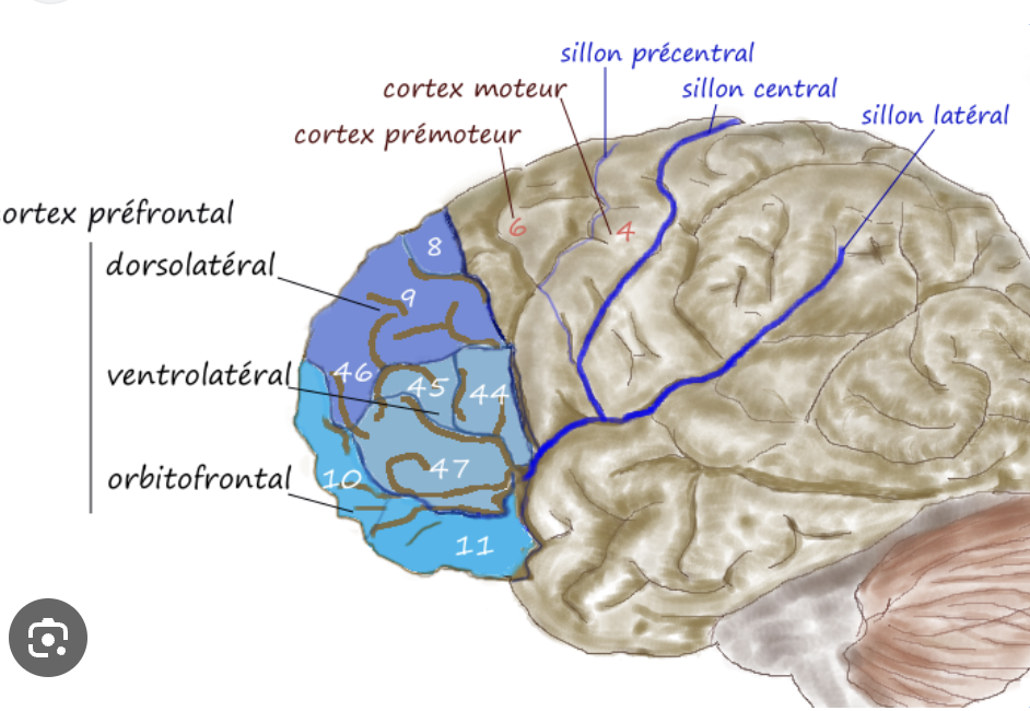
Define the prefrontal cortex anatomically
The PFC is a region of the frontal cortex (the most anterior part of the brain, anterior to central sulcus) that comprises several Brodmann areas and receives reciprocal projections from the thalamic medio-dorsal nucleus. It can be divided into medial PFC, dorso/ventrolateral PFC, and orbital PFC.
5
New cards
What is meant with saying the PFC “operates at the apex of the cortical hierarchy”?
The PFC is the latest stage of the cortical hierarchy and therefore integrates the information from previous areas (e.g., visual information) to then send projections to the premotor areas to elicit a behavioral response. It is therefore the last stage of sensory processing before a behavioral response is initiated.
6
New cards
*What are the three main functional territories of the fontal lobe?*
medial PFC, dorso/ventrolateral PFC, and orbital PFC
7
New cards
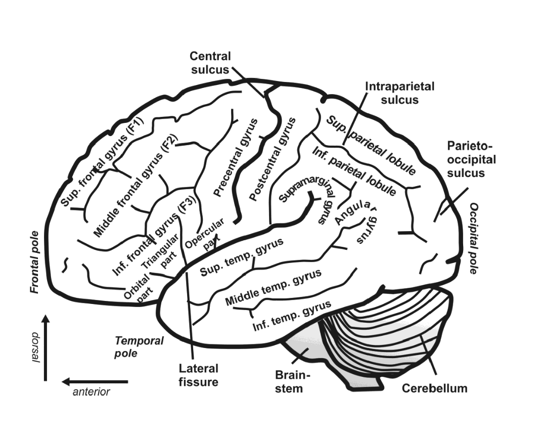
What are the names of the three frontal gyri, and where are they located on a lateral brain view?
*Superior frontal gryus (F1), middle frontal gyurs (F2), inferior frontal gyrus (F3)*
8
New cards
What is the thalamic nucleus that has intensive connections to the PFC?
From the thalamic nuclei, the mediodorsal nucleus has the most intensive connections to the PFC (\~80% of the connections).
9
New cards
How is the PFC connected to other (parietal & temporal) association cortical areas
\
\
The PFC is connected to the other association cortical areas through major bundles (fasciculi) of fiber projections. It has both input fibers from the parietal and temporal areas (for information on the internal milieu and cognition), as well as output fibers to these areas (goal-directed sequences, structures, programs).
10
New cards
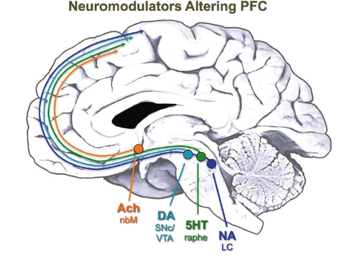
What are the four classical neuromodulatory systems of the brain? In which nuclei (and where) do these neuromodulator systems emerge?
1. Noradrenaline (NA) = Locus coeruleus (LC)
2. Dopamine (DA) = Substantia niga pars compacta (SNC), VTA, dorsol raphe
3. Serotonin (5HT) = dorsal and median raphe
4. Acetlycholine (Ach) = nucleus basalis of Meynert (nbM) in basal forebrain
11
New cards
Which cortical areas mature first, which ones last? What is the time course of myelination in humans? What does this tell about these cortical areas?
***Maturation means that they have their myelin sheets in place and function.***
1. primary and sensory motor cortices mature earliest (1-2 yo)
2. Parietal-temporal association cortices mature 2nd
3. PFC matures last (15yo +)
There is a structural gradient from the primary to the association cortices in the maturation of the brain. The primary sensory areas mature and are fully functioning first, the association cortices mature last.
12
New cards
What is the time course of myelination in humans? What does this tell about these cortical areas?
The myelination of sensory and motor areas is finished within the first year, whereas the association cortices mature over years with the PFC being the last to fully myelinate (\~ 16y).
It is sensible that the sensory areas mature first as their input is needed for the association cortices to mature. Also, this tells us that for cortices to mature, they are dependent on experiences. Sensory experiences are gathered early on while other experiences are gathered over longer periods of time.
It is sensible that the sensory areas mature first as their input is needed for the association cortices to mature. Also, this tells us that for cortices to mature, they are dependent on experiences. Sensory experiences are gathered early on while other experiences are gathered over longer periods of time.
13
New cards
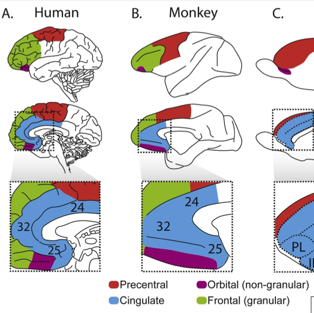
What is special about the PFC of primates (compared to rodents)?
The primate PFC is special in that it is granular, i.e., it has a clearly visible layer 4. This granular layer has only evolved in primates and is missing in other mammalian species such as rats.
BA 10-located at most anterior part of PFC (in humans) is very difficult to access in monkeys (because much smaller)
14
New cards
Compare human and NH primate visual and PFCs
Between diff primate species (including humans) there isn’t a significant difference in spine # in V1, V2(mild difference)
however, granular PFC shows a significant different in terms of the number of spines in prefrontal pyramidal cells of humans (sig. more) than in NH primates **indicating** the power of prefrontal cells.
however, granular PFC shows a significant different in terms of the number of spines in prefrontal pyramidal cells of humans (sig. more) than in NH primates **indicating** the power of prefrontal cells.
15
New cards
Which cortical areas/connections grew relatively bigger in the hominini lineage compared to monkeys/apes?
Specifically in primates, the dlPFC has recently evolved and is missing in other mammals. The dlPFC progressively expanded from prosimian, to simian primates and reached its largest extension in humans. In general, the PFC expanded disproportionally in Homininis compared to other primate species.
16
New cards
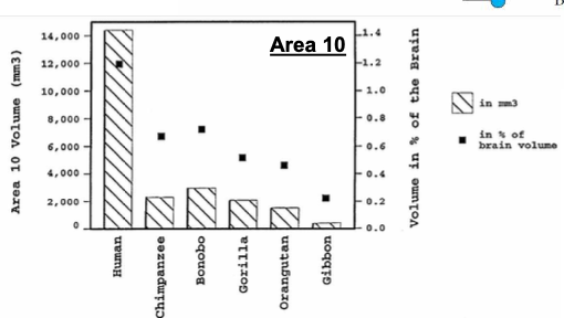
Which PFC area grew disproportionally large in humans compared to other primates?
\
\
In humans, area 10 (frontal pole) grew disproportionally large. This area is intensely connected with other neurons and has more excitatory synaptic connections (more connectivity = more processing power). This area is involved in various executive functions.
17
New cards
Name ten cognitive capabilities that may explain the expansion of the human PFC (relative to other primates).** \n **
* *theory of mind*
* *spoken language*
* *social cooperation*
* *tool use*
* *metacognition*
* *planning and prospection*
* *social and moral rules*
* *analogical reasoning*
* *imagination or simulation*
* *semantic associations*
\
* *spoken language*
* *social cooperation*
* *tool use*
* *metacognition*
* *planning and prospection*
* *social and moral rules*
* *analogical reasoning*
* *imagination or simulation*
* *semantic associations*
\
18
New cards
What is the name of the PFC equivalent in the avian brain, and what is the evidence that this structure is the “avian PFC”? How did this avian structure evolve, and from which telencephalic territories?
In the avian brain, the NCL (= neopallium caudolaterale) is the PFC equivalent. This area receives similar inputs as the PFC (dopamine, secondary sensory input areas, etc.). It is an analogous structure as it has evolved from a different part of the pallium (in mammals, the dorsal pallium gives rise to the neocortex, in birds the NCL evolved from the ventral pallium).