Intro to Patient Diagnostics
1/47
There's no tags or description
Looks like no tags are added yet.
Name | Mastery | Learn | Test | Matching | Spaced |
|---|
No study sessions yet.
48 Terms
Wilhelm Conrad Roentgen
discovered x-rays in 1895
first used dignostically in 1896
Attenuation
the reduction in the intensity of the X-ray beam as it passes through matter, caused by absorption or scattering of photons
more dense structures attenuate __________ meaning _______
less dense structures attenuate ___________ meaning ________
more meaning less blackening of the film
less meaning more blackening of the film
X-Ray (XR)
form of electromagnetic radiation or energy of extremely short wavelength
shorter wavelength = more penetration (energy) through tissue providing a better image
Radiolucent vs Radiopaque
Radiolucent
less dense objects
appears black on x-ray
ex: air in the lungs
Radiopaque
more dense
appears white on x-ray
ex: bones
Radiographic Densities (least to most)
gas (air), fat, soft tissue (water), bone (metal)
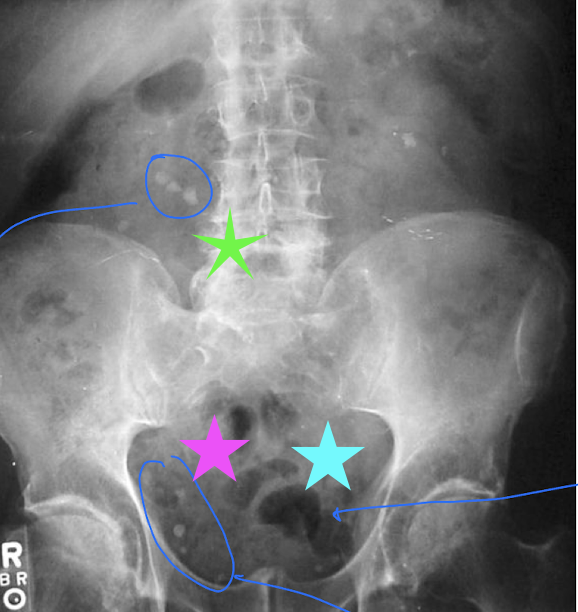
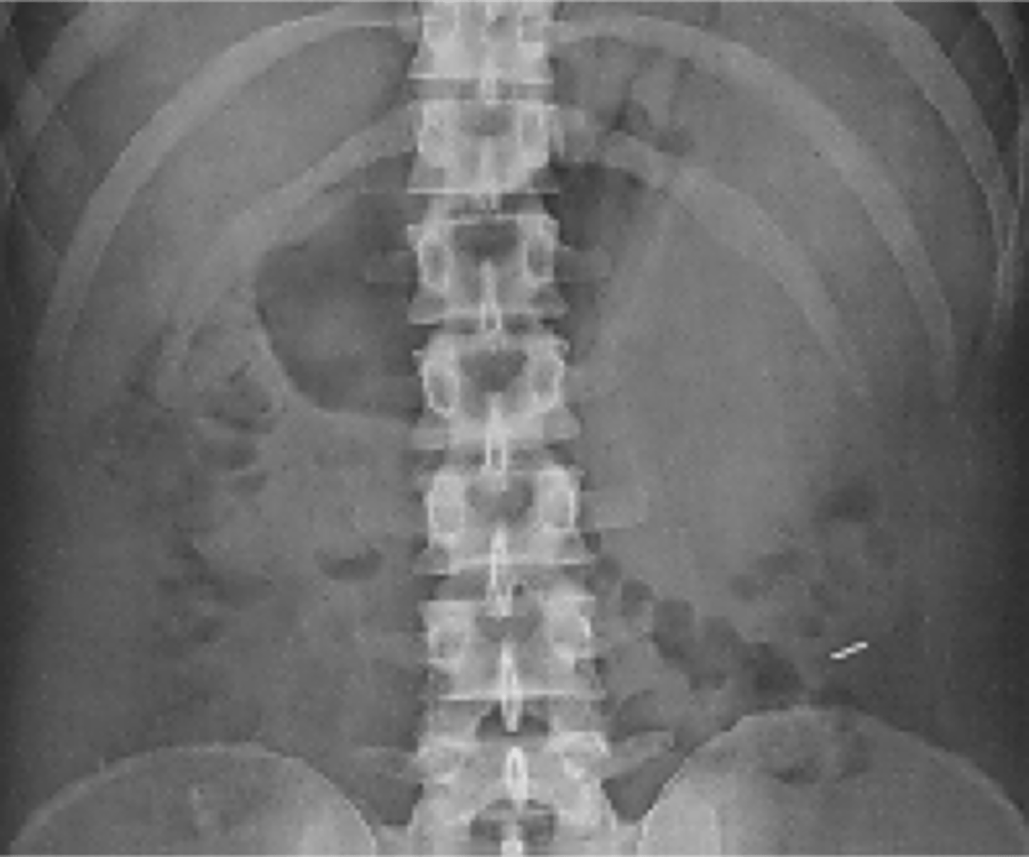
Identify what each of the colored stars indicate:
Green
Pink
Blue
Green: kidney stones - appear radiopaque because Ca+ presents that way
Pink: phleoboliths - Ca+ appears radiopaque
Blue: air bubbles

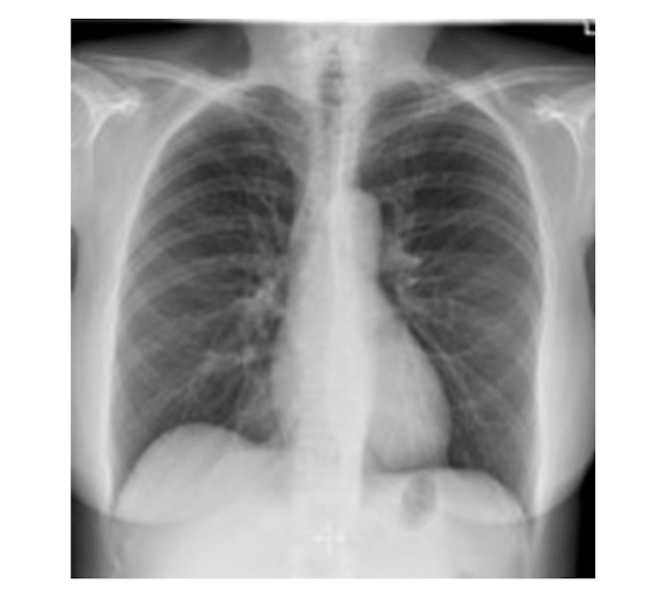
Identify the radiolucent and radiopaque figures in the X-ray image
Radiolucent (less dense, translucent)
air in the lungs
Radiopaque (more dense)
clavicle, heart, diaphragm, blood vessels (from density of blood)
What factors affect Image Quality in x-rays
thickness of part being examined - obese pt
motion
shorten exposure time to overcome
scatter
causes fog as the beam scatters
magnification
have the part of interest closest to the film
AP: anterior posterior where anterior side is to the film
PA: posterior anterior where the posterior side is to the film
distortion - object is not perfectly perpendicular to the beam
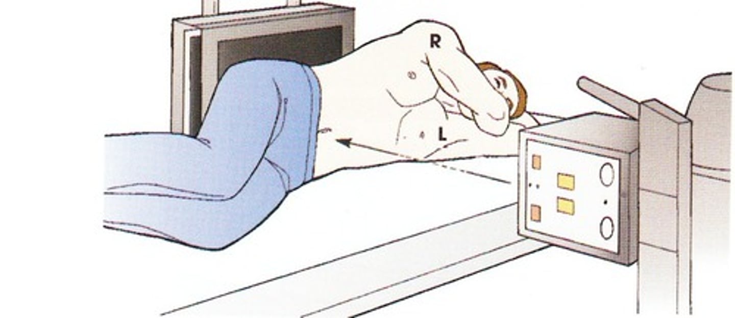
AP Film
Anterior-posterior
-where the film is placed behind the pt's back with the x-ray unit in front
-typically captured with portable x-ray when pt is incapacitated
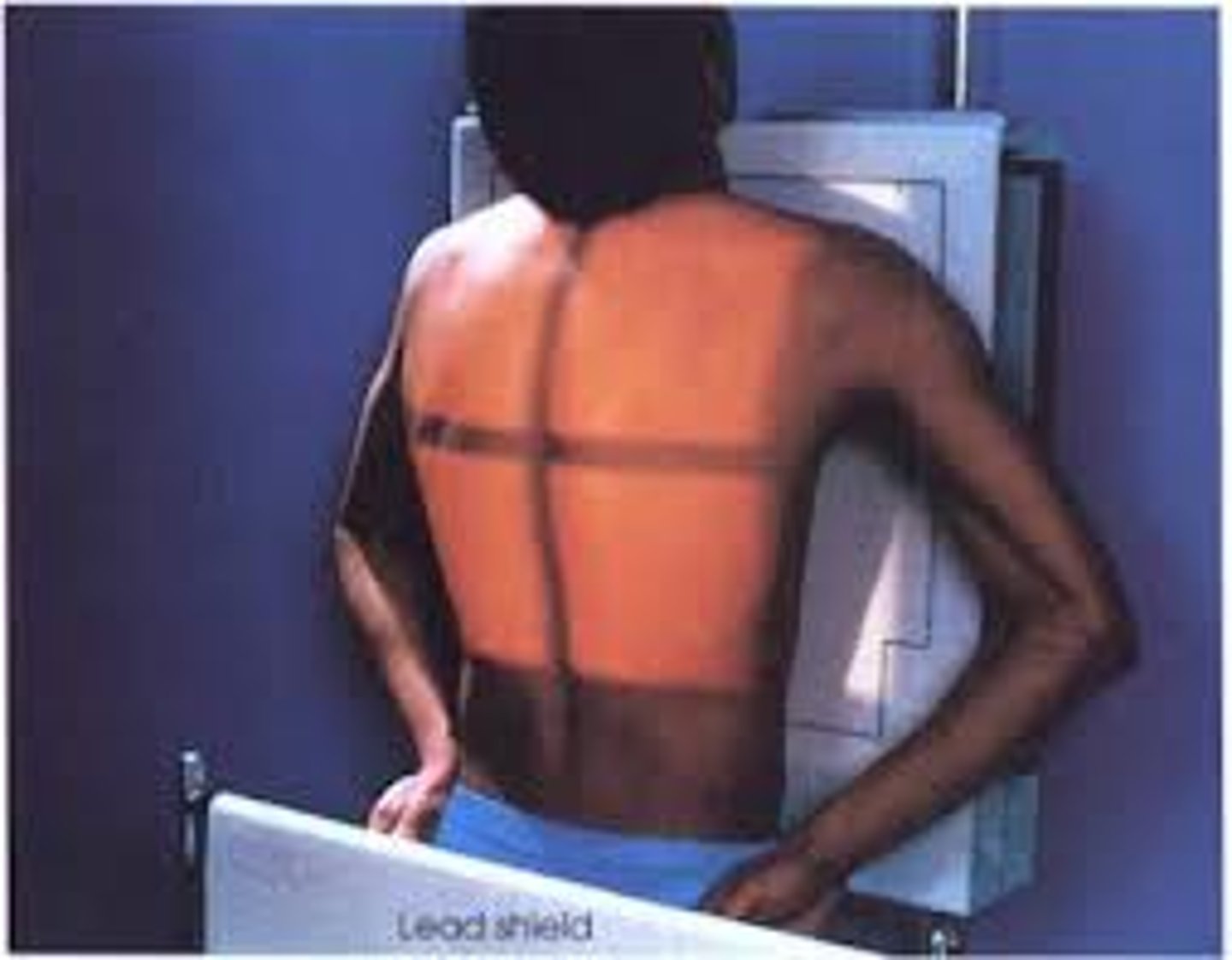
PA Film
posterior-anterior
-pt stands or sits upright with the chest against the film holder
-heart and lungs (normal chest x-ray)
Plain Film
no contrast
-skeletal
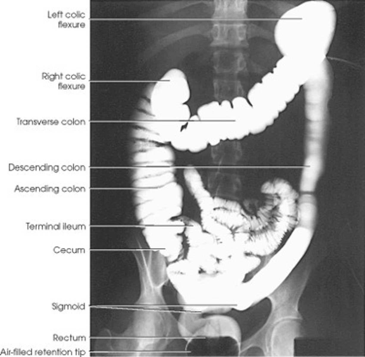
Contrast Studies and Examples
radiopaque contrast media administered
-GI tract (via swallow or enema)
-Urinary tract (urography, IV pyelogram/IVP) - inject contrast into the blood vessels, give time and it will pass to designated area to enhance visibility of structures in imaging.
-Blood Vessels (angiography)
-Vertebral Column (myelography) - contrast to CSF to light up column
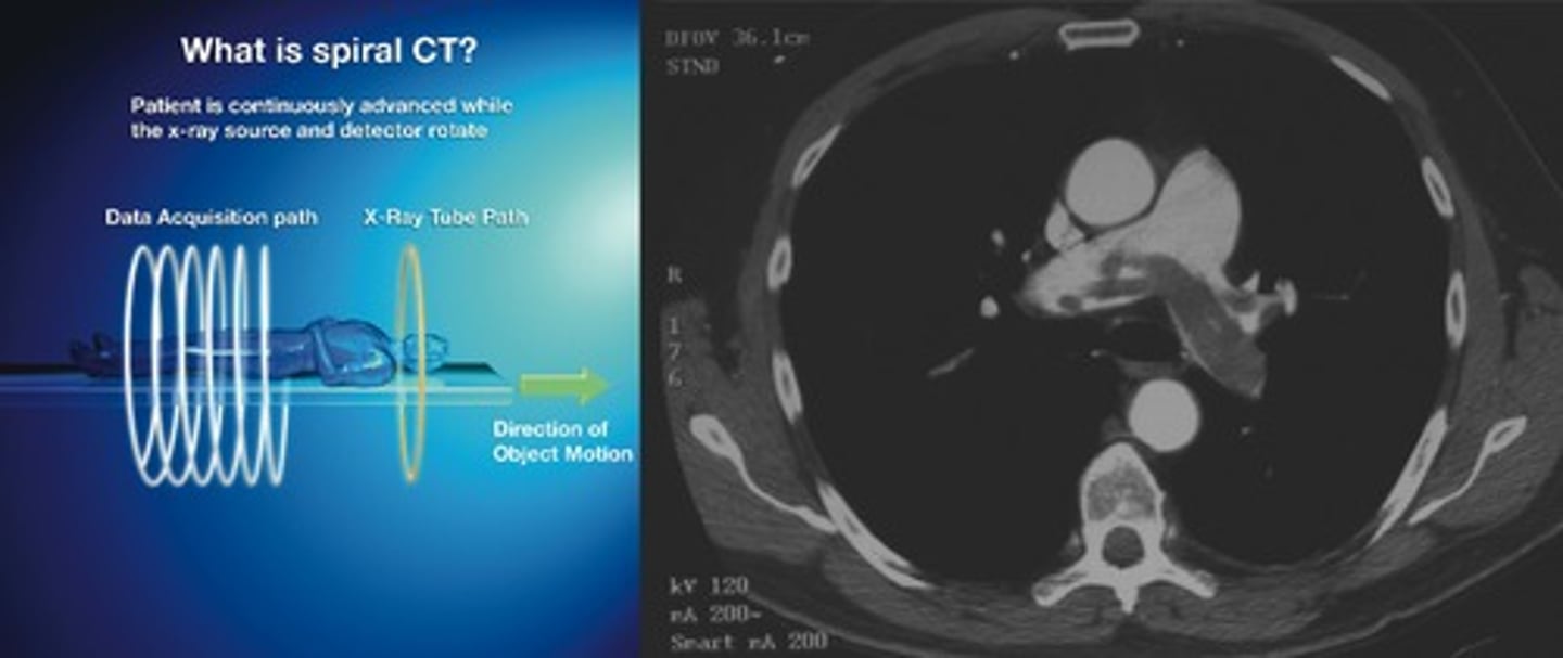
Computed Tomography (CT)
x-ray and detector system move through an arc of 360 degrees
-read as if you are standing at the patient's feet
-intensity of radiation is measured, analyzed assigning differing shades of gray based on attenuation
-computer reconstructs image
Traditional CT
-single-dimensional view
-approx. 2 mins
Spiral/Helical CT
-3D view
-20-30 secs
why should diabetics withold medication when IV contrast is used for a CT, specifically metformin (glucophage)?
it may cause acidosis when combined
a patient needs a screening for abdominal trauma, which form of imaging is the best?
computated tomography (CT)
CT Contrast
when hit with radiation, the x-ray beam is weakened by the uptake of the contrast
-appears radiopaque
-common forms: iodine, barium, gastrografin
what are some CT Contrast reactions
allergic reaction (good history): pretreat with steriods and diphenhydramine
contrast-induced nephropathy: check creatinine clearance and GFR
kidney may not be able to filter the contrast
Population Considerations for Contrast-induced Nephropathy
-kidney failure
-diabetics
-HTN
-elderly
-autoimmune disorders
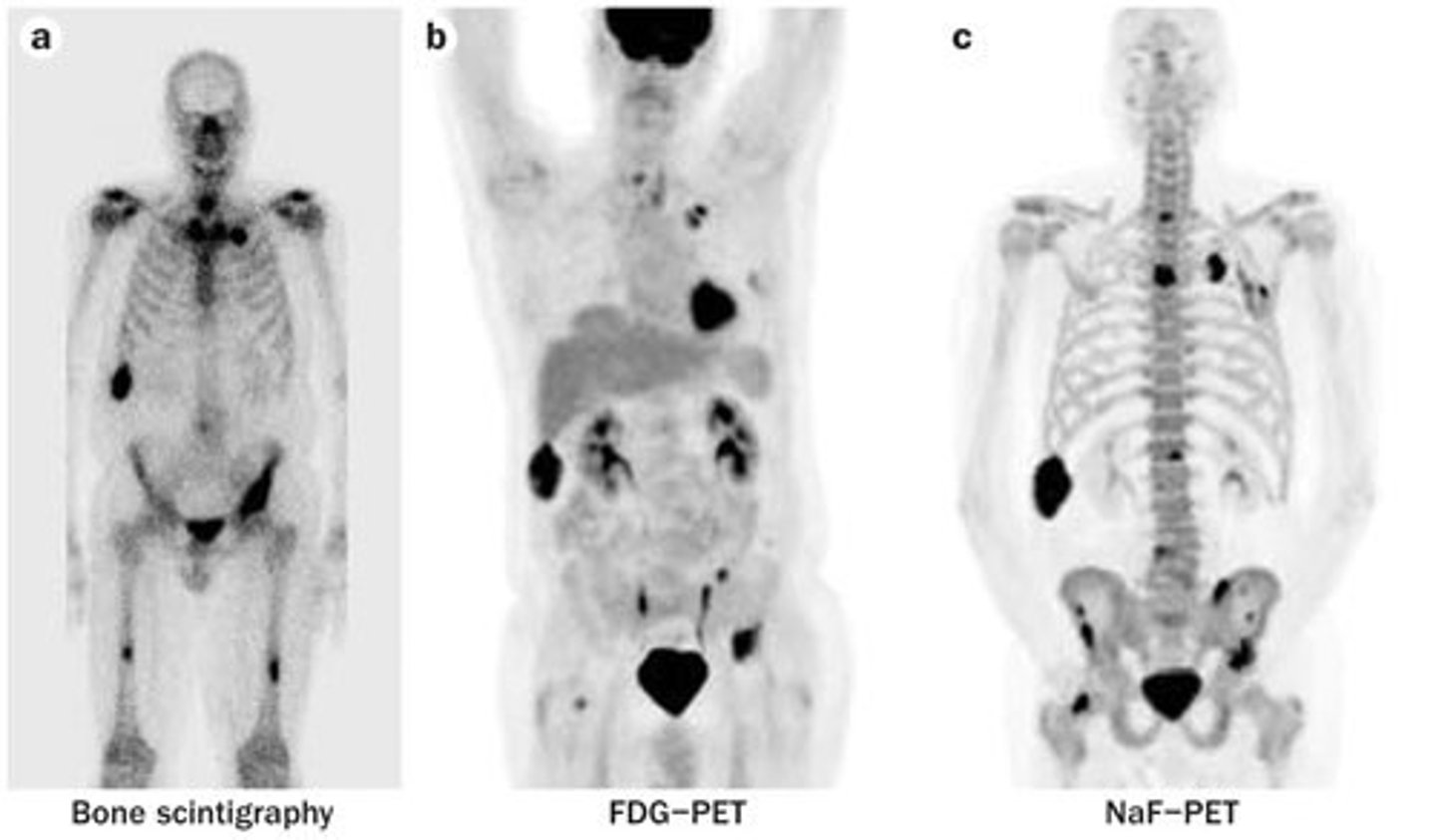
Nuclear Imaging
uses radioisotopes (most common technetium 99m)
-emits gamma rays, short half-life
Static vs Dynamic in nuclear imaging
"one shot" vs. series in sequential order
"hot" vs "cold" spots in nuclear imaging
increased uptake of radioisotopes vs. decreased uptake of radioisotopes
When would we want to use nuclear imaging?
helps diagnose things that are not acutely dangerous
maps cancer and where it has metastasized to
watching the body uptaking or not of radioisotope can watch for function and perfusion
_______ imaging is NOT for anatomy
nuclear
If we were to use nuclear imaging, what would we be looking for these these organs or areas?
Heart
Thyroid
Liver
Bone
Lung
Heart - myocardial perfusion, function, viability
Thyroid - nodules, cancer
Liver - masses, metastases, cholestasis
Bone - metastases, pain
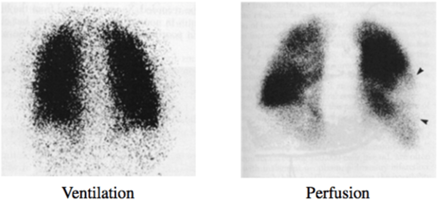
Lung - pulmonary embolism (quantification of perfusion)
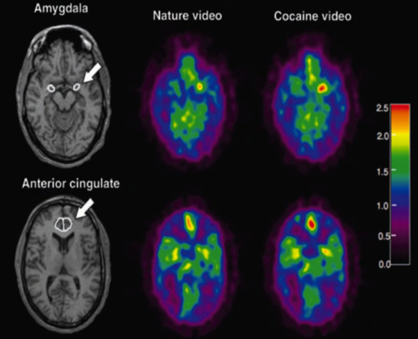
Positron Emission Tomography (PET) Scan (nuclear imaging)
high-energy particles emit positrons
type of radiographic imaging
VQ Scan (nuclear imaging)
inhalation/injection of radioisotope
-injection determines perfusion
-inhalation determines alveolar function
Physical Half-Life (nuclear imaging)
element would decay on its own
Biological Half-life (nuclear imaging)
normal physiological removal of the substance to which the isotope has been attached
Effective Half-life (nuclear imaging)
Formula combines the two to determine actual time the isotope remains "effective" in the body
When would we utilize PET/CT?
PET/CT combines metabolic and anatomical imaging.
perfusion + function and looking at anatomy
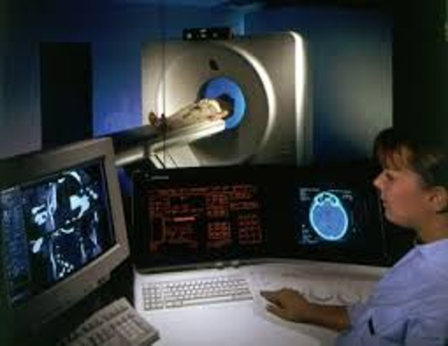
Magnetic Resonance Imaging (MRI)
magnetic fields and radio waves to produce computer-generated images that distinguish soft tissue types
non-invasive, no ionizing radiation
can produce images in any plane
Mechanism of MRI
nuclei of any atom with odd number of protons and neutrons behave like weak magnents in presence of a strong magnent
-most common is hydrogen ion
which form of imaging can produce images in any plane including sagital, coronal, and axial?
MRI
Indications for MRI
-intracranial abnormalities
-intraspinal abnormalities
-musculoskeletal abnormalities
-heart
-abdominal visceral abnormalities
-vascular abnormalities
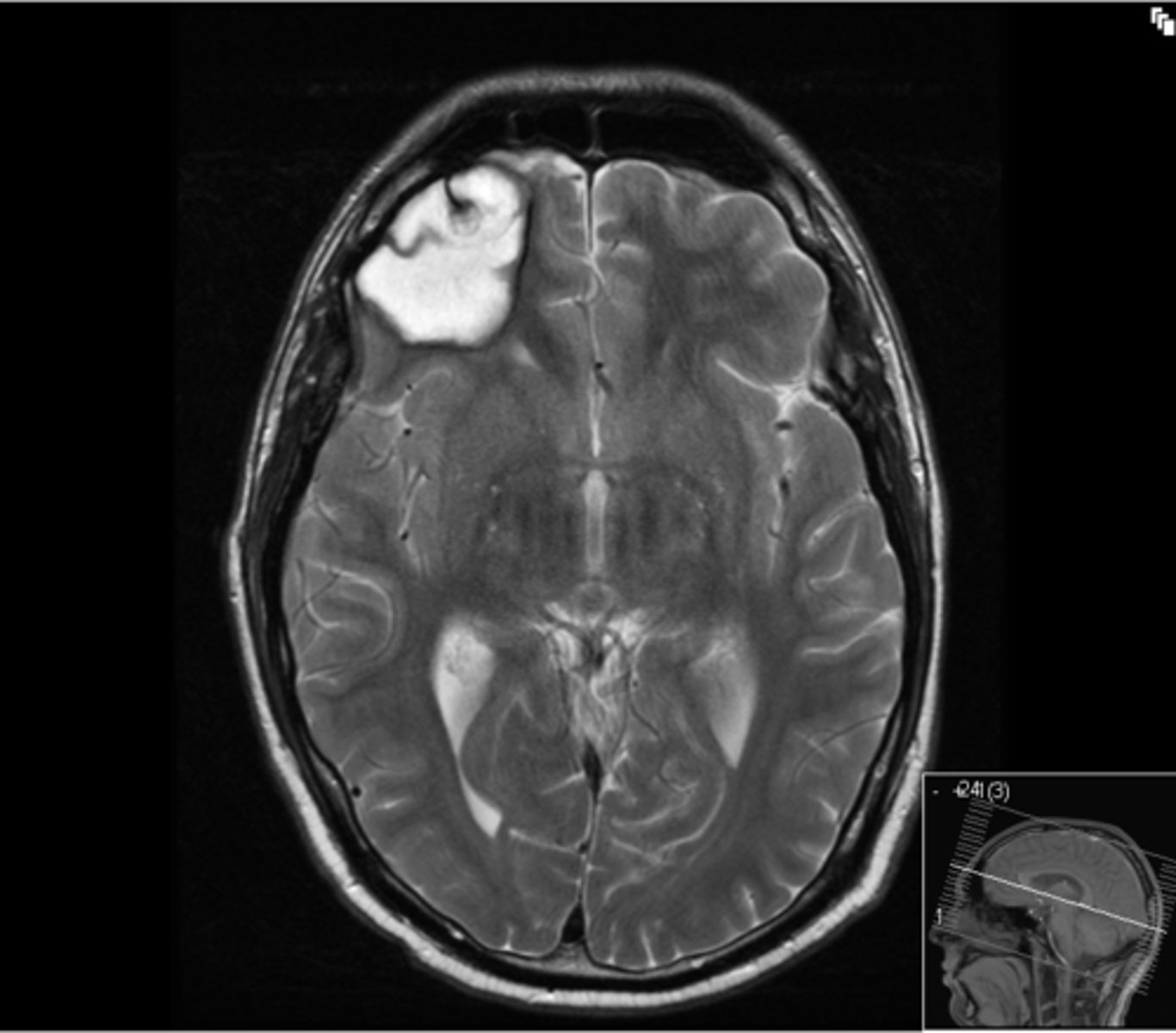
T1 MRI vs T2 MRI
T1: weighted scans are a standard basic scan, in particular differentiating fat from water - with water darker and fat brighter
T2: weighted scans similar to T1 where fat is differentiated from water but T2 fats shows darker than water
______ imaging is for detailed info rather than acute. Not for diagnosing but for needing a clear photo
MRI
Ultrasound
uses sonic energy (non-ionizing form of energy); echoes (reflections) of u/s beam from interfaces between tissues
-real-time
-demonstrates size, shape, and internal structure of organs/masses
Doppler Ultrasound is for …
-arterial stenosis
-venous occlusion
M-mode Ultrasound is used for…
echocardiography
can see movement of heart murmurs
Intervention Ultrasound is used for …
-fluid aspiration
-drainages (abscess)
-biopsies
-line placement
______ can be used for fetus anatomy, view ligament tears, tendon tears, cystic lesions, and even the eye
ultrasound
Where would we use Barium Contrast
GI tract
-oral or enema
-not if perforation is suspected (risk of peritonitis)
where would we use Iodine Contrast
used intravenously for x-ray and CT
-risk of contrast-induced neuropathy
PACS (picture archiving and communication system) was used to read medical imaging but is now _____ replacing the traditional view box and film. Also called “night hawk”
MIMPS - Medical imaging management and processing system
safer and more time efficient
If there is a GI tract perforation, and imaging is needed, how would we continue on getting a CT?
Water soluble contrast and IV hydration is needed for the patient