Brian and eyes labeling LAB
1/66
There's no tags or description
Looks like no tags are added yet.
Name | Mastery | Learn | Test | Matching | Spaced |
|---|
No study sessions yet.
67 Terms
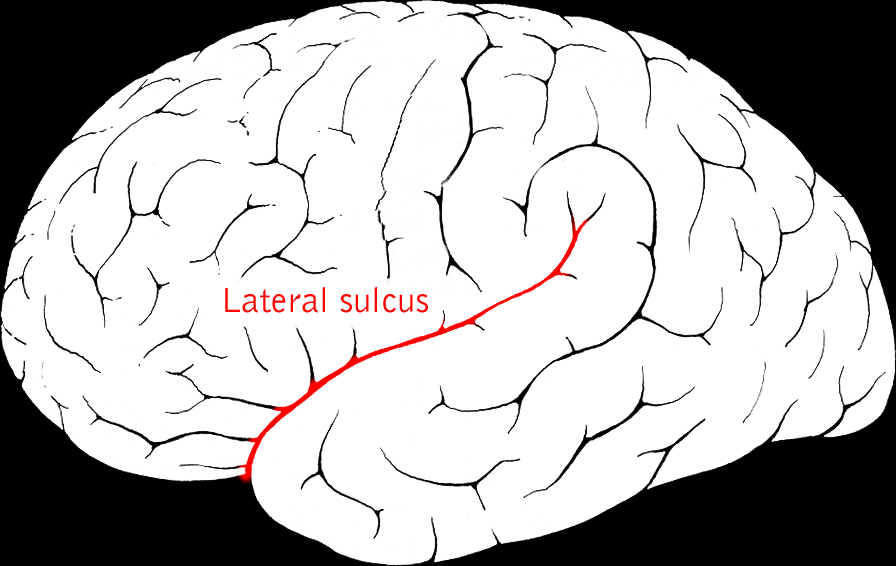
lateral fissure
separates the parietal lobe and frontal lobe
BETWEEN PRECENTRAL GYRUS
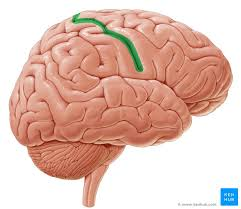
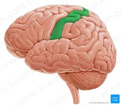
central sulcus
divid outline of precentral gyrus
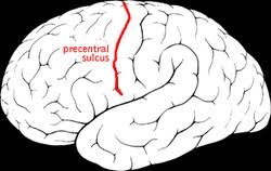
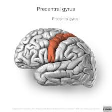
precentral sulcus
tongue shaped, middle line, looks like y shape on left side
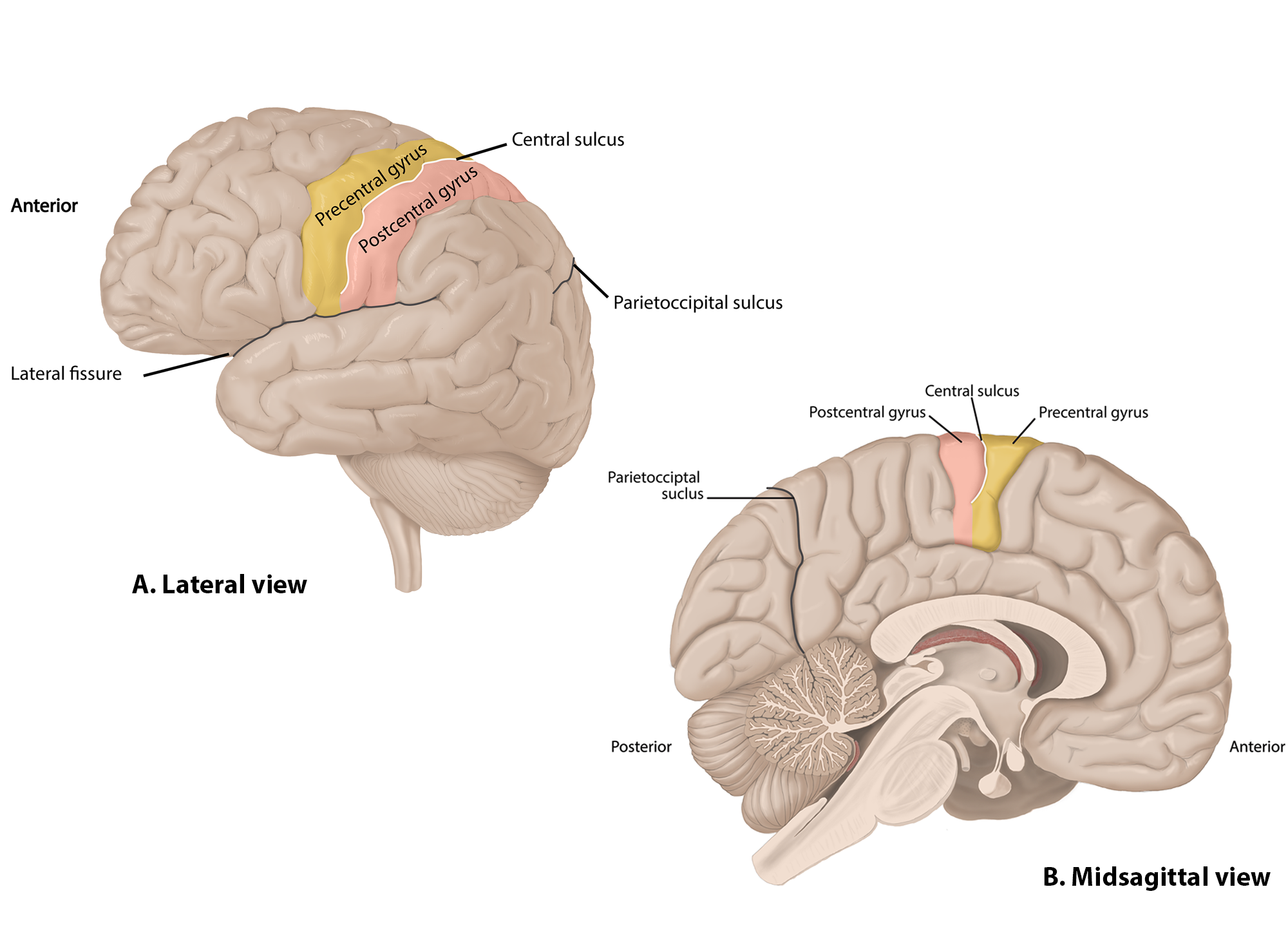
postcentral sulcus
to the left of postcentral gyrus
precentral gyrus
anterior postcentral sulcus anterior from y
postcentral gyrus
what is the order from left to right of the brain
post central sulcus
post central gyrus
precentral gyrus
precentral sulcus
folds (things that stick up)
gyri (gyrus)
lines on the brain
sulri (sulcus)
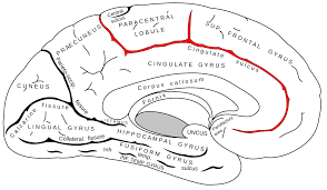
line above cingulate gyrus
cingulate sulcus
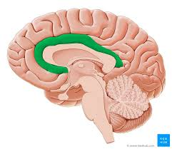
ring around corpas callosum
cingulate gyrus
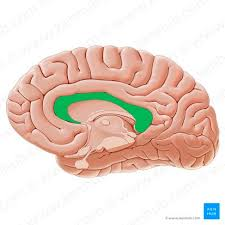
“c” shape, where axons, white matter
corpus callosum
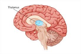
eye like structure
thalamus
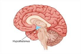
beak sturcture down and diagional from thalamus
hypothalamus
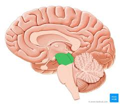
top of the brain stem, posterior to thalamus
midbrain
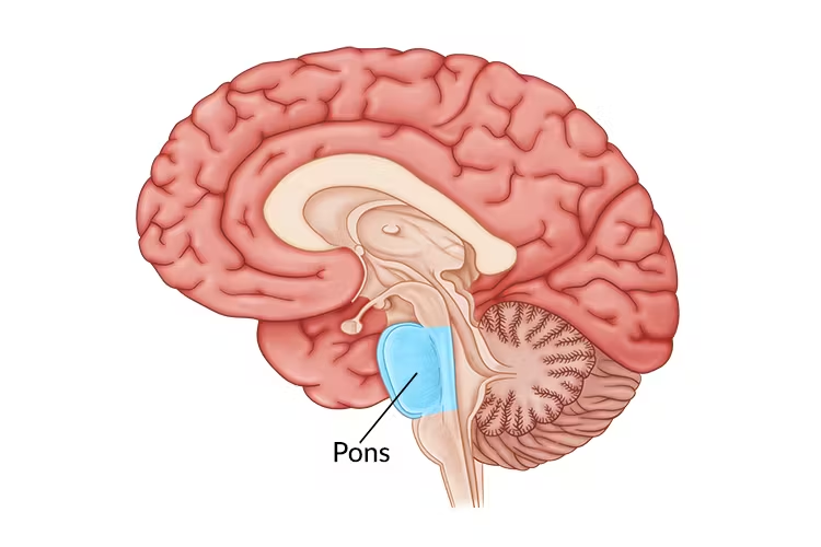
egg shaped, posterior from midbrain
pons
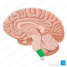
posterior of pons, stick
medulla
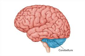
tree shaped
cerebellum
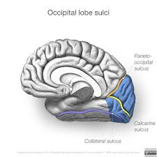
line of parietal/occipital lobe , anterior from cerebellum
parieto-occipital sulcus
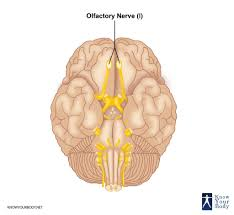
long, white nerve, medial in brain
CN I, olfactory nerve
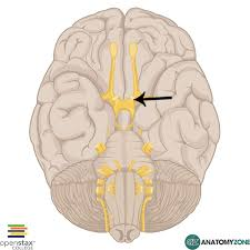
stump distal from CN 1, olfactory nerve
CN II Optic nerve

right above pons, line, inferior to CN II
CN III Oculomotor nerve

white wire that is sticking out
CN IV Trochlear Nerve
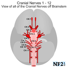
on top of pons
CN V trigeminal Nerve
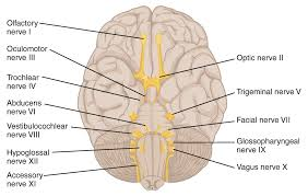
medial in brain, right by the 2 hemispheres
CN VI Abducens Nerve
2nd white line to the right of the hemisphere split
CN VII facial nerve
3rd line to the right of CN VII facial nerve
CN VIII vestibulocochlear nerve
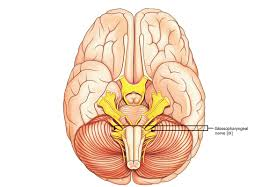
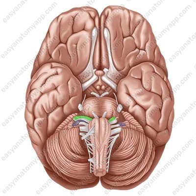
1st posterior from VII and VIII
CN IX Glossopharyngeal nerve
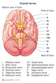
below CN IX
CN X Vagus nerve
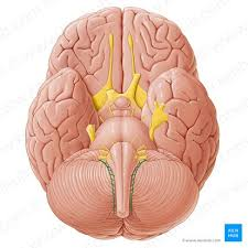
curve below CN X
CN XI Accessory nerve
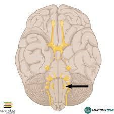
anterior from IX, X, on top of IX and X
CN XII Hypoglossal nerve

gray part, brown shaped horn at the top
Dorsal Gray horn

lateral side of the brown
lateral gray horn
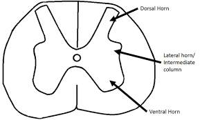
below dorsal gray horns
ventral gray horn
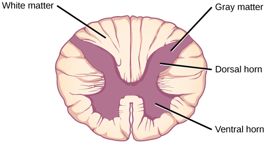
white around dorsal horn between horns
dorsal white matter
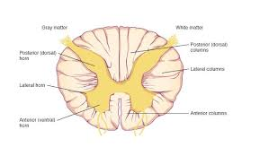
lateral white
lateral white matter
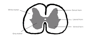
bottom white
ventral white matter
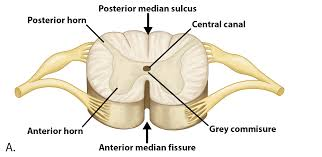
top line in middle
dorsal median sulcus
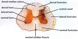
white part in the middle
ventral median fissure

hole in middle for fluid, black hole
central canal
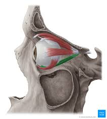
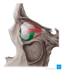
runs along top of eyeball
superior rectus
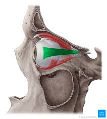
directly under eyeball
inferior rectus
muscle by nasal bones along the eyeball
medial rectus
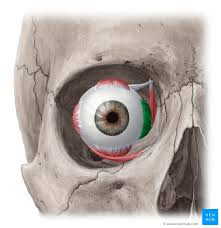
open side, closer to ear and zygomatic bone
lateral rectus
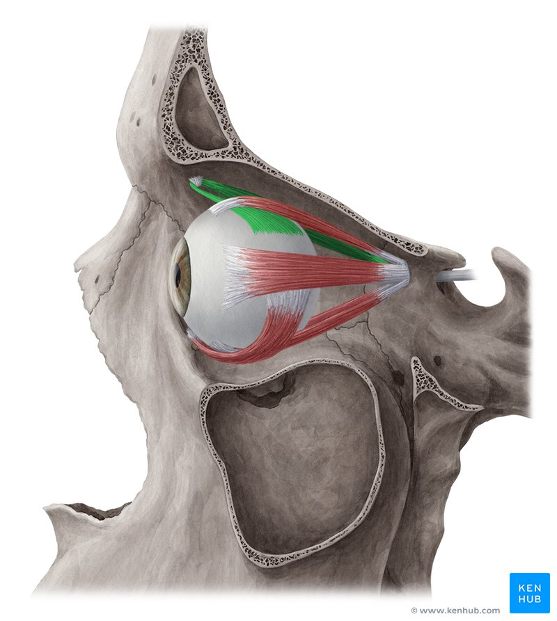
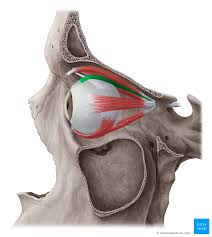
white line going to red by nose
superior oblique
connects inferior rectus and lateral under eyeball
inferior oblique
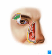
lateral, yellow above eye, tears form here
lacrimal gland
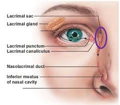
by nose, yellow, tears collected
lacrimal sac
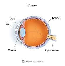
clear, outer layer of eyeball
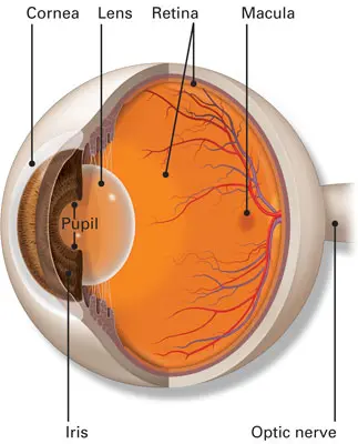
cornea
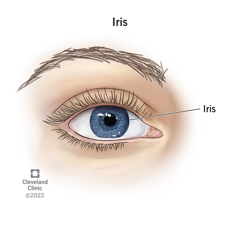
colored part of the eye
iris
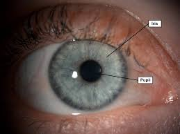
black hole in eye
pupil
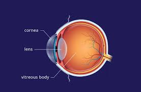
part behind iris
lens
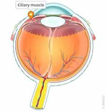
anterior from suspensory ligament, pediallike flower
ciliary muscle
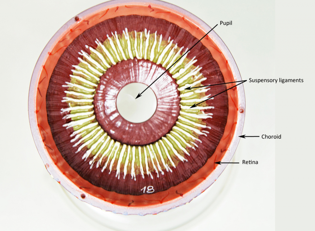
lines that touch lens
suspensory ligaments
oragne, inner coding of eye
retina
white that touches choroid, covers all eyeball
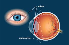
sclera
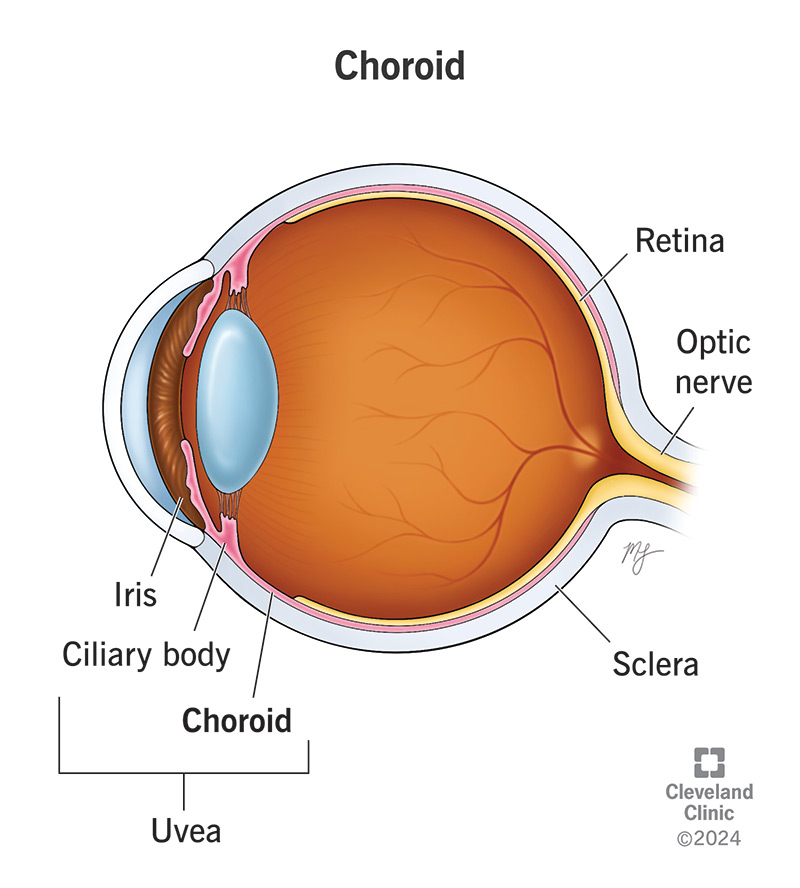
red rim outside retina
choroid
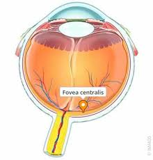
yellow dot on side by optic nerve
fovea centralis
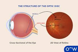
where optic nerve leaves eye
optic disc
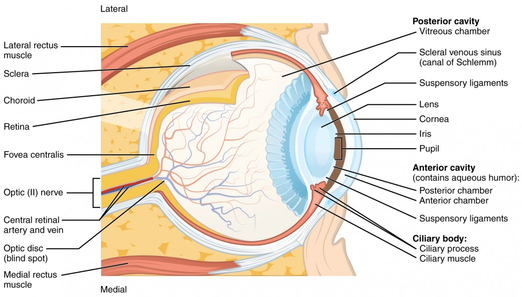
big clean ball in eye
posterior cavity
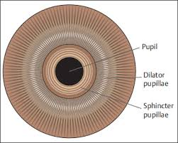
inner ring if pupil muscle that opens and closes eye
sphincter pupillae

outside muscle from pupil, enlarges pupil
dilator pupillae
what does the parietal lobe do in telencephalon
sensory processes
what does the temporal lobe do in telencephalon
hearing and smell
what does the frontal lobe do in telencephalon
motor processes
what does the occipital lobe do in telencephalon
Processing visual information and storing of memories
what does the Isula lobe do in telencephalon
Taste and memory of taste