Primary visual pathway (L8)
1/13
There's no tags or description
Looks like no tags are added yet.
Name | Mastery | Learn | Test | Matching | Spaced |
|---|
No study sessions yet.
14 Terms
Primary visual pathway from eyes to primary visual cortex in occipital lobe
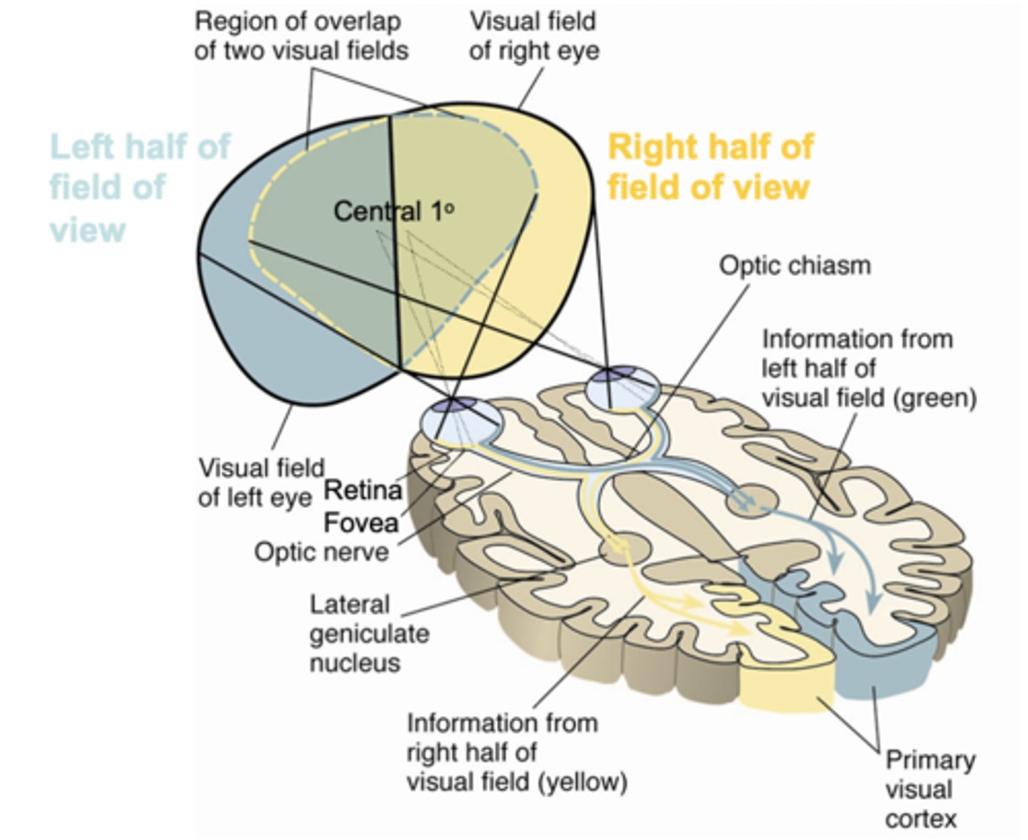
- Each eye has a visual field from which visual information (light) can enter the retina and excite component cells.
- Central area of field of view (sharpest vision + colour vision) excites the fovea.
- From the retina via the optic nerve crossing into the optic chiasm, visual information is transmitted further up to the primary visual cortex via the lateral geniculate nucleus (thalamic relay station).
- Left field of view ends up in right visual cortex (vice versa).
Information-processing stages in primary visual pathway
- Visual information processing is hierarchal; it happens sequentially across several stages.
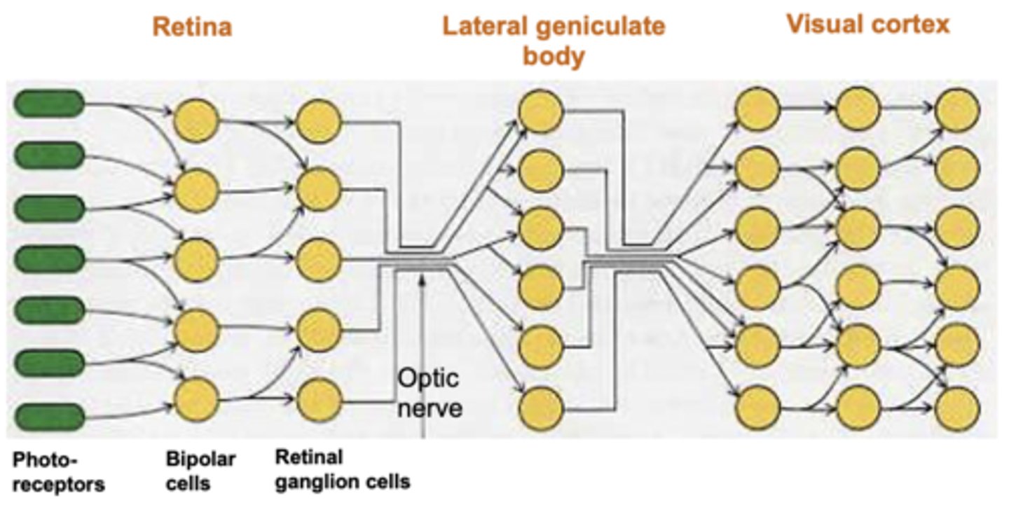
Photoreceptors
- Photoreceptors (rod s and cones) in retina detect presence or absence of light.
- Bipolar cells mediate between photoreceptors and ganglion cells.
- Photoreceptors and bipolar cells vary their voltage as they are stimulated (analogue signal) whereas all subsequent cells vary spike rate (all-or-nothing, digital signal).
- Photoreceptor detection of light is translated into excitation or inhibition of retinal ganglion cells via bipolar cells.
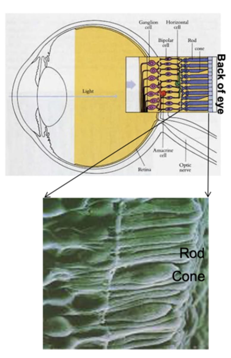
Rods
- More abundant (120m in human retina)
- No colour discrimination
- Sensitive in low light levels
- Higher density in periphery (don't look directly at dim stars)
- Can track high-rate changes (see flicker of 60Hz monitor from corner of your eyes)
Cones
- Less abundant (6 million in human retina)
- discriminate between 3 different wavelengths (S,M,L)
- Less sensitive to low light
- High concentration in fovea
- Cannot follow rapid changes (can't see 60Hz flicker when directly looking at monitor)
Receptive field of visual neurons
- Area of the retina/visual field in which visual stimulation will evoke a change in the firing rate of a given visual neuron.
- Substructure of a receptive field: description of how visual stimuli need to be presented in the receptive field of a visual neuron in order to evoke firing-rate changes.
Retinal ganglion neurons
- Receive input from multiple photoreceptors (via bipolar cells).
- ON-OFF centre-surround receptive fields.
- Light presented in 'ON' regions excites cells.
- Light in 'OFF' regions inhibits cells.
- ON and OFF regions are organised in 'centre-surround' fashion.
- Response rate of cell is based on the sum of stimulation in ON region - stimulation in OFF region.
- Neurons in the lateral geniculate body respond to visual stimuli in similar ways to retinal ganglion cells.
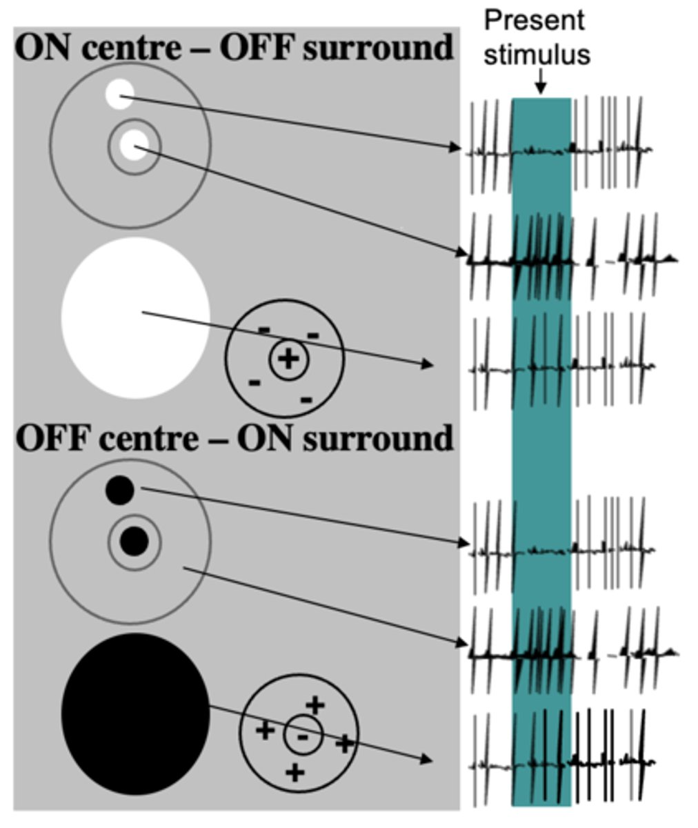
Functional significance of centre-surround fields
- We only need to respond to changes and boundaries (edges) → efficient.
- Luminance of features represented relative to their surround: helps preserve appearance of objects regardless of light levels in the environment → can also result in illusions.
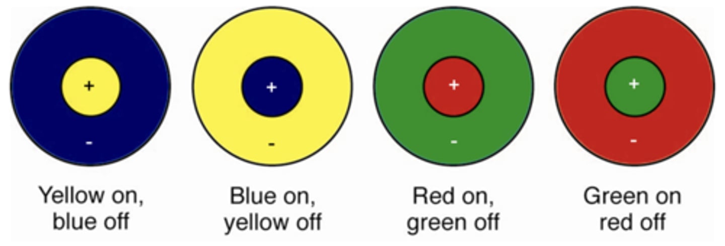
Colour sensitivity of retinal ganglion and LGN neurons
- Receive input from cones (differentially sensitive to different wavelengths) and are sensitive to colour.
- Colour-sensitive retinal ganglion and LGN neurons have receptive fields that show centre-surround colour opponency.
- Colour opponents either between yellow and blue, or red and green (e.g. when blue presented in periphery of receptive field, firing right is inhibited and where yellow presented in centre, firing rate increased → opposite effects when other way around: same for red and green).
- Colour opponency along with firing-rate adaptation (rebound effects) can explain negative afterimages.
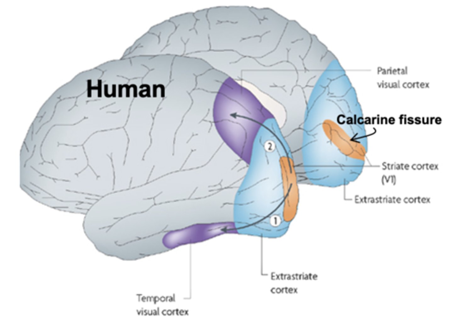
Primary visual cortex (striate cortex, V1)
- Sits around calcarine fissure, between the two hemispheres.
- Striate cortex that receives input from the lateral geniculate nucleus.
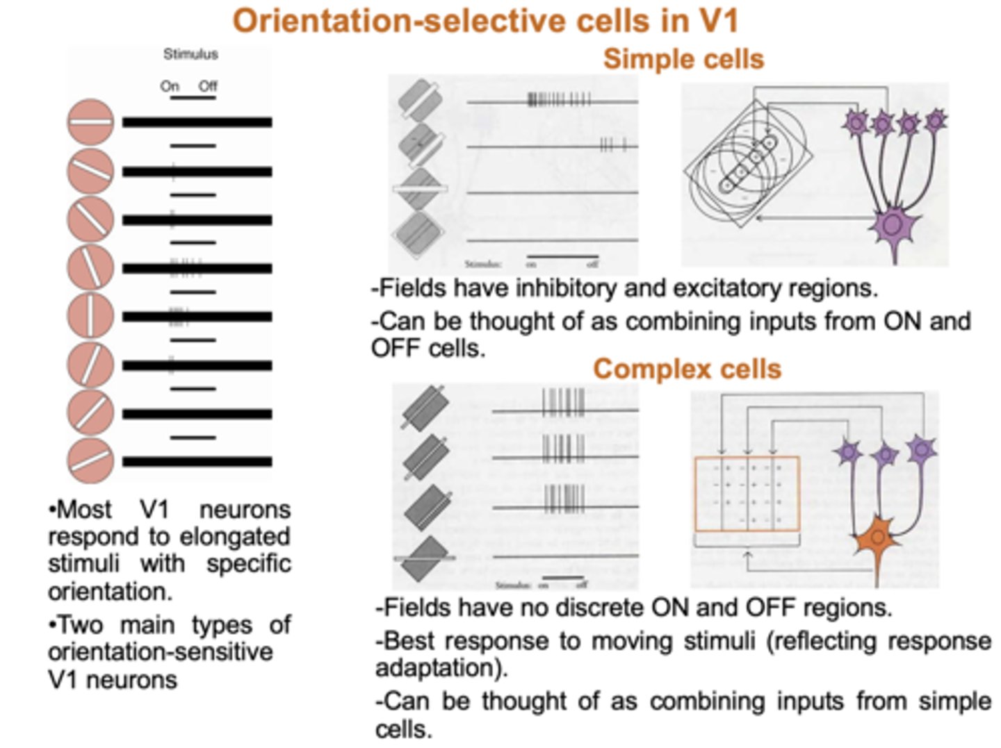
Orientation-selective cells in V1
- Respond to elongated stimuli of a particular orientation.
- Two main types of orientation V1 neurons: simple and complex cells.
- Simple cells: make up receptive fields of complex cells: have inhibitory and excitatory regions and ON and OFF regions.
- Complex cells: do not have discrete ON and OFF regions; wherever presented in receptive field, leads to excitation of the cell as long as stimulus presented in correct orientation, if not no response.
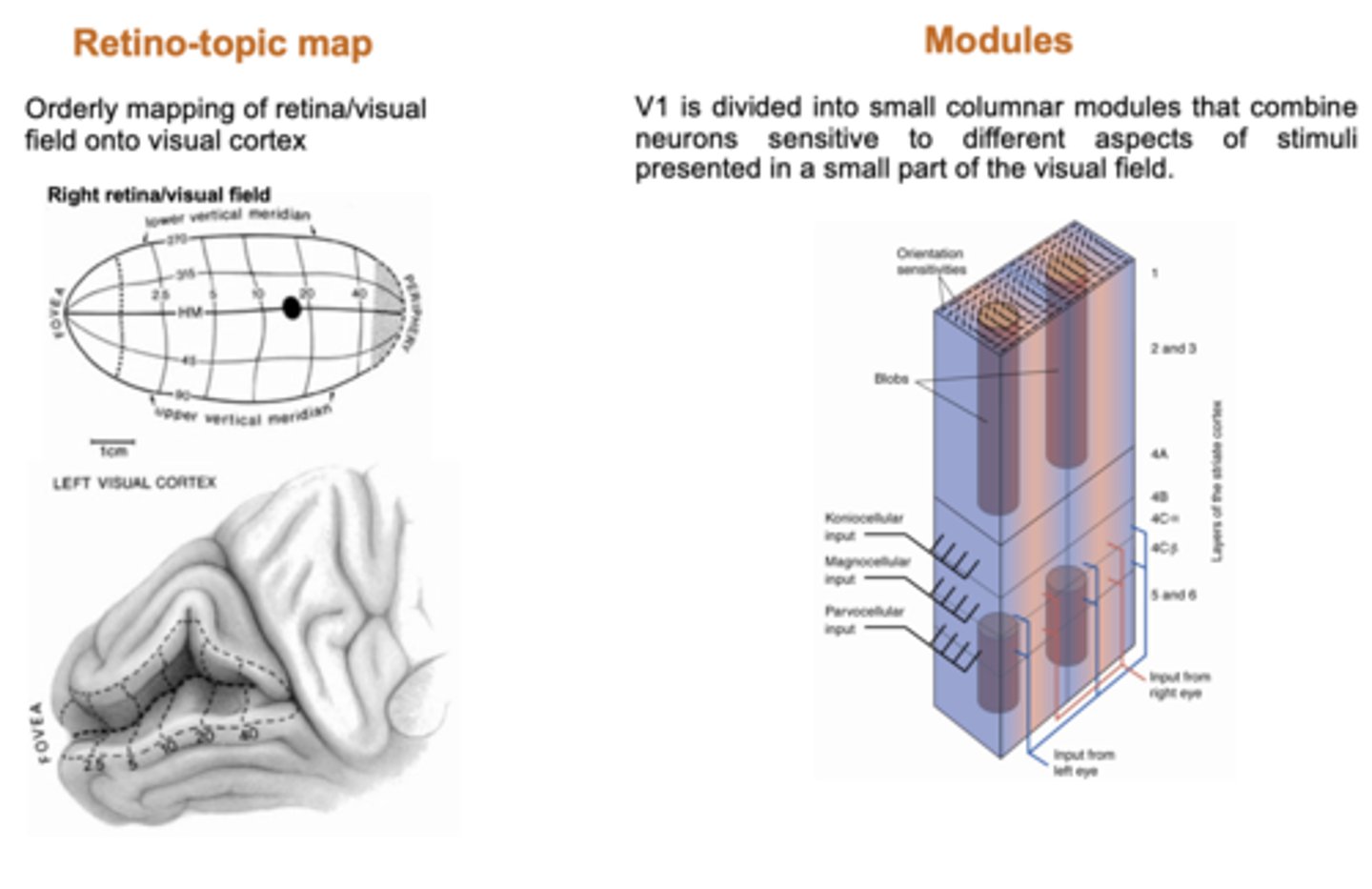
Maps and modules in V1
- Retino-topic map: orderly mapping of visual field onto visual cortex.
- Modules: V1 divided into small columnar modules that combine neurons sensitive to different aspects of stimuli presented in a small part of the visual field.
Further processing of visual information
- To result in perception and memory of the holistic visual properties of whole objects, visual information from modules in V1 need to combined and further processed.
- Processing takes place in visual association cortices (V2-V5m inferior temporal cortex, posterior parietal cortex) and other regions.
Blindsight
- Subjects with lesions to primary visual cortex and apparent 'blindness' can show appropriate response to visual stimuli of which they are not 'conscious'.
- Examples include looking (i.e. moving the eyes) or pointing towards visual stimuli; detection of movement etc.
- Blindsight highlights that there are additional visual pathways.
- Also highlights that brain can perform visual information processing which can guide subjects' behaviour without their conscious awareness.