Processing the Environment
1/76
Earn XP
Description and Tags
P/S
Name | Mastery | Learn | Test | Matching | Spaced | Call with Kai |
|---|
No analytics yet
Send a link to your students to track their progress
77 Terms
Visual cues
Depth, Form, Motion, Constancy
depth
how far or close an object is
motion
our ability to sense and interpret movement, encompassing both the movement of objects around us and our own body's movement
form
visually perceive objects in the world in response to the patterns of light that they caste on our retinas
Binocular Cues
retinal disparity
convergence
convergence
looking far way→ eyes are relaxed
looking closer to us → eyes contract
monocular cues
relative size
interposition
relative height
shading and contour
motion parallax
relative size
one object percieved to be bigger → think of it being closer to us
interposition
object in front is closer to us
relative height
higher object → further away
shading and contour
using light and shadow to interpret form
constancy
our perception of object doesn’t change even if it looks different on retina
size, shape, color
sensory adaptation
change in the sensitivity of your perception of a sensation
hearing
adapt to loud noises → inner ear muscles contract to protect ear drum
touch
sensory nerves saturated → temperature receptors densensitized
smell
can detect chemicals in air
over time desensitized to molecules
proprioception
sense that tells your brain where your body parts are in space and how they are moving, without needing to look
just noticeable difference
threshold where you notice a change in sensation
𝚫I
Weber’s law
𝚫I/I = k
ratio of the just noticeable difference over intensity is constant
𝚫I/I = k
the just noticeable distance and the intensity are directly proportional
absolute threshold
minimum intensity of stimulus that a person can detect half the time
more likely to detect it right when the intensity is higher
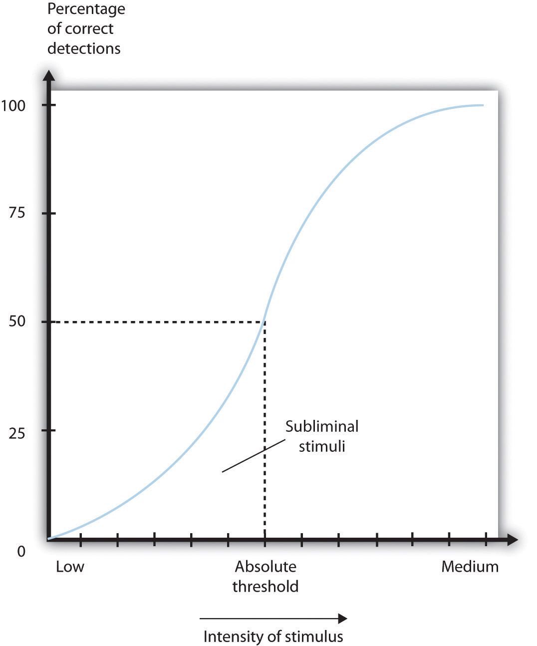
subliminal
stimuli we can’t detect 50% of the time
what influences detecting stimulus?
-expectations
-experiences
-motivation
-alertness
vestibular system
balance and spatial orientation
semicircular canals
posterior, lateral, and anterior canals are at 90 degrees to each other
filled with fluid called endolymph
cochlea
specialized auditory receptors that process sound and send info the brain
endolymph
shifts and allows us to detect what direction our head is moving in and strength of rotation
otolithic organs
utricle and saccule
information related to balance and spatial recognition
helps detect linear acceleration and head positions
how do otolithic organs help with detecting head position?
structures contain calcium carbonate crystals attached to hair cells with gel
if we move fast→ crystals move→pull on hair cells → action potential
what causes dizziness?
endolymph doesn’t stop spinning after stopping
signal detection theory
how we make decisions under conditions of uncertainty
how we discern between important stimuli and unimportant noise
at what point can a signal be detected
hit
stimulus present, guessed yes
miss
stimulus present, guessed no
false alarm
stimulus absent, guessed yes
correct rejection
stimulus absent, guessed no
d’ strategy
hit>miss → strong signal
hit<miss → weak signal
c strategy
conservative v.s liberal
conservative strategy
always no unless 100% sure
all correct rejections but some misses
liberal strategy
yes all the time
all hits but some false alarms
the signal distribution
the difference between the means of the two is d’
if signal shifts right → d’ is big and easy to detect
if signal shifts left → d’ is small and hard to detect
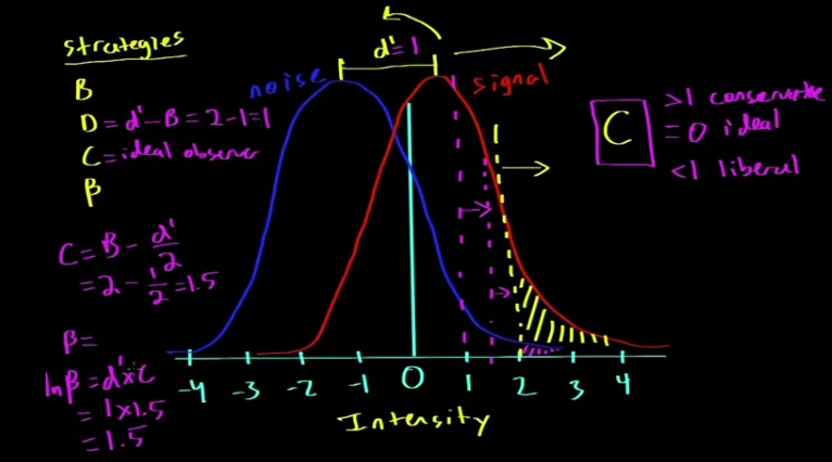

If strategy B choses a threshold 2
anything more than 2 is yes
less than 2 is no

strategy C
expressed via choice of threshold → threshold individual deems necessary for them to say yes
ideal observer, minimizes miss and false alarm
C = B-d’/2
When C = 0
participant is ideal observer
If <1 liberal
if >1 conservative
beta
beta = d’ x C
Bottom up processing
stimulus influences our perception
Top down
background knowledge influences perception
Gestalt principles
why we percejve things the way we do
Gestalt principle of similarity
items similar to one another are grouped together
Gestalt principle of pragnanz
reality is often organized reduced to simplest form as possible
ex: olympic rings perceived as five circles
Gestalt principle of proximity
objects that are close are grouped together
Gestalt principle of continuity
lines are seen as following the smoothest path
Gestalt principle of closure
objects grouped together are seen as a whole
Gestalt principle of common Fate
Elements that move in the same speed/direction are perceived as a group
conjunctiva
first layer that light hits
protects the cornea
cornea
transparent thick sheet of tissue
anterior 1/6th
anterior chamber
space filled with aqueous humor which provides pressure to maintain the shape of eye ball
what forms the ciliary body?
suspensory ligaments, attached to a ciliary muscle secretes the aqueous humor
posterior chamber
area behind the ciliary muscle
filled with aqueous humor
vitreous chamber
filled with vitreous humor, jelly-like substance to provide pressure to eyeball
retina
filled with photoreceptors
macula — special part of the retina rich in cones
fovea — completely covered in cones, no rods
choroid
pigmented black in humans, a network of blood vessels
it’s black because all the light is reflected
sclera
whites of the eye,
thick fibrous tissue that covers posterior 5/6th of the eyeball
attachment point of muscles
visual sensory information
sensation requires light to turn into a neural impulse by a photoreceptor
light
electromagnetic wave part of a large spectrum
Violet (400 nm) — Red (700 nm)
rods
for night vision
when light comes in → pupil → hits rod → rod is turned off → turns on bipolar cell → turns on a retinal ganglion cell → optic nerve → brain
cones
3 types: red (60%), green (30%), blue (10%)
all centered in fovea
rhodopsin
inside the rod there are a lot of disks stalked on top of one another with a lot of proteins on them
rhodopsin is a multimeric protein with 7 discs
contains a molecule called retinal which undergos a change conformation from bent to straight when light hits it
when retinal changes shape, rhodopsin changes shape
Phototransduction cascade
transducin (has 3 parts: alpha, beta, gamma) breaks from rhodopsin → alpha part comes to disk and binds to phosphodiesterase → phosphodiesterase takes cGMP and converts it to GMP → Na+ channels allow Na+ ions to come in → channels close as cGMP decreases as they need cGMP to bind to open → less Na+ enters the cell → cells hyperpolarize → turn off → glutamate is not released → doesn’t inhibit ON bipolar cells → bipolar cells turn on → activates retinal ganglion cell → sends signal to optic nerve
photoreceptors
specialized nerve that can take light and convert it to neural impulse
in rods there are optic discs (large membrane bound structures ) → proteins in membanes that fire APs to the brains
rods have rhodopsin while cones have photopsin
Differences between rods and cones
120 million rods v 6 million cones
cones are concentrated in the fovea
rods are 1000x more sensitive to light than cones (better at telling us whether light is present) (night vision)
cones are less sensitive but detect color
rods have slow recovery time compared to cones and takes a while to adjust to dark.
photoreceptor distribution in retina
where optic nerve connects to retina is a blind spot
rods found in periphery, cones found in foveo
If light hits peripher → goes through bundle of axons → energy lost
fovea light hits cones directly
Visual field processing
right side of body controlled by left side
all right visual fiels goes to the left side of the brain and vice versa
parvocellular pathway
form\shape of a stationary object
good at spatial resolution — able to capture fine details and boundaries of an object
poor temporal resoulution — can’t track motion
magno pathway
encode motion
high temporal resolution - can track motion
poor spatial resolution - can’t capture the boundaries of the object
no color
parallel processing
see all pathways at the same time
sound needs
pressurized sound wave
hair cell
sound waves
air molecules are pressurized and try