microbiology
1/95
There's no tags or description
Looks like no tags are added yet.
Name | Mastery | Learn | Test | Matching | Spaced | Call with Kai |
|---|
No analytics yet
Send a link to your students to track their progress
96 Terms
What does serotyping help differentiate?
Strains of the same species.
Does serotyping tell us if a strain is pathogenic?
No.
What does serotyping detect?
Surface antigens.
What types of antigens can serotyping distinguish between?
LPS, pilus types, flagellum types, and capsule types.
How does serotyping classify microorganisms?
Based on specific surface antigens.
What are examples of antigens detected in E. coli serotyping?
O antigen, H antigen, and K antigen.
What is the K antigen in E. coli?
The capsular polysaccharide.
What does the O antigen represent in E. coli?
The lipopolysaccharide (LPS).
What does the H antigen represent in E. coli?
Flagellar proteins.
What is the role of fimbrial antigens (F antigens)?
To help with adhesion to host tissues.
What is 16S rRNA found in?
The small subunit of the bacterial ribosome.
Why is 16S rRNA useful for bacterial identification?
It has conserved and variable regions.
What do conserved regions in 16S rRNA enable?
Primer binding in PCR.
Is 16S rRNA identical across all bacterial species?
No, it varies between species.
Why do we use the rRNA/ITS database for 16S analysis?
It specializes in 16S RNA sequences.
What does the expect value (E-value) indicate in sequence alignment?
The chance of a match occurring by chance.
What does a smaller E-value suggest?
A more reliable match.
What does a high % mortality in WT and restored fungal strains indicate?
That the strains are virulent.
Why is a restored strain used in virulence studies?
To confirm the knockout causes avirulence.
What can be concluded if a knockout fungal strain causes no mortality?
The knockout strain is avirulent.
How are bacteria traditionally classified, and what molecular methods are now preferred?
Traditionally by phenotype; now G + C content and rRNA sequences are more widely used for a molecular approach.
What are the classical techniques used to identify bacteria?
Techniques include Gram staining (to determine Gram-positive/negative bacteria) and API strips (several biochemical tests).
Why is 16S RNA ideal for bacterial identification?
It has conserved and variable regions, allowing evolutionary relationships to be studied while providing stable comparisons.
What is the purpose of PCR in bacterial identification?
PCR is used to amplify the genes encoding 16S ribosomal RNA, and the resulting sequences are compared.
What does serotyping group bacteria by?
It groups bacteria by surface antigens, often O antigen (LPS) and H antigen (flagellum).
Why is serotyping important in diagnosing infectious diseases?
It helps distinguish between bacterial strains, which is crucial for disease diagnosis.
What safety measures should be followed when working with a Bunsen burner?
Use a blue flame, work around the base of the burner, keep lids on, and flame tools and containers to maintain sterility.
How do you prepare a bacterial suspension from a plate?
Sterilize the loop, collect 2-3 colonies, sterilize the saline broth neck, place colonies in the bottle, mix evenly, and sterilize tools again.
What is a heat-fixed smear, and why is it used in Gram staining?
A thin bacterial suspension dried on a slide with heat to fix the cells for Gram staining.
Why must Gram stain be washed thoroughly?
To prevent incorrect interpretation of the staining results.
What is the purpose of a counterstain in Gram staining?
To stain structures that didn’t retain the primary stain, providing contrast.
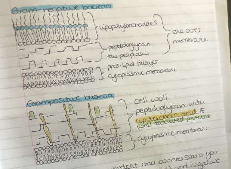
What are the typical appearances of Gram-positive and Gram-negative bacteria under a microscope?
Gram-positive appear longer and purple; Gram-negative appear shorter and pink.
What are API strips used for, and how should they be handled?
They test bacterial metabolism; handle with sterile techniques, fill wells properly, and reduce air bubbles by trickling suspension down the side of the wells.
What is the formula to calculate TM (melting point) of primers used in PCR?
TM=4×(G+C)+2×(A+T)T_M = 4 \times (G + C) + 2 \times (A + T)TM=4×(G+C)+2×(A+T).
What is the role of a mordant in Gram staining?
It fixes dye to the cell wall; iodine acts as a mordant, forming a crystal-violet-iodine complex trapped in Gram-positive peptidoglycan.
Why do Gram-negative bacteria lose the crystal violet stain? Why do Gram-negative bacteria lose the crystal violet stain?
Their thinner peptidoglycan layer and outer membrane fail to retain the crystal violet-iodine complex during decolorization.
What are lipoteichoic acids (LTAs), and where are they found?
LTAs are phosphate-rich polymers in Gram-positive bacteria; they are anchored to the cell membrane and extend through the peptidoglycan layer toward the surface
What were the observations of K12 and O157 E. coli when mixed with dynabeads?
K12 showed no aggregation; O157 showed minor clumps and signs of aggregation.
What components do the O, K and H antigens of bacteria detect?
O antigen detects LPS; K antigen detects capsule polysaccharides; H antigen detects flagellum.
Why is 16S RNA used for identifying bacteria, and how is it useful in PCR?
It has conserved regions for primer binding and variable regions for species differentiation, making it ideal for PCR amplification and comparison.
In questions about phylogeny, remember that thee smaller the numbers IN THE WHOLE LIST, the ____ recent their common ancestor
In questions about phylogeny, remember that thee smaller the numbers IN THE WHOLE LIST, the MORE recent their common ancestor
Why are primers required in a PCR reaction?
Primers are short single-stranded DNA sequences that bind to complementary regions on the target DNA. They provide a starting point for the DNA polymerase to synthesize the new DNA strand, as the enzyme cannot initiate DNA synthesis without a pre-existing strand to extend.
Using the sequence with the primers marked on it, work out how long you would expect the PCR product to be?
To calculate the length of the PCR product:
Identify the positions of the forward and reverse primers on the sequence.
Subtract the position of the 5’ end of the forward primer from the position of the 3’ end of the reverse primer.
Add 1 to include the full sequence length.
Example: If the forward primer starts at position 50 and the reverse primer ends at position 350, the product length would be:
350 - 50 + 1 = 301 base pairs (bp).
Would you expect the product to be exactly the same length from all micro-organisms? Explain your answer.
No, the PCR product is not always the same length for all microorganisms. The primers are designed to amplify conserved regions of the genome (like 16S rRNA) with variable regions in between. These variable regions differ in sequence and length across species, leading to PCR products of slightly different sizes. This variation is a key feature that allows for differentiation and identification of microorganisms.
How was the template DNA released from the bacterial cells?
The template DNA for PCR was released from bacterial cells by cell lysis techniques, such as:
Boiling the bacterial cells to break open the cell wall and release DNA.
Using enzymes (e.g., lysozyme) or chemical treatments (e.g., detergents like SDS) to disrupt the cell membrane.
Mechanical disruption (e.g., bead beating) in some cases to ensure all cells are lysed.
Why might it not be sufficient to identify only the species of bacteria in some cases?
Because disease-causing strains often belong to specific serotypes, indicating they express specific surface antigens relevant for diagnosis.
What does the serotype O157 indicate in E. coli?
It identifies a pathogenic strain, such as Enterohemorrhagic E. coli, associated with food poisoning outbreaks.
How does E. coli K12 differ from O157?
E. coli K12 is a relatively harmless laboratory strain, whereas O157 is pathogenic.
What tool did you use in the practical class to investigate the serotype of E. coli?
Antibodies immobilized onto magnetic Dynabeads®
What is a Dynabead®?
A magnetic bead coated with a specific antibody against a bacterial surface antigen.
What happens when Dynabeads® are mixed with a bacterial suspension or soluble antigen?
Binding occurs between the antibody on the bead and the antigen in the sample.
How are bound materials separated from unbound materials when using Dynabeads®?
A magnetic field is applied to separate the beads, and unbound material is washed away.
What does the light micrograph of E. coli K12 mixed with Dynabeads® coated with anti-O157 antibody show?
Bacteria and beads are evenly distributed, indicating no specific interaction.
What does the light micrograph of a different E. coli strain mixed with Dynabeads® coated with anti-O157 antibody show?
Beads are aggregated into large clumps, indicating a specific reaction between the O antigen and the antibody.
Why are Dynabeads® useful for identifying bacterial serotypes?
They enable separation and detection of specific antigen-antibody interactions using magnetic fields.
Which step in the Gram stain procedure differentiates the two types of bacteria?
The decolorization step, where gram-positive bacteria retain the crystal violet-iodine complex and appear purple, while gram-negative bacteria lose the stain and take up the counterstain, appearing pink.
What is the difference observed between E. coli K12 and E. coli O157 when mixed with Dynabeads?
E. coli K12 mixed with anti-O157 Dynabeads shows even distribution with no aggregation, while E. coli O157 shows large clumps, indicating specific interaction between the O antigen and the antibody.
What cellular component is the O antigen detected by the O175 antiserum?
The O antigen is part of the lipopolysaccharide (LPS) on the outer membrane of the bacterial cell.
What cellular component is the H antigen in E. coli O157?
The H antigen is the flagellum, a structure used for bacterial motility.
What cellular component is the K antigen?
The K antigen is the capsular polysaccharide, a protective layer surrounding the bacterial cell.
Why was variations in API numbers the case?
Variations in API numbers can arise from inconsistent handling, slight differences in metabolic activity of the strain, or human error in interpreting test reactions. Biochemical variability between strains can also contribute.
What is the importance of several different API numbers giving the same identification?
It highlights that different biochemical profiles can still lead to the same organism identification, demonstrating variability within a species while emphasizing the robustness of the API database.
Have a look at identifications that don't fit with the rest for a particular organism. Can you determine which tests caused the identification to be wrong?
Identifications that do not match the majority for a given organism often result from aberrant test results. Identifying which specific test(s) deviate (e.g., a positive result where others are negative) helps pinpoint inconsistencies.
What does the %id mean in API analysis?
%id represents how closely the organism's biochemical profile matches the database's profile for the stated taxon relative to other taxa. Higher %id indicates a stronger match.
What does the T index indicate?
The T index measures how closely the organism's biochemical reactions match the most typical reactions for the stated taxon. A T value closer to 1.0 indicates a closer match to the typical profile.
Can an identification be highly accurate (%id) but still have a low T index?
Yes, an identification can have high %id (indicating it matches the taxon relative to others) but a low T index if there are several atypical reactions that deviate from the typical profile for that taxon.
What does the Quality Phrase in API analysis indicate?
The Quality Phrase evaluates the quality of the identification by integrating the %id and T index, providing a qualitative summary of the reliability of the stated taxon.
What does PCR allow in DNA cloning or sequencing?
PCR allows a lot of control for DNA cloning/sequencing in a test tube by amplifying a specific DNA sequence using oligonucleotide primers.
What is the key capability of PCR?
PCR allows amplification of specific DNA sequences.
Why does PCR work for amplifying DNA?
PCR works because DNA polymerase synthesizes a complementary copy of a template strand and requires a short double-stranded region with a free 3' hydroxyl to begin synthesis.
What is the role of synthetic oligonucleotides in PCR?
Synthetic oligonucleotides act as primers, binding to the 3' end of the target DNA sequence to provide a double-stranded region with a free 3' hydroxyl for DNA polymerase to begin synthesis.
What defines where synthesis begins in a PCR reaction?
The position of the primer defines where synthesis begins.
What are the steps in the first PCR cycle?
1. Heat to 94°C to denature the DNA strands.
2. Cool to around 55°C to allow primers to anneal.
3. Add Taq polymerase and dNTPs for synthesis.
What happens to the template DNA in the first PCR cycle?
The initial template strands are separated by heating, allowing primers to bind to each strand and DNA polymerase to synthesize complementary copies.
What is produced during PCR cycle 2?
The initial template molecules and new strands from cycle 1 are separated by heating, creating four single strands. Primers bind to these strands, and DNA polymerase synthesizes new complementary copies.
How do the products of PCR cycles 1 and 2 differ in growth?
Initial template strands grow linearly across cycles, while new DNA molecules synthesized during later cycles grow exponentially.
Why is a high temperature (90–94°C) used repeatedly during PCR?
The high temperature separates DNA strands by breaking hydrogen bonds between them (denaturation).
Why are heat-stable DNA polymerases used in PCR?
Most DNA polymerases denature at high temperatures, but heat-stable polymerases like Taq polymerase remain functional, allowing repeated cycles of denaturation and synthesis.
What is the typical order of steps in a PCR reaction?
The steps are:
1. Denaturing (separating DNA strands).
2. Annealing (primers bind to target DNA).
3. Extending (DNA polymerase synthesizes complementary DNA).
4. Repeat these steps for amplification.
What are the standard temperature and duration conditions in PCR?
Denaturation at 92°C for 30 seconds, primer annealing at 55°C for 1.5 minutes, and extension at 72°C for 1 minute.
How can PCR be used in genetic tests?
PCR is used to detect disease-specific mutations, such as in sickle cell anemia, by amplifying and analyzing specific DNA regions.
What mutation is involved in sickle cell anemia?
In sickle cell anemia, the GAG codon (coding for glutamic acid) mutates to GTG (coding for valine), altering a restriction enzyme site (MstII).
How is PCR used to detect the sickle cell mutation?
PCR amplifies the region around the MstII site. The amplified product is digested with the MstII enzyme. Presence or absence of enzyme cutting determines the mutation status.
What is the result of MstII digestion in individuals without the sickle cell mutation?
MstII cuts the PCR product at the recognition site, producing two fragments.
What is the result of MstII digestion in individuals with the sickle cell mutation?
MstII does not cut the PCR product because the mutation disrupts its recognition site, resulting in one uncut fragment.
How does the MstII digestion pattern appear in heterozygotes for sickle cell anemia?
Heterozygotes show three bands: two fragments from the chromosome without the mutation and one uncut fragment from the chromosome with the mutation.
What must you know before amplifying a gene like human insulin using PCR?
You need to know the DNA sequence of the target region, primer sequences flanking the gene, reaction conditions (e.g., temperatures), and required reagents (e.g., Taq polymerase, dNTPs).
What are oligonucleotides?
Oligonucleotides are short (up to 50 bases) single-stranded synthetic DNA molecules.
What are oligonucleotides used for?
Oligonucleotides are used as primers for PCR and sequencing, probes for blotting, and linkers for cloning
What should a good pair of primers avoid?
A good pair of primers should not be complementary to each other to prevent ligation in the PCR mix.
What is the typical length of a good primer?
Good primers are typically 17–30 nucleotides long
Why should primers not be shorter than 17 nucleotides?
Shorter primers may lack uniqueness in long genomes, increasing the risk of nonspecific binding.
Why should a primer have a G-C content of around 50%?
G-C content around 50% ensures a high melting temperature (T.M.), which requires higher temperatures to denature, improving primer stability. Too much more would mean too high a T.M.
What should be avoided in primer design regarding nucleotide runs?
Long runs of a single nucleotide should be avoided to prevent unstable binding and secondary structure formation
What are the advantages of PCR cloning?
Advantages include:
- No selection needed after cloning.
- Ability to clone a precise region regardless of pre-existing restriction sites
What is a disadvantage of PCR cloning?
A disadvantage is that some sequence information of the target DNA must be known beforehand.