Adv Lesson 1/2 - Intracardiac Electrograms/Basic EP Study
 Call Kai
 Learn
Learn Practice Test
 Spaced Repetition
 Match
Match Flashcards
 Knowt Play
1/46

There's no tags or description
Looks like no tags are added yet.
Name | Mastery | Learn | Test | Matching | Spaced | Call with Kai | Chat |
|---|
No analytics yet
Send a link to your students to track their progress
47 Terms
physiologic recorder
It Is the central piece of equipment in the EP lab and source of patient data.
programmable stimulator
Used for introducing a complex sequences of paced beats during an EP study
qaudripolar catheters and the EP study they’re used for
atrium, ventricule, and the His bundle
decapolar catheters and the EP study they’re used for
vicinity of the CS (left atrial and left ventricular) signals
HRA catheter specifcs
quad cath placed in lateral high RA close to SA node
HIS cather specifcs
quad cath placed in basal RV next to Bundle of HIS
looks at conduction in low septal RA, AV node, and into Purkinje system
RVA cath specifics
quad cath placed at apex of RV
CS cath specifics
deca cath placed in CS along AV groove
provides info about activation on left side of heart
electrogram configurations
unipolar or bipolar
unipolar sensing
positive pole in contact with cardiac tissue and negative pole is outside the heart
signal represents the electrical activity at he distal tip electrode
clinical applications to unipolar sensing
localization and timing of focal arrhtyhmias
unipolar is less influenced by what and more influenced by what
less influenced by cath position
more influenced by far field signal than bipolar sensing
signal interpretation of unipolar sensing
deflection becomes negative as the wave front moves away from the anode
unipolar origin signal
by nearly a complete steep negative deflection with little to no R wave
bipolar sensing
Both (cathode) and (anode) are within the heart in close proximity
represents the electrical activity that passes between the distal tip electrode (cathode) and the proximal ring electrode (anode)
clinical applications of bipolar sensing
to establishing block across an ablation line
bipolar is less influenced by what and more influenced by what
MORE influenced by catheter position than unipolar but LESS influenced by far field signal
PR interval (interval, physiologic event, normal value)
measured from the beg of P wave to beg of Q wave
time electrical impulse takes to travel from SA node to ventricles
120-200 ms
QRS (interval, physiologic event, normal value)
measured from beg of Q to end of S
ventricular depolarization
<120ms
QT (interval, physiologic event, normal value)
measured from beg of Q to end of T
reflects the total duration of depolarization and repolarization
<440ms in men and <460ms in women
ST (interval, physiologic event, normal value)
measured from the end of S to beg of T
represents the initial, slow phase of ventricular repolarization
segment is usually an isoelectric line, an elevated ST segment can mean myocardial infarction
PA interval (interval, normal value)
beg of P to the atrial sig on the HRA
25-55ms
AH interval (interval, physiologic event, normal value)
beg of His channel to the His deflection
time taken for cardiac sig to travel over the AV node
50-120ms
HW interval (interval,normal value)
His deflection on the His channel to the earliest ventricular activation
35-55ms
what kind of activation is seen in CS electrodes during NSR
concentric activation
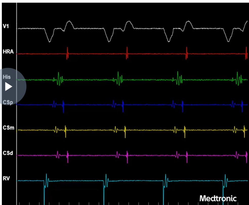
normal retrograde conduction
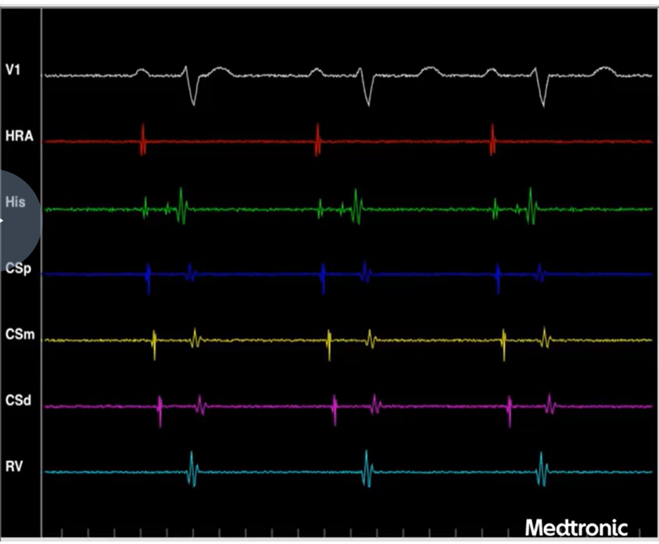
normal antegrade conduction
atrial pacing maneuvers
SNRT, Decremental Pacing, Extra Stimulus
SNRT
sinus node recovery time; test designed to asses Sinus node automaticity and analyze overdrive suppression
overdrive suppression
the temporary slowing of automaticity seen when an automatic focus is exposed to rapid, extrinsic external stimuli
mechanism of SNRT
a quadripolar catheter placed in the high right atrium near the sinus node is paced at rates faster than the intrinsic heart rates for at least 30 seconds
sinus node automaticity recovers to the rate before pacing, and the return cycle length is slower because of overdrive suppression.
SNRT = interval in the HRA from the last paced complex to the first intrinsic atrial beat
(burst pacing)
SNRT values
normal SNRT = <1500ms
normal Corrected SNRT = <550ms
Decremental atrial pacing
used for wenckebach point; When the atria are paced at higher rates (decremental pacing) the slowing down of conduction through the AV node is demonstrated by prolongation of PR interval followed by a dropped atrial beat.
AV node slows down conduction because of
1. Small diameter of the Nodal myocytes (conduction velocity is a function of cell diameter)2.
2. Complex arrangement of cells- nodal myocytes are separated by extensive connective tissue (leading to slower conduction)2.
3. Poor electrical connections between cells- fewer gap junctions2.
AV wenckebach point is used for
1) Assessing AV node function, 2) induction of a tachycardia, 3) uncovering dual AV nodal physiology characterized by AH jump which we will explore in future lessons and 4) inadvertant induction of AF
normal AV wenckbach values
<450ms
atrial extra stimulus pacing
performed by delivering an extra systole ( S2 )at the end of the drive train of 6-8 beats (S1), this pacing maneuver is useful for
1. Evaluating the antegrade conduction properties of the normal A-V conduction system or a by-pass tract if present
2. Testing decremental conduction properties of the AV-node and uncovering dual AV nodal physiology. AH interval will progressively be longer as we shorten the S1-S2 coupling interval in normal individuals. Typically, when a slow pathway is present, there is a sudden increase in the AH interval as conduction blocks in the fast pathway and conducts more slowly over the slow pathway. This sudden increase in the AH interval (>50 ms) with a 10 ms shortening of the S1-S2 coupling interval is often called a jump3.
3. As the S1-S2 coupling interval is shortened it also helps evaluate the AV nodal effective refractory period (AVNERP) and the atrial effective refractory period (AERP). Normal AVNERP is 230-425 ms and normal AERP is between 170-300 ms3. The effective refractory period (ERP) is the longest S1-S2 interval that fails to capture or depolarize the tissue of interest1.
4. Inducing arrhythmias, this pacing maneuver is often used to induce AVNRT or AVRT.
ventricular pacing maneuvers
ventricular decremental pacing and extra stimulus pacing
ventricular decremental pacing purpose
establish the retrograde wenckebach cycle length of the AV conduction system.
ventricular decremental pacing mechanism
Pacing begins 100 ms faster than the intrinsic rate and is decreased every 10-20 ms every few beats until 1:1 VA conduction is no longer maintained seen in HRA
why isn’t the ventricle paced faster?
The ventricle is not paced faster than 250 ms because VT or VF maybe induced at higher rates of pacing
how to know if there is ventricular capture?
when every ventricular pacing spike is immediately followed by a wide QRS complex
the following is important to observe during decremental pacing in ventricles
did the pacing capture the ventricles?
is there VA conduction
what is the pattern of retrograde atrial activation
does VA time prolong with increased pacing rates
what is indicated if VA time is not prolonged?
there is a by-pass tract
concentric activation pattern
During incremental ventricular pacing notice the atrial activation pattern. In normal individuals, the first atrial signal after V pacing will appear in the His-channel, suggesting that the signal is travelling over the AV node from the ventricle to the atrium. The atrial signal next manifests on the CS proximal electrodes and displays a proximal to distal activation pattern.
eccentric activation pattern
the first atrial signal manifests in the CS distal electrode and travels from the distal to the proximal CS electrode suggesting the presence of a left-lateral by-pass tract
ventricular extra stimulus pacing purpose
by delivering progressively shorter coupling intervals with each pacing sequence. This pacing maneuver is useful for evaluating,
1. Evaluating the retrograde conduction properties of the normal A-V conduction system or a by-pass tract if present3. V-A conduction time will progressively lengthen due to decremental properties of the AV node with decreasing S1-S2 coupling interval.
2. As the S1-S2 coupling interval is shortened it also helps evaluate the retrograde AV nodal effective refractory period (retrograde AVNERP) and the ventricular effective refractory period (VERP). In most individuals retrograde block occurs at longer coupling intervals than antegrade block. Normal VERP is 170-290 ms.
3. Inducing arrhythmia, AVNRT or AVRT can be induced using this maneuver.