Topic 3.2: Muscoloskeletal - Skeletal system and bones
 Call Kai
 Learn
Learn Practice Test
 Spaced Repetition
 Match
Match Flashcards
 Knowt Play
1/49

There's no tags or description
Looks like no tags are added yet.
Name | Mastery | Learn | Test | Matching | Spaced | Call with Kai | Chat |
|---|
No analytics yet
Send a link to your students to track their progress
50 Terms
functions of skeletal system - list (6)
Support -> structural framework for the body -> attachment point for tendons of muscles
Protection - > encloses visceral organs
Movement -> skeletal muscles attach to bone -> contraction pulls on bone to produce movement
Mineral homeostasis -> stores minerals like NaCl and Ca2+ which can be released into blood on demand
Blood cell production -> red bone marrow within bones produces RBCs
Triglyceride storage -> yellow bone marrow stores triglycerides
division of skeletal system (2)
axial → bones of head, neck and trunk (~80)
appendicular → bones of limbs including pectoral and pelvic girdle (~126)
composition of bone (4)
form of connective tissue → ECM surrounding widely separated cells
35% organic components -> eg. collagen
65% inorganic hydroxyapatites -> eg. calcium phosphate, calcium carbonate
Composition allows bones to be strong and resilient -> minerals resist compression, collagen resists tension
compact bone - summary
dense and forms outer shell of all bones
Surrounds spongey bone or medullary cavity
Hollow pillars of bone matrix containing nerves and blood vessels
Composed of repeating structural units (osteons) -> consist of concentric lamellae arranged around central Haversian canal
Blood vessels and nerves = contained within Harversian canal
spongey bone - summary (5)
also called trabecular
always inside outer layer of compact bone
Not organised into osteons -> consist of lamellae which are arranged into thin lines (traveculae)
Organised into lines of compressive stress and tensile stress -> aligned to assist with weight bearing
Filled with red or yellow bone marrow as well as medullary cavity
periosteum - summary (2)
dense layer of vascular connective tissue that envelopes bones except at surface of joints
Attached to underlying bone via perforating fibres of collage -> extend into bone's extracellular matrix
general structure of bones (3)
compact bone sandwiching spongey bone
periosteum surrounding except at surface of joints
classifications of bones - list (4)
long bones
short bones
flat bones
irregular bones
classifications of bones - long bones (2 + examples)
longer than they are wide
bear a lot of weight
femur and humerus
classifications of bones - short bones (3 + examples)
similar width to length
stability and support with little movement
Sesamoid bones = special type that forms in a tendon
carpal bones of wrist
classifications of bones - flat bones (2 + examples)
thin, flattened usually a bit curved
protect vital organs and provide surfaces for large muscle attachment
sternum and skull bones
classifications of bones - irregular bones (1 + examples)
don’t fit into other categories because of complicated shapes
vertebrae
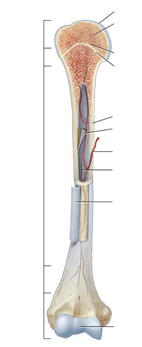
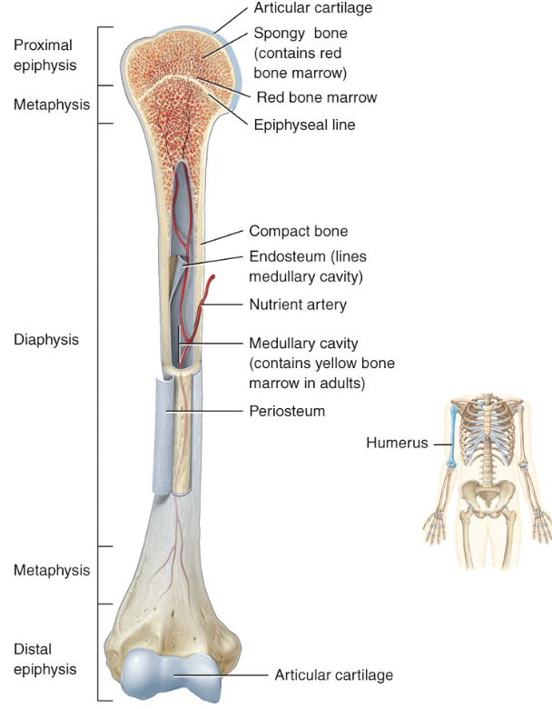
gross anatomy of long bones - left
proximal epiphysis
metaphysis
diaphysis
metaphysis
distal epiphysis
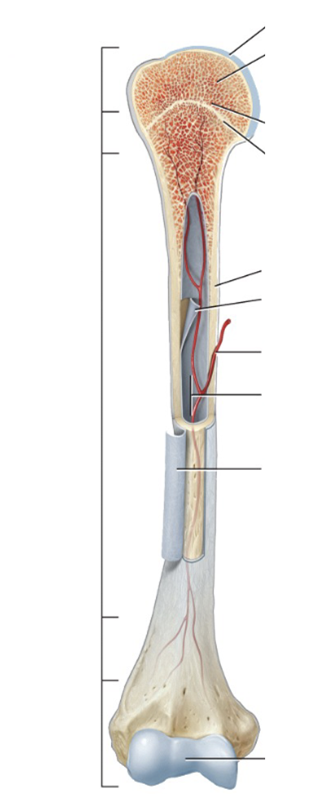
gross anatomy of long bones - top right
articular cartilage
spongey bone → red bone marrow
red bone marrow
epiphyseal line
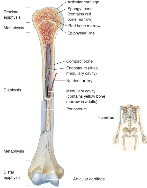
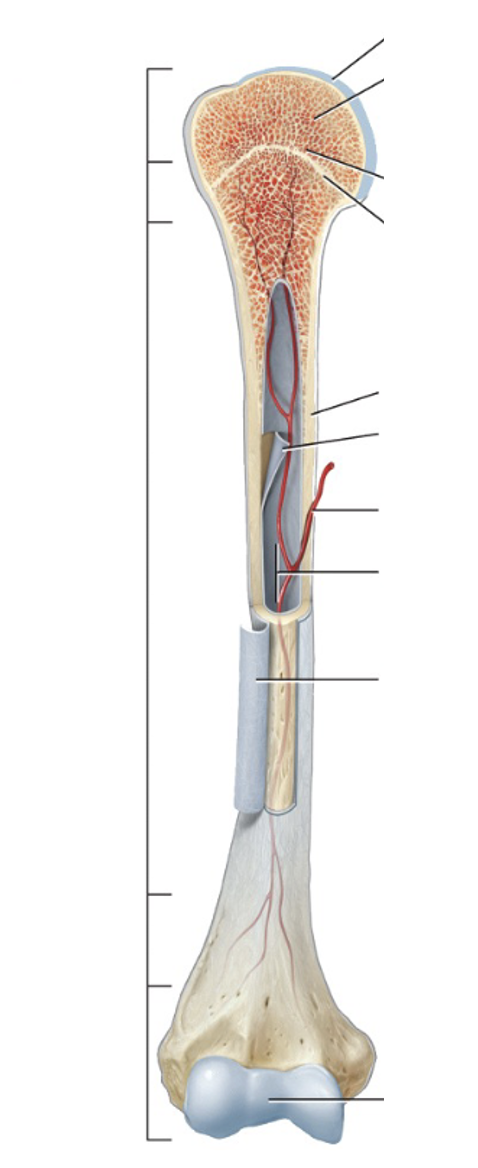
gross anatomy of long bones - bottom right
compact bone
endosteum
nutrient artery
medullary cavity → contains yellow bone marrow in adults
periosteum
articular cartilage
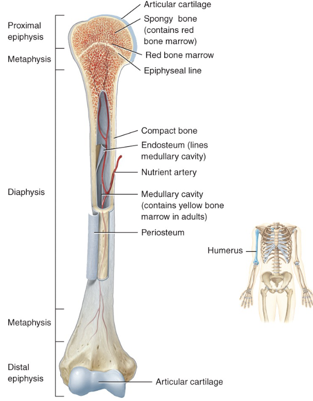
main structural regions of long bones (3)
Diaphysis = shaft of long bone -> long axis of bone
Epiphysis = proximal and distal ends
Metaphysis = regions between epiphysis and diaphysis
main structural regions of long bones - metaphysis in adults vs children
epiphyseal growth plate of hyaline cartilage in growing bone
epiphyseal line in adults
anatomy of long bones - membranes (2)
periosteum → outer double-layer fibrous covering entire surface except at joints
endosteum → thin membrane that lines medullary cavity, internal bone surfaces, trabecular and canals of compact bones
anatomy of long bones - vasculature (2)
Marrow cavity (medullary) = hollow space within diaphysis that contains yellow (fat) marrow and blood vessels
Nutrient artery = located outside of periosteum but heads into centre of bone towards medullary cavity
anatomy of long bones - articular cartilage def
hyaline cartilage covering the joint surface
blood supply of bone tissue and periosteum (3)
Periosteal arteries supply periosteum and outer compact bone -> enters through many small canals and perforates into compact bone
Nutrient artery enters via nutrient foramen at centre of diaphysis -> enters medullary cavity, courses
Ends of long bone = supplied by epiphyseal and metaphyseal arteries
nerves of bone tissue and periosteum
nerves accompany blood vessels
veins carry blood away from bone and accompany arteries → rich sensory nerve supply to periosteum
very sensitive to tearing and tension
bone growth and remodelling cells (2)
Osteoblasts = bone-building cells
Synthesise and secrete collagen fibres, organic components, to built extracellular matrix of bone
Initiate calcification
Osteoclasts = break down bone ECM -> bone resorption
Release lysosomal enzymes and acids to digest proteins and mineral components of ECM
ossification - def
bone formation
bone development from mesenchyme processes - list (2)
intramembranous ossification → directly from mesenchyme
endochondral ossification → from cartilage derived from mesenchyme
intramembranous ossification - summary (2)
Mesenchymal models of bone form during the embryonic period
Direct ossification occurs in foetal development
endochondral ossification - summary (2)
Cartilage models of bones form from mesenchyme during foetal period
Bone replaces cartilage
intramembranous ossification - steps (4)
Development of ossification centre
Chemical signalling tells mesenchymal cells to cluster together and differentiate
Osteoblasts secrete organic extracellular matrix
Calcification
Calcium and other mineral salts are deposited and ECM calcifies
Formation of trabecular
ECM develops into trabeculae that fuse to form spongey bone around blood vessels
Development of periosteum and compact bone
Mesenchyme at periphery of the bone develops into the periosteum
Periphery of spongey bone will become compact bone tissue
intramembranous ossification - example bones (5)
Flats bones of skull
facial bones
mandible
medial part of clavicle
hardening of fontanelles
endochondral ossification of long bones - steps (6)
Development of cartilage model
Mesenchymal cells develops into chondroblasts -> form hyaline cartilage model
Growth of cartilage model via cell division of chondrocytes
Development of primary ossification centre
At mid-region of cartilage model, cartilage hypertrophies and calcifies → periosteal capillaries grow into calcified cartilage to supply interior
Nutrients supplied by capillaries initiates primary ossification centre → becomes diaphysis
Development of medullary
As primary ossification centre grows towards end of bone, osteoclasts breakdown some of newly formed bone -> forms medullary cavity
Development of secondary ossification centres
Branches of epiphyseal artery enter still forming bone of proximal and distal epiphyseal -> blood supply triggers development of secondary ossification centre in ends of long bones
Formation of articular cartilage and epiphyseal plate
Spongey bones remains in interior bone rather than being broken down by osteoclasts
Bone growth occurs from the articular cartilage in the epiphyseal plate
bone growth in adolescence to adulthood (3)
Bone growth occurs in length and diameter until ~25 years -> only in diameter past 25
To avoid altering joint, bone growth occurs from diaphyseal side of epiphyseal plate -> avoid changing shape of epiphysis
Epiphyseal plate begins like as epiphyseal cartilage that allows bone to grow in length and ossifies once bone length is achieved
longitudinal bone growth - summary steps (2)
Interstitial growth of cartilage on epiphyseal side of epiphyseal plate via chondrocyte proliferation
Replacement of cartilage on diaphyseal side with bone via endochondral ossification
epiphyseal plate - zones (4)
resting cartilage
proliferating cartilage
hypertrophic cartilage
calcified cartilage
epiphyseal plate - zones of resting cartilage
Small, scattered chondrocytes
cells don’t function in cell growth but act as anchor for epiphysis to already formed bone
epiphyseal plate - zones of proliferating cartilage (3)
Chondrocytes slightly larger than those in resting cartilage and undergo interstitial growth
Cells arranged like stacks of coins
Divide and replace those that die at diaphysis side
epiphyseal plate - zones of hypertrophic cartilage
Mainly matured chondrocytes arranged into columns
epiphyseal plate - zones of calcified cartilage (2)
Very thin (usually one cell thick) and consists of dead chondrocytes -> ECM has calcified so chondrocytes dead
Osteoclasts dissolve calcified cartilage whilst osteoblasts and capillaries invade area -> bone
appearance of epiphyseal plate in juvenile vs adult bones
appears as darker bone in juvenile bones
epiphyseal cartilage no longer visible in adults (faint line remains)
appositional bone growth - summary (3)
Growth in width/ thickness
new bone deposited on outer surface whilst older bone lining medullary cavity destroyed
Medullary cavity enlarges as bone increases in thickness -> osteoblasts become osteoclasts and destroy bone
bone remodelling rates (3)
differ between bones and parts of same bone
5-7% of bone mass is recycled every week
Spongey bone replaced every 3-4 years
Compact bone replaced every 10 years
bone remodelling - summary process
combination of bone deposit by osteoblasts and bone resorption by osteoclasts
bone remodelling - purpose (2)
to maintain proper bone proportions during growth
change shape in response to mechanical stress
factors affecting bone growth and remodelling (4)
vitamins and minerals → calcium hardens bone ECM and Vit D increases calcium absorption
exercise → weight bearing activities stimulate osteoblasts
hormones → estrogen and testosterone stimulate bone deposition by osteoblasts
age → demineralisation due to decreasing hormone levels as activity of osteoclasts outpaces osteoblasts
types of bone markings - list (3)
sites of muscle and ligament attachment
projections that help form joints
depressions and openings for passage of nerves and vessels
types of bone markings - sites of muscle and ligament attachment
Created by stress of muscles pulling on bone
Eg. gluteal tuberosity on femur -> attachment for the gluteus maximus
types of bone markings - projections that help to form joints
Special shapes that connect together whilst still allowing movement
Eg. radius and ulna articulate with humerus but still allow flexion and extension of elbow
types of bone markings - depressions and openings for passage of nerves and vessels
Lots in skull for cranial nerves, carotid arteries and jugular veins passing in and out of skill
Eg. foramen magnum (occipital bone) -> passageway for spinal cord
types of bone markings - sites of muscle and ligament attachment examples (8)
tuberosity
crest
trochanter
line
tubercle
epicondyle
spine
process
types of bone markings - projections that help to form joints examples (4)
head
facet
condyle
ramus
types of bone markings - depressions and openings for passage of nerves and vessels examples (7)
grove
fissure
foramen
notch
meatus
sinus
fossa