Family Medicine: Cardiology
 Call Kai
 Learn
Learn Practice Test
 Spaced Repetition
 Match
Match Flashcards
 Knowt Play
1/109

There's no tags or description
Looks like no tags are added yet.
Name | Mastery | Learn | Test | Matching | Spaced | Call with Kai | Chat |
|---|
No analytics yet
Send a link to your students to track their progress
110 Terms
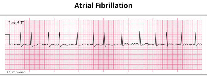
Atrial Fibrillation
Irregularly irregular rhythm with disorganized atrial activity and absence of P waves
-RF: HTN, valvular disease, CAD, HF, hyperthyroidism, OSA
-Dx: EKG, ECHO, CBC, TSH, CMP, BNP
-Tx: unstable needs immediate synchronized cardioversion, stable patients can be rate controlled first with beta blocker (metoprolol) or CCB (diltiazem), rhythm control for symptomatic/HF/young patients, anticoagulation with DOAC
CHADS-VASC
What scoring system is used to determine if a patient with A. Fib needs to be anticoagulated?
-Score of greater than 2 for men or 3 for women
Atrial Flutter
Regular rhythm with a sawtooth pattern (flutter waves) and a narrow QRS complex, making an atrial rate of around ~250-300 and ventricular rate ~150 with a 2:1 block
-Presentation: palpitations, tachycardia
-RF are the same as A. Fib
-Dx: EKG, ECHO / TEE to rule out thrombus before cardioversion
-Tx: cardioversion if unstable, stable can be rate controlled, catheter ablation is highly effective

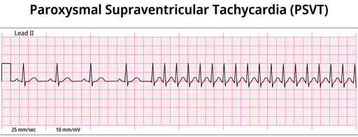
Paroxysmal Supraventricular Tachycardia
Narrow-complex tachycardia with no discernable P waves, with rates typically 150-250 bpm
-Presentation: patient of any age with sudden onset of palpitations and lightheadedness, hemodynamically stable
-Tx: stable can be treated with vagal maneuvers then IV adenosine 6 mg rapid push, cardioversion if unstable, long term can be managed with AV nodal blockers like CCBs and beta blockers, catheter ablation if symptomatic
Sick Sinus Syndrome
Dysfunction in the sinus node’s automaticity and impulse generation, which can present as brady-tachy syndrome or sinus arrest
-Most often in elderly with syncope, fatigue, or palpitations
-Tx: permanent pacemaker if symptomatic

Sinus Arrhythmia
Normal, minimal variation in the SA node’s pacing rate in associated with the phases of respiration. The heart rate frequently increases with inspiration and decreases with expiration
-No treatment required, physiologic finding

First Degree
What type of AV block is being described?
-Prolonged conduction through the AV node, making the PR interval > 200 ms but consistent beat-to-beat
-Usually benign and caused by increased vagal tone, inferior MI, medications, Lyme disease, and aging
-Tx: no tx required, monitor
Mobitz 1 (Wenckebach)
What type of AV block is being described?
-Progressive PR prolongation until a P wave is blocked, then the cycle resets (Longer, longer, longer, drop)
-MCC is inferior MI, increased vagal tone, medications
-Tx: usually nothing is indicated, atropine if symptomatic bradycardia, rarely needs pacing

Mobitz 2
What type of AV block is being described?
-Constant PR interval with sudden, unpredicted dropped QRS. The block is at or below the Bundle of His, making it a high risk lesion (Some dropped, some get through)
-High risk of progression to complete heart block
-Tx: permanent pacemaker

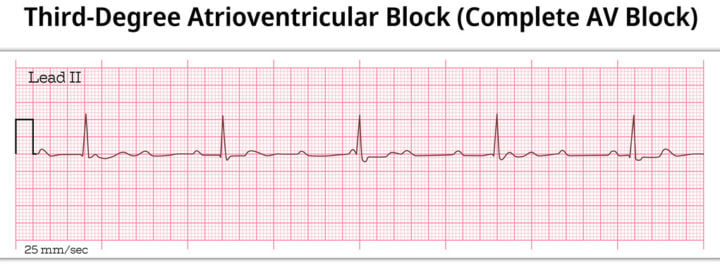
Third Degree
What type of AV block is being described?
-Complete failure of conduction from atria to ventricles, where the P waves and QRS complexes march independently (Ps and Qs don’t agree)
-Tx: transcutaenous pacing immediately if unstable, permanent pacemaker required
Junctional Rhythm
Rhythm originating in the AV junction when the SA node fails or slows, with a rate typically around 40-60 bpm or 60-100 bpm
-EKG shows narrow QRS, no preceding P wave or inverted P waves in II, III, aVF
-MCC is digoxin toxicity
Premature Junctional Contraction
Early ectopic beat originating in the AV junction
-EKG shows narrow QRS complex, no P wave or inverted P wave
-Dx: EKG
-Tx: observation if asymptomatic, symptomatic bradycardia requires a permanent pacemaker, reverse digoxin toxicity
PAC
Early atrial beat with an abnormally shaped P wave, typically with a narrow QRS complex
-Common triggers are caffeine, stress, alcohol, hyperthyroidism, electrolyte abnormalities

PVC
Early wide “bizarre” QRS without a P wave, often followed by a compensatory pause
-Common, usually benign
-Can be bigeminy, trigeminy, couplets, or triplet

Ventricular Tachycardia
Three or more consecutive ventricular premature beats, with a wide QRS complex and tachycardia
-Considered sustained if > 30 seconds
-Tx: immediate defibrillation if symptomatic, stable can have an IV antiarrhythmic (amiodarone) or cardioversion

Ventricular Fibrillation
Erratic rhythm with no discernable waves
-Hemodynamically unstable → pulseless, leads to sudden cardiac death within minutes if untreated
-Tx: immediate defibrillation + CPR per ACLS protocol, Epi 1 mg every 3-5 minutes, amiodarone 300 mg IV after second shock

Torsades de Pointes
Polymorphic ventricular tachycardia that appears to be twisting around a baseline, which is associated with prolonged QT interval
-Tx: IV magnesium sulfate, correct electrolytes and DC all QT prolongated medications

Dilated Cardiomyopathy
Most common cardiomyopathy, characterized by LV dilation + systolic dysfunction
-Causes: idiopathic (MC), ischemic, viral
-Dx: ECHO shows reduced EF, dilated LV, BNP is elevated, CXR shows cardiomegaly
-Tx: ACE/ARB/ARNI + beta blocker + spironolactone + SGLT2

Hypertrophic Cardiomyopathy
Cardiac abnormality that leads to the muscle in the wall of the heart growing and thickening to the point that it blocks blood flow exiting the heart
-Autosomal dominant condition due to mutations in beta-myosin
-Dx: ECHO shows asymmetric septal hypertrophy with dynamic LVOT obstruction
-Presentation: dyspnea, angina, syncope with a high pitched mid-systolic murmur at the LLSB that increases with standing/Valsalva
-Tx: beta-blockers or verapamil for symptoms, avoid vasodilators, surgical myomectomy is gold standard
Restrictive Cardiomyopathy
Stiff, non-compliant ventricles with impaired diastolic filling
-MCC cardiac amyloidosis and sarcoidosis
-Presentation: Kussmaul sign (JVP rises with inspiration)
-Dx: ECHO shows thickened walls and diastolic dysfunction, endomyocardial biopsy is definitive, avoid digoxin
Acyanotic
Are these congenital heart diseases cyanotic or acyanotic?
-Left-to-right shunt
-VSD, ASD, PDA, coarctation of the aorta
VSD
Hole forms in the wall that separates the right and left ventricles of the heart, which allows too much blood to be pumped in the lungs
-Murmur: holosystolic at LLSB with an associated thrill
-Presentation: large presents with CHF symptoms and poor feeding, smalls with a loud murmur
-Dx: ECHO
-Tx: small can be observed, large need surgical/catheter closure
ASD
Small hole forms between the atria, which is the most common CHD presenting in adults
-Presentation: fixed, widely split S2 that does not vary with respiration + systolic flow murmur at LUSB
-Tx: percutaneous transcatheter closure
PDA
Failure of closure of the fetal ductus arteriosus, which is common in premature infants
-Murmur: continuous “machinery” murmur at left infraclavicular area, bounding pulses, wide pulse pressure
-Tx: give premature infants indomethacin to close the PDA, full term needs catheter procedure
Coarctation of the Aorta
Narrowing of the aorta, just distal to the left subclavian artery
-Presentation: upper extremity HTN with diminished/delayed femoral pulses, BP differential between arms and legs
-Dx: CXR shows rib notching
-Tx: catheter based balloon angioplasty + stenting

Cyanotic
Are these congenital heart defects cyanotic or acyanotic?
-Right to left shunt, causing deoxygenated blood to reach systemic circulation
-Tetralogy of Fallot, Transposition of the Great Arteries, Truncus Arteriosus
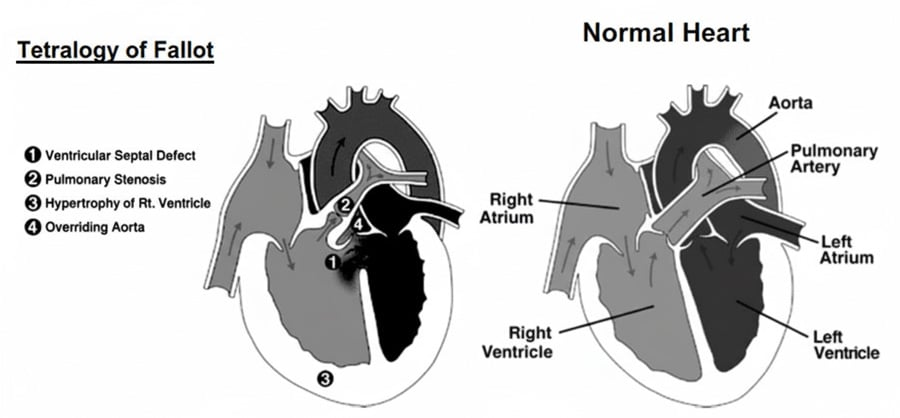
Tetralogy of Fallot
Most common cyanotic CHD, which is a combination of pulmonary stenosis, RV hypertrophy, overriding aorta, and VSD
-Presentation: paroxysmal cyanosis, hyperpnea, irritability triggered by crying or feeding which is relieved by squatting
-CXR: boot-shaped heart
-Tx: complete surgical repair
Transposition of Great Arteries
Aorta arises from RV, PA arises from LV, leading to two non-communicating circuits
-Incompatible with life without a mixing lesion (ASD, VSD, PDA)
-Dx: CXR shows “egg on string” sign
-Tx: prostaglandin E1 immediately to maintain PDA
Unstable Angina
New-onset, rest, or worsening angina
-No rise in troponin, due to partial occlusion of coronary artery
-Dx: 12 lead EKG within 10 minutes of presentation, check troponin I or T, CXR, ECHO
-Tx: morphine, oxygen, nitrates, aspirin, beta-blocker, ACE inhibitor, statin, heparin + ticagrelor
NSTEMI
ST-segment depression or T-wave inversion with elevated biomarkers
-Due to partial occlusion
-Dx: 12 lead EKG within 10 minutes of presentation, high sensitivity troponin I or T
-Tx: MONA
STEMI
ST-segment elevation > 1 in 2 contiguous leads or new LBBB
-EKG diagnosis, activate the cath lab, do not wait for troponin
-Tx: MONA, emergent PCI within 90 minutes, fibrinolysis within 30 minutes
Stable Angina
Chest pain that is predictable, relieved by rest and/or nitroglycerin, which is indicative of stable, flow-limiting atherosclerotic plaque
-Presentation: classic exertional substernal pressure/tightness ± radiation to left arm, jaw, neck. This is reproducible with exertion or emotional stress
-Dx: EKG, stress testing for diagnosis and risk stratification, coronary CT angio
-Tx: lifestyle modifications, aspirin 81 mg daily, statin, beta blocker, nitro PRN
Prinzmental Angina
Coronary artery vasospasm causing transient ST-segment elevations, which typically occurs at rest or in the early morning
-Triggered by smoking, cocaine, amphetamines, ergot alkaloids, stress, cold exposure and is associated with Raynaud phenomenon and migraines
-Dx: transient ST elevation that responds to nitro, normal troponin, coronary angiography
-Tx: CCBs are first line, smoking cessation
Heart Failure
LV remodeling produces dilation, thinning, and mitral valve incompetence
-Presentation: exertional dyspnea that progresses to be at rest, orthopnea, nonproductive cough, fatigue, nocturia. Can have crackles/rales, JVD, edema, S3 (HFrEF) or S4 (HFpEF)
-Dx: ECHO, BNP, CXR. A BNP less than 100 makes HF unlikely, EKG, CXR
-Tx: ARNi + beta-blocker + MRA + SGLT2, avoid CCBs
Class I
What class of heart failure is being described?
-No limitation of physical activity
Class II
What class of heart failure is being described?
-Slight limitation of physical activity, comfortable at rest
Class III
What class of heart failure is being described?
-Marked limitation of physical activity, comfortable at rest
Class IV
What class of heart failure is being described?
-Unable to carry on any physical activity without discomfort, symptoms of angina at rest
CCBs, NSAIDs, TZDs
What three classes of drugs should be avoided in HFrEF?
Primary Hypertension
Resting BP > 130/80 on at least two readings on at least two separate visits with no identifiable cause
Elevated
What is this BP classified as?
120-129 / < 80
Stage 1
What is this BP classified as?
130-139/80-89
Stage 2
What is this BP classified as?
>140/>90
< 130/80
Per the ACC/AHA blood pressure guidelines, what is the target BP in someone with comorbidities or diagnosed with hypertension?
Lifestyle changes, reassess
What are the two pillars of treatment for someone with elevated BP?
-In 3-6 months after starting
Medication
What should be started in a patient with Stage 1 HTN with CVD, DM, CKD, or a PREVENT risk > 7.5%?
2 medications
What should be started in a patient with stage 2 HTN?
-Preferably in a single-pill fixed-dose combination
ACE/ARB, CCB
What types of medications for HTN can be started in non-black patients?
Thiazides, CCBs
What two classes of blood pressure medications are most effective for black patients?
ACE/ARB
What HTN medication classes should be started in a patient with DM and proteinuria, HFrEF, or post-MI?
ACE
What class of anti-HTN medications can cause cough, angioedema, and hyperkalemia?
-Contraindicated in pregnancy
Hyperkalemia
What electrolyte imbalance is associated with spironolactone use?
Spironolactone
What agent can be added to resistant hypertension (uncontrolled on 3 drugs, including a diuretic)?
Secondary Hypertension
HTN with an identifiable, often correctable, cause. This should be suspected if onset < 30 or > 55 years, sudden onset, resistant HTN, HTN with hypokalemia, or organ damage out of control to HTN duration
-Causes: aldosteronism, apnea, bad kidneys, coarctation of aorta, cushing syndrome, drugs, endocrine, renal artery stenosis
-Dx: BMP, UA, thyroid, aldosterone-to-renin ratio, plasma metanephrines for pheochromocytoma
-Tx: underlying cause treatment
Hyperaldosteronism
What is the most likely cause of secondary hypertension in this patient?
HTN + hypokalemia + metabolic alkalosis
Fibromuscular Dysplasia
What is the most common cause of renal artery stenosis in a young woman?
-Tx: balloon angioplasty
Hypertensive Urgency
Very high BP (> 180/120) without target-organ damage
-Tx: restart or intensify long-acting oral antihypertensives, close outpatient follow up
Clonidine
What antihypertensive agent should be avoided in patients due to its risk of rebound HTN with a missed dose?
Hypertensive Emergency
Severe HTN with signs of damage to target organs, such as retinal hemorrhages, papilledema, encephalopathy, AKI, intracranial hemorrhage, aortic dissection, pulmonary edema, or MI
-Tx: admit to ICU for titratable IV infusion to lower MAP by no more than 25% in the first hour, then to <160/100 over 2-6 hours, then to normal over the next 24-48 hours
Orthostatic Hypotension
Sustained drop in SBP > 20 or DBP > 10 within 3 minutes of standing
-Presentation: lightheadedness, dizziness, presyncope/syncope, blurred vision, weakness
-Common Causes: volume depletion, medications, DM, Parkinson disease, endocrine, elderly
-Dx: orthostatic vital signs
-Tx: non-pharmacologic is first line with medication review/compression/slow position changes, Midodrine if refractory or Fludrocortisone
Vasovagal Syncope
MCC of syncope, triggered by exaggerated parasympathetic outflow + sympathetic withdrawal, which causes bradycardia and vasodilation
-Triggers: prolonged standing, pain, emotional stress, sight of blood, venipuncture, defecation
-Prodrome of lightheadedness, nausea, diaphoresis, pallor, blurred vision, warmth
-Dx: clinical, tilt-table if recurrent
-Tx: reassurance and education, avoid triggers, counter-pressure maneuvers, increased salt and fluid intake
20
At what age should you start screening all adults for lipid disorders?
40-75
Per the USPSTF, lipid testing is tied to the decision to start a statin in adults of what age range, rather than a fixed screening age?
Statin
What should be started in these patients regardless of calculated risk score?
-Any form of ASCVD → prior MI, ischemic stroke, PAD
-LDL > 190
-DM in those 40-75 years old
-CKD stage 3-4 or HIV in those 40-75
PREVENT
What calculator is used to determine the 10 year ASCVD risk?
-Low = < 3%
-Borderline = 3-4.9%
-Intermediate = 5-9.9%
-Higher = > 10%
Lipoprotein A
What should be measured once in adulthood?
-Greater than 125 nmol/L marks higher risk long-term
Coronary Artery Calcium Score
What is used to reclassify borderline/intermediate patients when the statin decision is uncertain?
55, 85
What are the LDL-C and non-HDL goals for a patient who is very high risk?
70, 100
What are the LDL-C and non-HDL goals for a patient who is high risk?
<100
What is the LDL-C goal for a patient who is borderline/intermediate?
Ezetimibe
What is the first PO medication to add for a patient on a statin if they are still not at goal?
Pancreatitis
A triglyceride level above 500 puts a patient at risk of what complication?
Fibrate + Omega 3s
What two medications should be given to a patient once their triglycerides are above 500?
Flushing
Niacin can be used as a supplement for triglyceride lowering. What is the most common side effect associated with it?
-Can be eased by taking ASA before
Aspirin
What can be initiated for primary prevention of cardiovascular disease in adults 40-59 with a > 10% 10-year risk but should be avoided in adults > 60?
Acute Pericarditis
Inflammation of the pericardial sac, which is often viral or idiopathic
-Presentation: sharp/pleuritic chest pain that is worse supine and relieved by sitting forward/leaning, pericardial friction rub
-Dx: EKG shows ST elevation widespread and PR depression but can be clinical
-Tx: NSAIDs + Colchicine BID for 3 months
Dressler Syndrome
Acute pericarditis post-MI
Aortic Stenosis
Obstruction of LV outflow of blood moving from the left ventricle into systemic circulation, which is due to a narrowing of the aorta
-MC in older patients or bicuspid AV in younger patients
-Presentation: angina, syncope, heart failure
-Murmur: systolic ejection crescendo-decrescendo at the RUSB/2nd intercostal space with radiation to the neck, split S2 that increases with squatting and expiration and decreases with hand grip
-Tx: valve replacement
Aortic Regurgitation
Aortic valve does not close fully during diastole, causing leakage of blood backward through the aortic valve into the LV during diastole
-Presentation cardiogenic shock (acute), wide pulse pressure (chronic)
-Murmur: soft, high-pitched, blowing diastolic murmur along the LLSB that increases with squatting, sitting, leaning forward, and handgrip
-Tx valve replacement, vasodilators
Mitral Stenosis
Obstruction of flow from the left atrium to left ventricle due to a narrowed mitral orifice
-Presentation: shortness of breath and CHF
-Murmur: diastolic low-pitched rumbling murmur with opening snap at the apex with the patient in the LLD position that increases with squatting, expiration, and LLD position
-Tx: diuretics, rate control, anticoagulation with warfarin, percutaneous balloon mitral valvuloplasty
Mitral Regurgitation
Mitral valve does not close fully, causing leakage into the LA during systole
-Presentation: syncope and shortness of breath
-Murmur: blowing holosystolic murmur at apex radiating to the axilla that increases with handgrip, squatting, expiration
-Tx: diuretics, ACEi, mitral valve replacement
Mitral Valve Prolapse
Leaflets of the mitral valve bulge or prolapse back into the left atrium during systole
-Associated with connective tissue disorders
-Murmur: late systolic murmur with a mid-systolic click which increases with standing, valsalva, and handgrip
-Tx: reassurance, beta blocker if symptomatic
Pulmonary Stenosis
Narrowing of the pulmonic valve, leading to obstruction of blood flow from the RV
-Presentation: dyspnea, fatigue, chest pain, palpitations
-Murmur: harsh systolic ejection crescendo-decrescendo murmur with widely split S2 at the LUSB that radiates to the left shoulder and neck, split S2. Increases with squatting and inspiration
-Tx: mild can be observed, mod-to-severe needs percutaneous balloon valvuloplasty
Pulmonic Regurgitation
Leakage of blood back through the pulmonic valve, which is most commonly caused by pulmonary hypertension
-Murmur: high-pitched, early diastolic decrescendo murmur at the LUSB and increases with inspiration
-Tx: address underlying pulmonary HTN
Tricuspid Stenosis
Narrowing of the tricuspid valve, impairing the RA and affecting RV flow
-MCC is rheumatic heart disease
-Presentation: mid-diastolic rumbling at the LLSB with an opening snap, increases with inspiration and squatting
-Tx: diuretics for symptomatic congestion
Tricuspid Regurgitation
Backward flow of blood from RV to RA during systole, which is most commonly caused by RV dilation
-Murmur: high-pitched holosystolic murmur at the LLSB that increases with squatting and inspiration
-Tx: treat underlying cause, diuretics, surgery
Rheumatic Heart Disease
Chronic valvular damage following acute rheumatic fever
-Most commonly affects the mitral valve
-Dx: clinical (Jones Criteria) + evidence of strep infection
-Tx: PCN V PO x 10 days or IM benzathine PCN G
Joints, Heart, Nodules, Erythema Marginatum, Sydenham chorea
What are the major parts of the JONES criteria?
-Need 2 major or 1 major + 2 minot criteria
-Minor: fever, elevated ESR/CRP, prolonged PR interval, arthralgia
Aortic Aneurysm
Tender, pulsatile abdominal mass
-RF: male, age > 65, smoking, family history, HTN
-Presentation (ruptured): sudden severe abdominal/back pain, pulsatile mass, hypotension
-Dx: abdominal US
-Tx: surveillance if < 5.5 cm, larger than that requires surgical repair
One-time abdominal US
What is the suggested screening for an abdominal aortic aneurysm in men 65-75 who have ever smoked?
Marfan Syndrome
What are thoracic aortic aneurysms often associated with?
Aortic Dissection
Tear in the aortic intima → blood dissects into media → creates false lumen
-Presentation: sudden, severe “tearing/ripping” chest or back pain with pulse deficits, BP differential between arms, new murmur
-RF: HTN
-Dx: widened mediastinum on CXR, CT angio is definitive
-Tx: surgical repair if type A, IV esmolol/labetalol to lower BP, type B uncomplicated needs medical management

Type A
Stanford classification of an AAA that involves the ascending aorta
Type B
Stanford classification of an AAA that involves only the descending aorta
Acute Limb Ischemia
Sudden arterial occlusion, causing ischemia distal to obstruction
-MC source of embolism is cardiac without anticoagulation (hx of A. Fib)
-Presentation: pain, pallor, pulselessness, paresthesias, paralysis, poikilothermia
-Dx: CTA
-Tx: IV heparin immediately, catheter thrombolysis vs embolectomy vs amputation
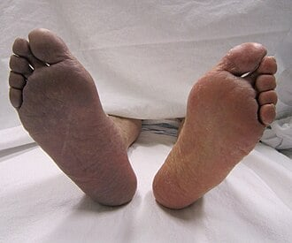
Compartment Syndrome
After treating a patient with acute limb ischemia, what complication should you monitor for?
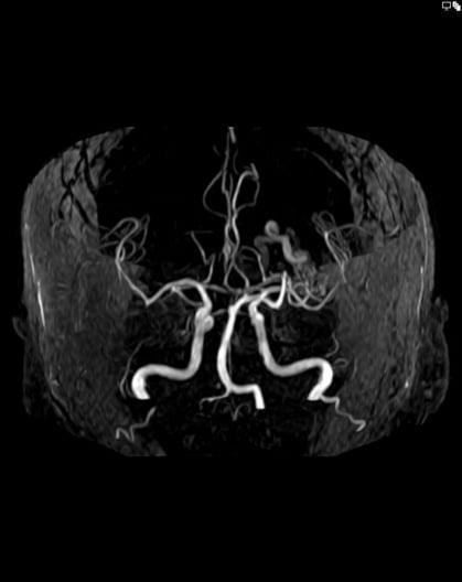
AVM
Abnormal direct connection between arteries and veins, bypassing the capillary bed and causing arteriovenous shunting
-Types: pulmonary, cerebral, hepatic, peripheral
-Dx: CT angiography
-Tx: embolization (pulm), brain surgery (cerebral)
Pulmonary AVM
Connection between arteries and veins in the lungs, causing a right-to-left shunt and hypoxemia that is refractory to oxygen
Atherosclerosis
Underlying pathology of most cardiovascular disease
-Patho: endothelial injury → LDL deposition in intima → oxidation → macrophage uptake → fatty streak → fibrous plaque → atheroma
-RF: smoking, HTN, DM, hyperlipidemia, obesity, sedentary lifestyle, diet
-Dx: lipid panel, BP monitoring
-Tx: lifestyle modifications, stain therapy, BP control, glycemic control
Deep Vein Thrombosis
Thrombus formation in deep veins, MC in the lower extremities
-Virchow’s triad → stasis + endothelial injury + hypercoagulability
-RF → prolonged immobility, surgery, malignancy, pregnancy, OCP/HRT use, thrombophilia, hx of VTE
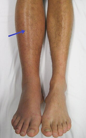
-Presentation: unilateral leg swelling, pain, warmth, erythema, Homans sign
-Dx: duplex ultrasonography, Wells DVT score
-Tx: DOACs first-line, LMWH if malignancy related, IVC filter if contraindication