Looks like no one added any tags here yet for you.
Excoriation
Raw linear lesion breaking epidermis
Macule
< 5mm diameter colored, flat, lesion
Patch
5mm diameter colored, flat, lesion
Papule
Elevated, domed or flat-topped lesion < 5mm in diameter
Nodule
5mm elevated lesion
Plaque
Elevated flat-topped lesion usually > 5mm in diameter
Pustule
Pus-filled, raised, discrete lesion
Vesicle
Fluid-filled raised lesion < 5mm in diameter
Bulla
5mm fluid-filled raised lesion
Wheal
Elevated, itchy, transient, erythemic/blanched lesion
Scale
Dry, plate-like formation
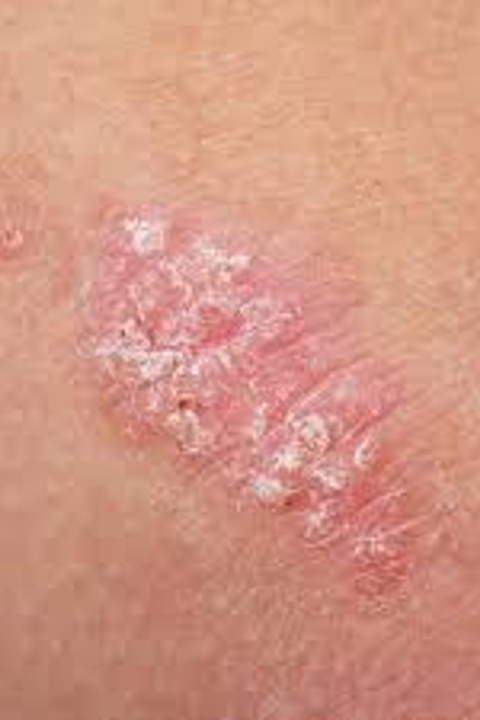
Psoriasis
Autoimmune disorder causing itchy, red scaling skin
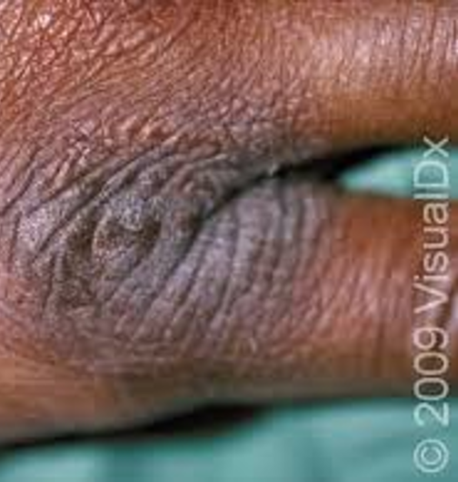
Lichenification
Thickened, rough skin caused by repeated rubbing
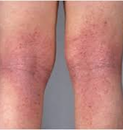
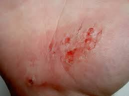
Eczema
Dry, itchy patches of skin aka dermatitis caused by contact with an environmental trigger
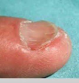
Onycholysis
Separation of the nail plate from the nail bed
Koilonychia
Flattening of the nail plate
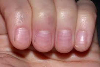
Muehrcke Nails
Alternating bands of white running parallel to the lunule
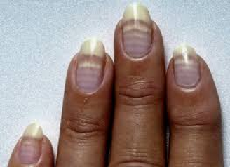
Mee’s Lines
Multiple alternating light/white bands running parallel to the lunule
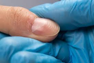
Beau’s Lines
Horizontal ridges on the nails
Abrasion
Scrape which may cover a small or large area
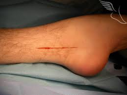
Incision
Linear cut by a sharp object
Laceration
Jagged tear in skin caused by a dull object or solid impact
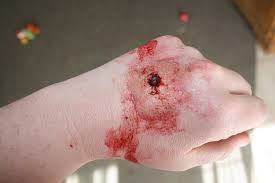
Puncture
Penetrating wound caused by slender, sharp object
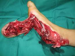
Avulsion
Mechanical force rips chunks of flesh out
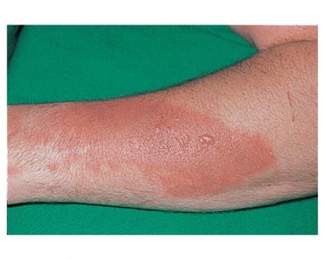
First-degree burns
Only epidermis is damaged; skin is red and swollen
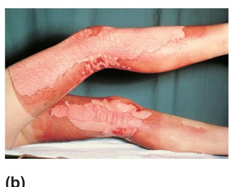
Second-degree burns
Epidermis and upper dermis damaged; skin is red with blisters
Third-degree burns
Destroys entire skin layer; burned area is gray-white or black
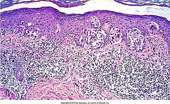
Melanoma
2% of skin cancers but causes most skin cancer deaths
Basal Cell Carcinoma
Most common skin cancer; arises in stratum basale, rarely metastasizes
Squamous Cell Carcinoma
Second most common skin cancer; more common in men than women
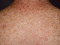
Ephelis (freckles)
Most common childhood pigmented lesion; small tan-red or light-brown macules caused by sun exposure
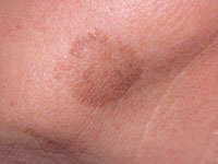
Lentigo
Small tan-brown macules or patches that do not darken with sun exposure
Nevi
Congenital skin lesions; important to differentiate from melanoma
rules of nine
anterior & posterior head & neck: 9%
anterior & posterior upper limbs: 18%
anterior & posterior trunk: 36%
anterior & posterior lower limbs: 36%
perineum: 1%
critical burn
>25% 2nd
>10% 3rd
3rd face, hands, feet
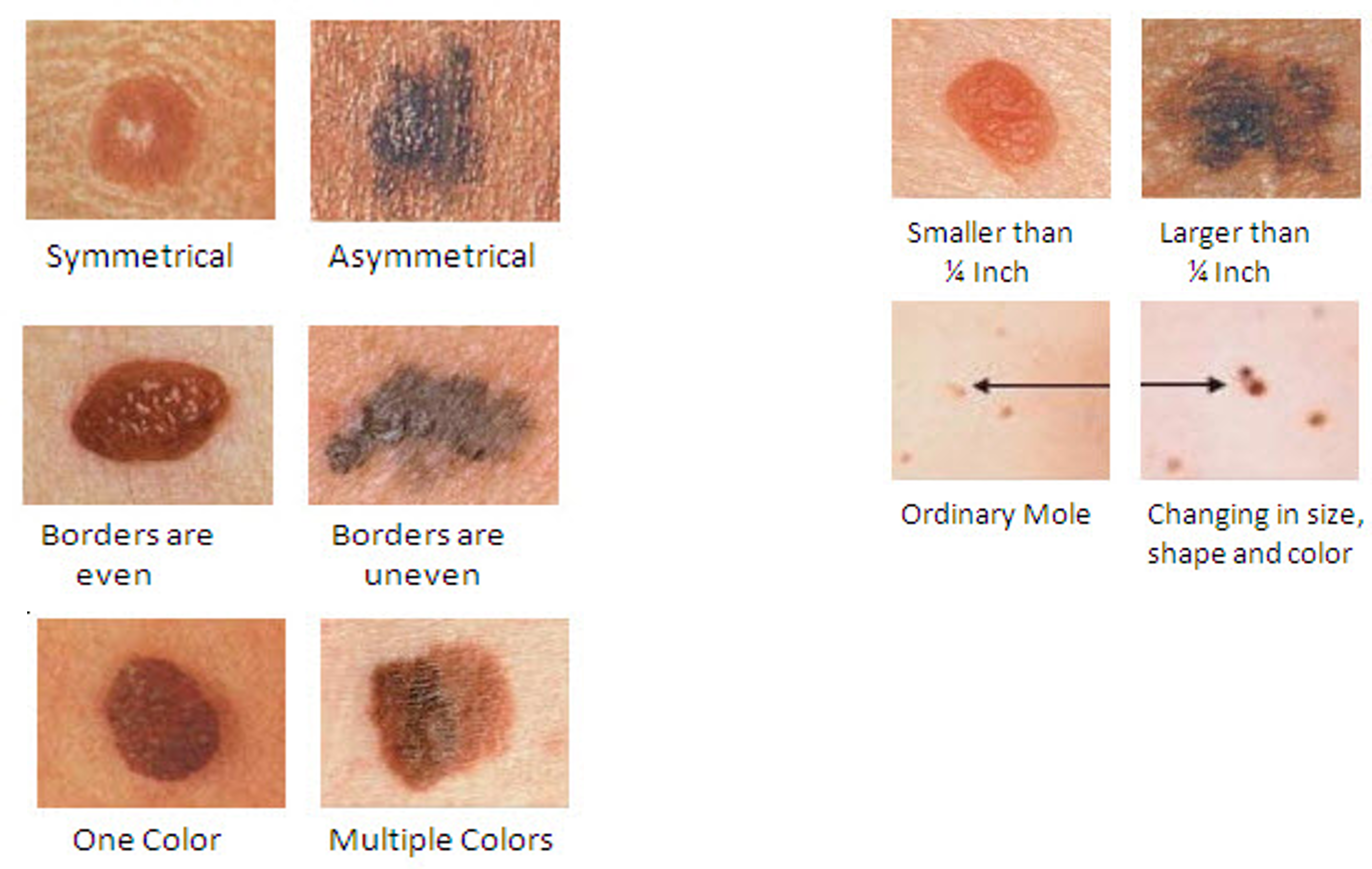
asymmetry, border irregularity, color, diameter, evolution
ABCDE Rule
D >6mm
ugly duckling
Developed by Grob et al in 2008, this asserts that nevi tend to resemble each other on an individual basis, so any deviation is diagnostic of melanoma.
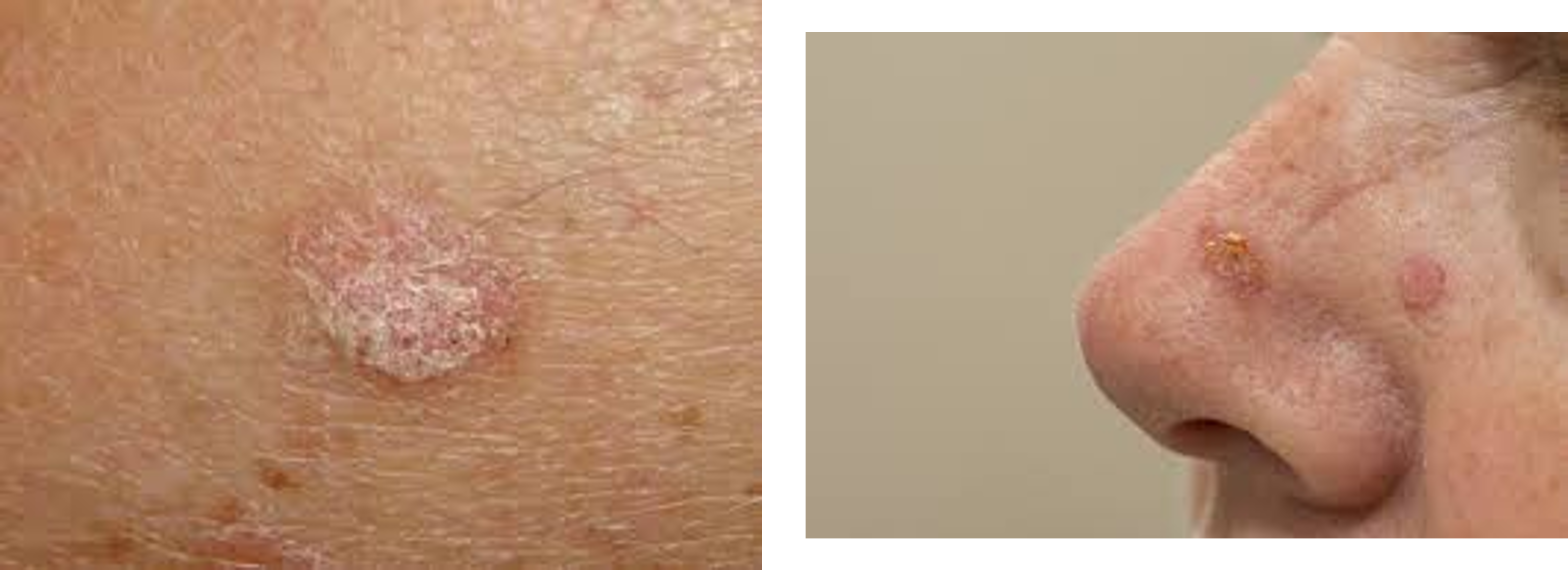
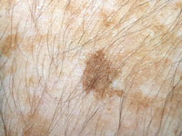
Actinic Keratoses
Also known as Solar or Senile keratoses, rough, scaly patches on sun-exposed skin. They are considered precursors to skin cancer, particularly squamous cell carcinoma. These lesions occur due to prolonged ultraviolet (UV) light exposure and are most common in fair-skinned individuals over the age of 40. Actinic keratoses can vary in color, from tan to red to brown, and may feel dry or itchy. Regular monitoring and potential treatment, such as cryotherapy or topical medications, are recommended to manage these lesions.
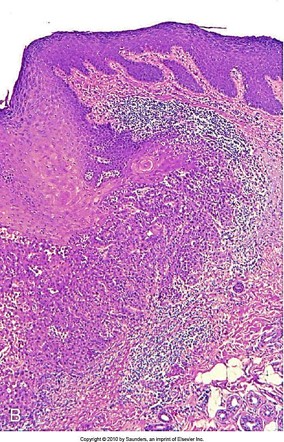
tongue, squamous cell carcinoma
Structure’s name and corresponding disorder
simplex
solar
tanning bed
profusa
junctional nevi
compound nevi