Pathology of Haemic & Lymphoid System
1/120
There's no tags or description
Looks like no tags are added yet.
Name | Mastery | Learn | Test | Matching | Spaced | Call with Kai |
|---|
No analytics yet
Send a link to your students to track their progress
121 Terms
What are the 3 reactions of circulating blood cells to injury?
Decreased survival
Altered distribution
Changes in structure or function
Term for Decreased Erythrocyte (RBC)
Anemia
Term for Decreased Reticulocyte (Immature RBC)
Reticulopenia
Term for Decreased Leukocyte (WBC)
Leukopenia
Term for Decreased Neutrophil
Neutropenia
Term for Decreased Lymphocyte
Lymphopenia
Term for Decreased Monocyte
Monocytopenia
Term for Decreased Eosinophil
Eosinopenia
Term for Decreased Basophil
Basopenia
Term for Decreased Platelets
Thrombocytopenia
Term for increased Erythrocyte
Erythrocytosis
Term for increased Reticulocytes
Reticulocytosis
Term for increased Leukocytes
Leukocytosis
Term for increased Neutrophils
Neutrophilia
Term for increased Lymphocytes
Lymphocytosis
Term for increased Monocytes
Monocytosis
Term for increased Eosinophils
Eosinophilia
Term for increased Basophils
Basophilia
Term for increased Platelets
Thrombocytosis
A broad term for Erythrocytes with ABNORMAL SHAPES, w/c can be subclassified based on specific shape changes.
Poikilocytes
T or F: Some specific shapes of RBC have unique diagnostic significance, while others are more non-specific.
True
The diagnostic significance of poikilocytes depends on these 3 factors:
Number
Shape
Context
True or False: Few misshapen RBC in blood from normal or ill animals may not hold a diagnostic value.
True
What are the 8 Types of Pokilocytes
D E C A D E S S
Dacryocyte
Echinocyte
Codocyte
Acanthocyte
Drepanocyte
Elliptocytocyte
Schizocyte
Spherocyte
This type of Poikilocyte is spherical w/ about 3-12 blunt tip of club-shaped spicules of varying lengths on their surface. These spicules are asymmetric or irregular.

Acanthocyte
This type of Poikilocyte is spiculated, & is a.k.a. crenated or “BURR“ cells. This has uniformed sharp spicules w/c are evenly spaced.
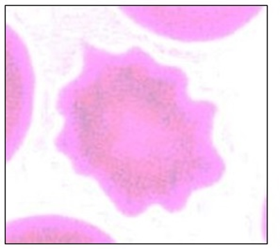
Echinocyte
This type of poikilocyte is linked to liver disease and lipid metabolism disorders.
Acanthocyte
This type of poikilocyte can be an artifactual change seen in stored or “old“ blood
Echinocyte
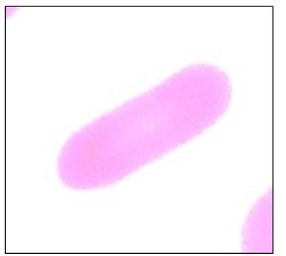
This type of Poikilocyte is a.k.a. the “Teardrop cell” & is associated with fibrotic states of marrow, hemolytic anemia, & drug reactions such as phenothiazine & chloramphenicol.
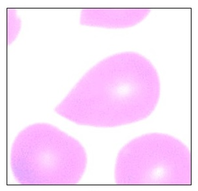
Dacryocyte
What are the 3 things Dacryocyte can be associated with?
Fibrotic marrow
Hemolytic Anemia
Drug reactions with PhenoThiazine & ChoramPhenicol
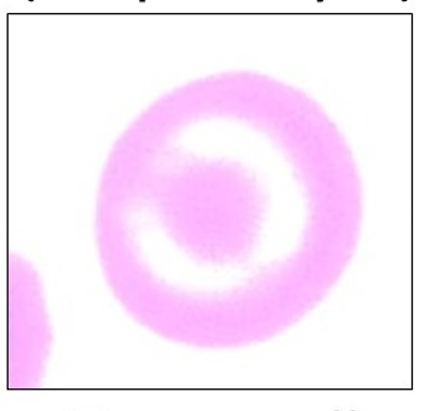
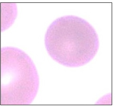
This type of Poikilocyte is a.k.a. “Target Cells” or Leptocytes. It has a normal central pallor, resembling a Bull’s eye
Codocyte
This type of Poikilocyte develops from excess membrane & decreased HgB (hemoglobin). It is also associated w/ Iron deficiency Anemia, Obstructive liver disease & Cirrhosis
Codocyte
What are the 3 diseases codocytes can be associated with?
Iron deficiency anemia
Obstructive liver disease
Cirrhosis
This type of Poikilocyte is a fragmented RBC that may be due to mechanical injury or through the trapping of RBC in the circulation by fibrin. (Ex. Thrombosis)
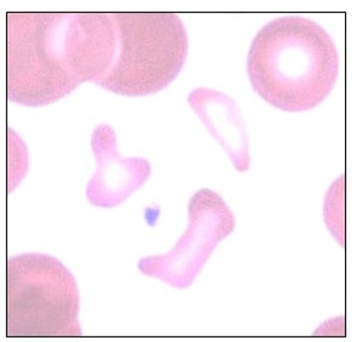
Schizocyte
This type of Poikilocyte is common in DIC (Disseminated Intravascular Coagulation), & iron deficiency anemia
Schizocyte
This type of Poikilocyte is normal in young ruminants
Schizocyte
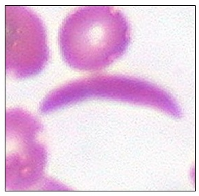
This type of Poikilocyte is a.k.a. “Sickle cell” & is normal in deers.
Drepanocyte
Drepanocytes are poikilocytes associated with 2 things w/c are:
Blood parasites (Malaria & Trypanosomiasis)
Recessive gene defect in humans
This poikilocyte is elongated & has 3 types which distinctions are not clinically relevant, though some forms may occur more frequently in certain diseases.
Elliptocytes
What are the 3 types of Elliptocytes?
Type 1: Slightly oval-shaped & is formerly known as ovalocyte
Type 2: Rounded- oval shape
Type 3: Elongate elliptical
This type of poikilocyte indicates the presence of red cells with increased central thickness.
Spherocyte
What are the 2 causes of spherocytosis?
Membrane depletion
Accelerated RBC aging (As a result of a pathological process such as Immunohemolytic Anemia)
What are the 2 terms that refer to an increase in Red Cell Mass
Polycythemia
Erythrocytosis
What are the 2 things that can happen if there is polycythemia / erythrocytosis?
Hypervolemia
Hyperviscocity
What are the 4 subsequent disease manifestations of hypervolemia and hyperviscosity resulting from erythrocytosis / polycythemia?
Polyuria
Polydipsia
Neurologic abnormalities
Signs of Thrombosis or Mucosal hemorrhage
This is defined by an excess of peripheral RBC, HgB, & Hematocrit or PCV, beyond reference intervals.
Polycythemia / Erythrocytosis
What are the 5 histories relevant to erythrocytosis?
C R E M D
Cardiac ds. signs
Respiratory ds. history
Endocrine ds. signs
Medication history
Dehydration risk factors
A history of vomiting, diarrhea, polyuria, hypodipsia, and inappetence points to which specific risk factor when evaluating a patient for erythrocytosis?
Dehydration risk factor
A history of chronic cough, increased respiratory effort, brachycephalic obstructive airway syndrome (BOAS) points to which specific risk factor when evaluating a patient for erythrocytosis?
Respiratory disease history
A history of exercise intolerance, exertional pelvic limb weakness, syncope, or cyanosis (suggesting R-L shunting) points to which specific risk factor when evaluating a patient for erythrocytosis?
Cardiac disease signs
A history of polyuria, polydipsia, haircoat changes, & weight changes points to which specific risk factor when evaluating a patient for erythrocytosis?
Endocrine disease signs
A history of taking EPO (Erythropoietin)-stimulating drugs, diuretics, & SGLT2 (Sodium-Glucose Cotransporter 2) inhibitors points to which specific risk factor when evaluating a patient for erythrocytosis?
Medication history
What are examples of Erythropoietin-stimulating drugs?
Darbepoetin
Molidustat
What are examples of SGLT 2 inhibitor drugs?
Bexagliflozin
Velagliflozin
What are the 2 Types of Erythrocytosis / Polycythemia?
Relative erythrocytosis
Absolute erythrocytosis
This type of erythrocytosis is caused by decreased plasma volume (e.g. dehydration) & is most common in veterinary patients where RBC mass is normal.
Relative erythrocytosis
This type of erythrocytosis is where there is true increase in RBC mass due to disease.
Absolute erythrocytosis
What are the 2 Types of Absolute erythrocytosis?
Primary erythrocytosis
Secondary erythrocytosis
This type of Absolute erythrocytosis is a.k.a. Polycythemia Vera
Primary erythrocytosis
This type of absolute erythrocytosis is an autonomous, uncontrolled overproduction of RBC within the bone marrow that occurs independently of erythropoietin (EPO) stimulation.
Primary erythrocytosis or Polycythemia vera
What are the 2 causes of secondary erythrocytosis?
Appropriate - increased EPO release in response to hypoxia
Inappropriate - increased EPO, hormones, or cytokine release despite normal oxygen levels
This type of absolute erythrocytosis is an absolute overproduction of RBC that is driven by excessive stimulation from EPO.
Secondary erythrocytosis
What are the 3 endocrine-related diseases resulting to secondary erythrocytosis?
Hyperthyroidism in cats
Acromegaly in cats
Hyperadrenocorticism in dogs
What are the 3 diseases that cause inappropriate type of secondary erythrocytosis?
Renal neoplasia
Non-neoplastic renal lesions
Other neoplasms
What are the 4 diseases/situations that cause appropriate type of secondary erythrocytosis?
Lung disease
Heart disease
High altitude
Hemoglobin disorder
What are the underlying biological processes or pathways (mechanisms) that cause Anemia?
RBC loss (hemorrhage)
RBC destruction (hemolysis)
Insufficient RBC production (bone marrow suppression)
T or F: The origin of anemia can often be identified through examination & diagnostic methods.
True
Many of the clinical signs related to anemia are attributable to ________.
Hypoxia
What system is primarily impacted by anemia?
Circulatory system
What organs are primarily affected by anemia?
Heart
Kidneys
In asymptomatic patients or those presenting with unrelated illness, during what routine diagnostic procedure is anemia most frequently identified as an incidental finding?"
Complete Blood Count (CBC)
What are the 4 criteria used to classify anemia?
Cell size
HgB content
Etiology
Bone marrow response
What three terms are used to describe red blood cell sizes when classifying anemia?
Normocyte - normal
Macrocyte - abnormally large size
Microcyte - abnormally small size
What 2 terms are used to describe red blood cells based on HgB content when classifying anemia?
Normochromic - normal color
Hypochromic - below normal
What are the 4 primary causes used to classify anemia?
Blood loss
Hemolysis
Bone marrow diseases
Deficiencies in:
iron
copper
cobalt
folic acid
B12
What hallmark cells are looked for on a blood smear to determine a patient's bone marrow response to anemia?
Immature RBC (Reticulocytes)
When classifying anemia based on how the bone marrow is reacting to the shortage of red blood cells, what are the 2 types of bone marrow response?
Regenerative
Non-regenerative
8 Clinical consequences of anemia
Pallor
Weakness
Tachycardia
Tachypnea
Heart murmur
Hypotension
Cardiomegaly
Left ventricular hypertrophy
7 Clinical signs of blood loss
External bleeding
Hematuria
Melena (Stool that contains digested blood)
Hematochezia
Petechiae
Ecchymoses
5 Clinical signs of hemolysis
Icterus
Hepatomegaly
Splenomegaly
Lymphadenomegaly
Pigmenturia
2 Clinical signs of decreased RBC production
Usually nonspecific & related to the underlying disease
Chronic kidney disease signs: vomiting, anorexia, weight loss
Regenerative anemia occurs when the bone marrow responds appropriately to a loss or destruction of RBCs by increasing the production of immature cells (reticulocytes) to compensate. What are the 2 primary situations that account for this response?
Hemorrhage (Blood loss)
Hemolysis (RBC destruction)
Based on onset & duration, what are the 2 types of hemorrhage?
Acute
Chronic
What are the 4 possible causes that result to acute loss?
Trauma
Perforation
Rupture
Coagulopathies (e.g. rodenticide toxicity)
A type of hemorrhage that may eventually become nonregenerative (e.g. GI bleeding)
Chronic loss
What are the 3 main causes of RBC destruction (hemolysis)
Non-immune membrane damage
Phagocytosis by neoplastic macrophages
Immune-Mediated Hemolytic Anemia (IMHA)
2 examples of non-immune membrane damage that cause hemolysis
Disseminated intravascular coagulation (DIC)
Caval syndrome
A serious, life-threatening blood disorder where the body’s clotting proteins become abnormally active.
Disseminated intravascular coagulation (DIC)
A severe, life-threatening stage of canine heartworm disease. It occurs when a large mass of adult heartworms migrates into the right side of the heart and the vena cava. This mass physically blocks blood flow and interferes with the heart's valves.
Caval syndrome
An example of phagocytosis by neoplastic macrophages that causes hemolysis
Hemophagocytic histiocytic sarcoma
A condition in which the body’s immune system attacks & removes its own RBC, leading to severe anemia.
Immune-Mediated Hemolytic Anemia
This type of anemia occurs when the bone marrow factory fails to produce enough new red blood cells to replace the old ones that are naturally dying out.
Nonregenerative anemia
2 scenarios where we see a non-regenerative anemia on a blood panel.
Preregenrative phase
Lack of RBC production
2 Causes of lack of RBC production leading to nonregenerative anemia
Medullary causes (Bone marrow disease)
Extramedullary causes (Systemic disease)
What are the 2 types of medullary causes for a lack of RBC production?
Primary - originates within the bone marrow
Secondary - external factors suppresses marrow function
Extramedullary causes for a lack of RBC production refer to systemic diseases or conditions where the bone marrow itself is physically healthy, but it stops producing RBCs. What is a condition that results to this?
Chronic Kidney Disease (CKD): This is the most common extramedullary cause. If the kidneys are damaged, EPO production drops, and the bone marrow never receives the "start" signal to manufacture RBCs.
What are the 3 compartments where leukocytes concentrate?
Bone marrow pool
Circulating pool
Marginal pool
One of the compartments where leukocytes await differentiation. (This also includes the lymph nodes and spleen)
Bone marrow
One of the compartments where leukocytes are carried at the circulation
Circulating pool
One of the compartments where leukocytes are present at the margins of blood vessels, binded at the endothelium
Marginal pool