AP II midterm (physio)
 Call Kai
 Learn
Learn Practice Test
 Spaced Repetition
 Match
Match Flashcards
 Knowt Play
1/123

There's no tags or description
Looks like no tags are added yet.
Name | Mastery | Learn | Test | Matching | Spaced | Call with Kai | Chat |
|---|
No analytics yet
Send a link to your students to track their progress
124 Terms
pulmonary circulation
pump blood from heart to lungs only (right side)
systematic circulation
pump blood from heart to body (left side)
size of the heart
250-350gram
location of the heart
within mediastinum
Base of the heart
in line with 2nd rib
Apex of the heart
in line with 5th intercostal space
Interatrial septum
separate L&R atrium
Interventricular septum
separate L&R ventricle
heart valves’ function
ensure unidirectional blood flow
Atrio-Ventricular (AV) valves contain […]
Bicuspid valve (left)
Tricuspid valve (right)
→ prevent backflow into atria
Semilunar valves contain […]
aortic valve
pulmonary valve
→ prevent backflow into ventricles
Chordae tendineae’s function
prevent AV valves opening backwards during ventricular contraction
Papillary muscles’ function
prevent AV valves from prolapsing during ventricular contraction
Coronary arteries
carry blood away from the heart
Collateral blood vessels’ function
stimulate angiogenesis (blood vessels formation)
Veins’ function
carry blood toward the heart
Fibrous pericardium
protect the heart
Parietal layer of Serous pericardium
form the outer lining
Pericardial cavity
Reduces friction as the heart beats by allowing it to move smoothly
Visceral layer of Serous pericardium
attach directly to the heart’s surface
Myocardium
contain contractile cells to form pumping function
Endocardium
release substances to prevent blood clots
creat blood clots if damaged
AV valves opening
atrial pressure > ventricular pressure
→ blood flows into ventricles.
AV valves closing
ventricular pressure > atrial pressure
→ prevents blood flowing back into the atria
Semilunar valves opening
ventricular pressure > artery pressure
→ blood leaves the ventricles
Semilunar valves closing
artery pressure > ventricular pressure
→ prevents blood flowing back into ventricles
Pericarditis
problem with heart filling
→ can reduce cardiac output
Pericarditis’ symptoms
chest pain, friction pain, cough, fever
Pericarditis’ causes
viral/ bacterial infection
idiopathic (occurs on its own)
myocardial infarction (heart attack)
Pericarditis’ treatment for infections
antibiotics, non-steroidal, anti-inflammatory
Pericarditis’ treatment for cardiac tamponade
drain fluid from cavity via syringe
Myocarditis
problem with heart’s pumping ability (contraction)
Myocarditis’ symptoms
chest pain, heart failure, arrhythmias
Myocarditis’ causes
viral/ bacterial infections
autoimmune reaction
Myocarditis’ treatment
antibiotics, positive inotropes, diuretics
Endocarditis
problem with valves
→ blood flow backward, reduce the heart’s efficiency
Endocarditis’ symptoms
leaky valves, heart failure, blood clot on valves’ leaflets
Endocarditis’ causes
infective and non-infective cause
Endocarditis’ treatment
antibiotics, valve replacement surgery
Angia Pectoris
temporary chest pain caused by reduced blood flow/oxygen to the heart muscle
Myocardial Infarction (heart failure)
heart muscle death due to artery occlusion and prolonged oxygen deprivation
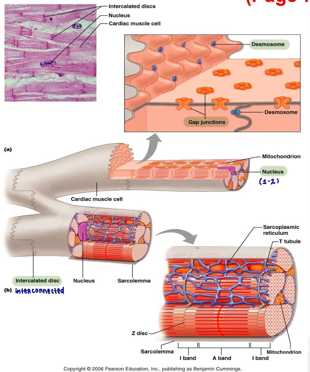
Mitochondria
produce ATP (energy) for heart muscle contraction
Intercalated disc (in heart muscle cells)
connect cardiomyocytes
→ help them contract together
Intercalated disc contains:
desmosomes
gap junction
Desmosomes’ function
hold cardiomyocytes together during heart contractions
Gap junction’ function
allow ions (electrical signals) to pass between cells
The heart’s functional syncytium
all fibers contract together as a single, coordinated unit
How is skeletal muscle contraction initiated?
Motor neurons trigger contraction
How is cardiac muscle contraction initiated?
AV nodes generate AP that triggers contraction
How does skeletal muscle increase contraction force?
Recruit more muscle fibres
How does cardiac muscle increase contraction force?
Increase calcium concentration
→ increase contraction force
Tetanus
continuous muscle contraction from rapid, repeated stimulation
Why skeletal muscle can go into tetanus phase?
short refractory period → can be stimulated again quickly
Why cardia muscle CAN’T go into tetanus phase?
already have long Plateau phase to rest
SA node (sino-atrial)
specialized cells generate APs for heart contraction
AV node (atrioventricular)
electrical relay station
→ pause impulse for 0.1s
AV bundle (atrioventricular)
carry AP from atria (AV node) → ventricles
Bundle branches
bifurcate from AV bundle
→ carry impulses through IV septum
SA node’s initial rate
70-100 bpm
AV node’s initial rate
40-60 bpm
AV bundle’s initial rate
20-40 bpm
Heart block
Impulse from atria to ventricles is delayed/blocked
→ damage to AV node
Bundle branch block
Impulse is delayed/blocked in a bundle branch
→ uncoordinated ventricular contraction
Ectopic foci
another place generates impulses instead of SA node
→ backup nodes take over when primary node is damaged
Ventricular extrasystoles (palpitation)
Extra ventricular heartbeat
→ caffein, nicotine, stress, exercise, etc.
Vagus nerve → heart
From parasympathetic center
decrease AP generation
slow HR (innervate SA, AV node)
Sympathetic cardiac nerve → heart
From sympathetic center
increase AP generation
increase HR (innervate SA, AV, contractile cells)
Sympathetic stimulation to increase HR
increase Sodium (Na+) and Calcium (Ca2+) permeability
Parasympathetic stimulation to decrease HR
Release ACh
→ increase Potassium (K+) permeability
→ decrease Calcium (Ca2+) permeability
Refractory period
The period when cardiac cell can’t generate another AP
→ prevent tetanus in heart muscles
Plateau phase
when Ca²⁺ enters while K⁺ (potassium) leaves
→ keeping membrane potential elevated
Systole
heart contraction (pumps out blood)
→ depolarization
Diastole
heart relaxes and fills up blood
→ repolarization
ECG function
reflects the electrical changes in the heart over one cardiac cycle
ECG helps to determine:
HR, heart rhythm
Sinus rhythm
normal HR - 75 bpm
Sinus bradycardia
low HR ~35-40 bpm
Sinus tachycardia
high HR >100 bpm
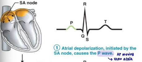
P wave represents:
atrial depolarization (systole)
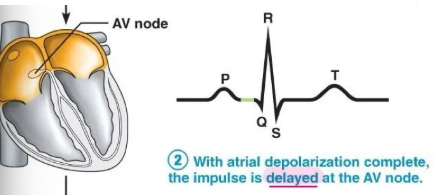
P-Q interval represents:
atrial depolarization → ventricular depolarization
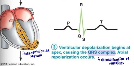
QRS complex represents:
ventricular depolarization (systole)
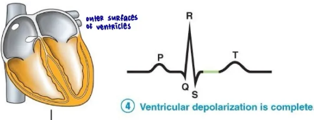
S-T segment represents:
ventricular depolarization → repolarization
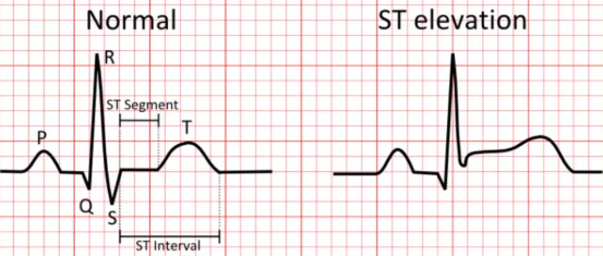
elevated S-T segment represents:
myocardial infrarction
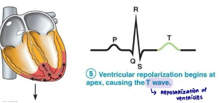
T wave represents:
ventricular repolarization
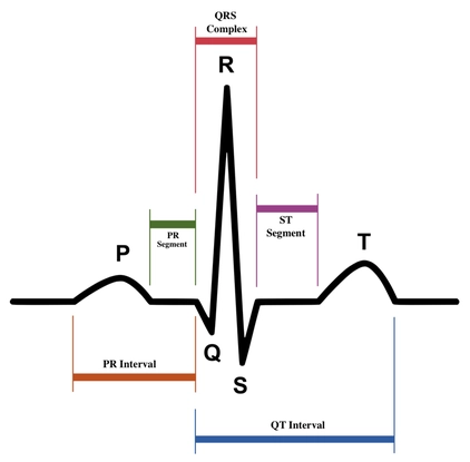
Q-T interval represents:
venticular depolarization → repolarization
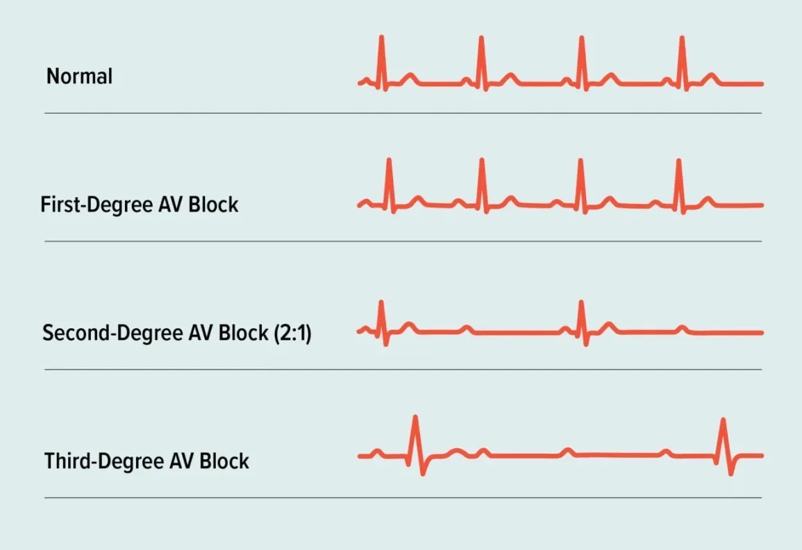
Heart block (ECG)
Impulse is blocked at AV node
→ slow, irregular heartbeat
→ prolonged P-R interval
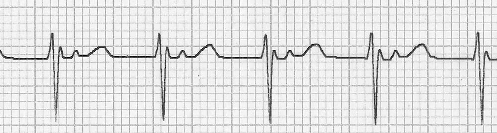
Junctional rhythm (ECG)
AV node takes over damaged SA node
→ slower heartbeat
→ no P wave
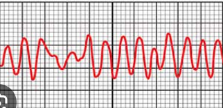
VF (ventricular fibrillation) - ECG
ventricles can’t pump blood effectively
→ rapid heartbeat
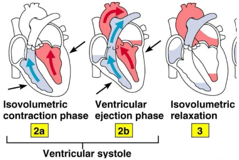
Isovolumetric contraction phase
ventricles start contracting
→ ventricular pressure rising up
→ all valves closed
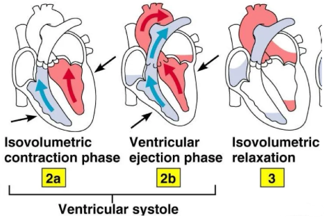
Isovolumetric ejection phase
ventricular pressure > arterial pressure
→ semilunar valves open
→ blood is pumped out
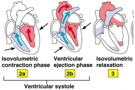
Isovolumetric relaxation
ventricles relax after the pump
→ pressure falls down
→ all valves shut
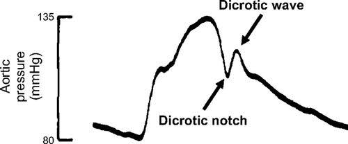
Dicrotic notch
small drop in aortic pressure
→ aortic valve closes
→ pressure drops
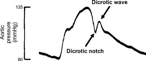
Dicrotic wave
small rise in aortic pressure after the notch
→ aorta recoils
→ pressure rises slightly
EDV (end-diastolic volume)
blood volume in ventricle before ventricular contraction
ESV (end-systolic volume)
remaining blood volume in ventricles after contraction
SV (stroke volume)
amount of blood pumped out per heartbeat
= EDV-ESV
Increased SV cause:
increase contraction force
decrease ESV
Contractility
the heart’s ability to contract properly
Contraction force
actual strength of the heart’s contraction
‘Lub’ heart sound (1st)
AV valves closure