Cerebrum Structures & Functions
1/62
There's no tags or description
Looks like no tags are added yet.
Name | Mastery | Learn | Test | Matching | Spaced |
|---|
No study sessions yet.
63 Terms
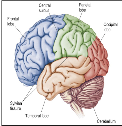
What lobes of the brain does the Sylvian fissure separate?
Frontal, parietal, and temporal lobes
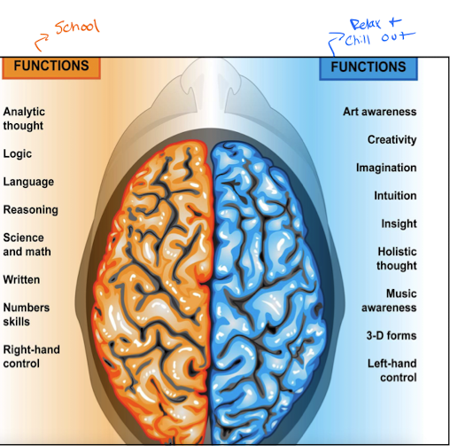
What are the functions of the left side of the brain (hemispheric specialization)?
Analytic thought
Logic
Language
Reasoning
Science and math
Written
Number skills
Right-handed control

What are the functions of the right side of the brain (hemispheric specialization)?
Art awareness
Creativity
Imagination
Intuition
Insight
Holistic thought
Music awareness
3-D forms
Left-handed control
What is the dominant hemisphere in all right-handed and most left-handed people?
The hemisphere in which language functions are localized, so the left hemisphere.
What is the function of the frontal lobe?
Mediates:
Cognition
Expressive language
Motor planning
Mathematical calculations
Working memory
Prefrontal lobes mediate executive functions, self-insight, and regulation.
Impulsive decision making
What is the function of the parietal lobe?
Sensory detection
Perception
Interpretation
What is the function of the temporal lobe?
Hearing
Comprehension of language and long term memory
What is the function of the occipital lobe?
Interpretation of visual stimuli from the optic pathoways.
What is the structure that is a portion of the cerebral cortex, lies deep in the lateral fissure, and is identified as the 5th lobe?
Insula
Which lobe is the Wernicke’s area located?
Temporal lobe
Specifically where on the temporal lobe is Wernicke’s area located?
Superior temporal gyrus
Which hemisphere is Wernicke’s area located?
In the dominant hemisphere, so the left hemisphere for most people.
Each cerebral hemisphere is concerned primarily with sensory and motor processes on the body on the ipsilateral or contralateral side?
Contralateral
Each cerebellar hemisphere is concerned with processes occurring on the ipsilateral or contralateral side of the body?
Ipsilateral
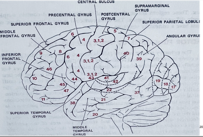
What area on the brain is the primary motor area (M1) located?
What does this area do? What tracts originate here?
Area 4; pre-central gyrus
Where voluntary motor activity is initiated; corticospinal tracts originate here
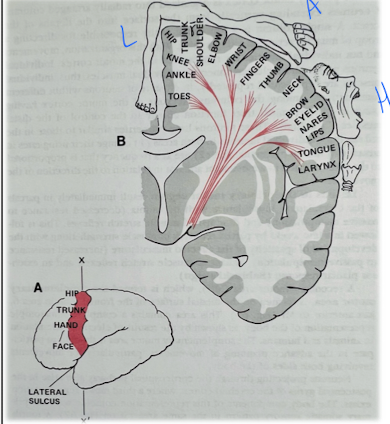
T/F: the homunculus of the brain does not change.
False; it changes with the person’s use of the body part.
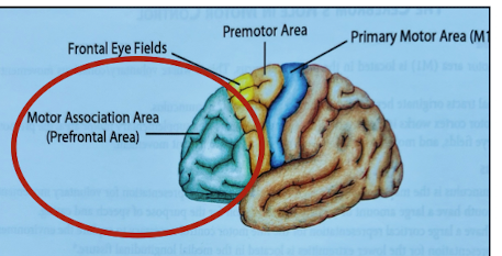
What is the significance of the prefrontal area?
It is the motor association area
What area of the brain has a role in cognitive planning of movement and possess a lot of someone’s personality?
Motor association area (AKA prefrontal area)
Which area on the brain is the premotor cortex located? List specifically which aspect of the hemisphere.
Area 6; Anterior to M1 on the lateral aspect of the hemisphere.
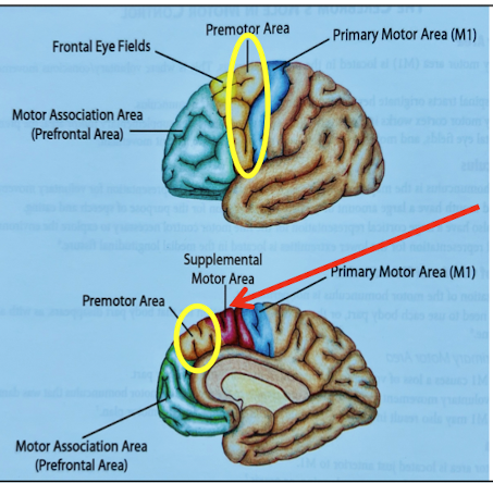
What is the role of the premotor cortex (area 6)?
Role in motor planning/praxis* and selects movements in response to external cues
*Plan and execute movement.

What area of the brain is the supplementary motor area? List specifically where it is on the hemisphere.
Area 6 on the medial aspect of the hemisphere.
What is the role of the supplementary motor area?
Advanced planning of movement, particularly involving both sides.
It selects movements based on remembered sequence of movement.
A lesion to the supplementary motor area may cause what?
Akinesia (absent or reduction in voluntary movements)
What is ideational praxis?
Loss of the ability to conceptualize and plan a motor sequence action. The perception of an object’s intended purpose is lost.
Ex. Someone had a stroke and they use a fork as a hair brush or difficulty sequencing mutli-step tasks.
Ideational praxis is caused by damage to what area of the brain?
Motor association area (AKA prefrontal area)

What is ideomotor planning?
Deficit of the execution on an action upon request. Often can execute spontaneous actions such as gestures and one-step tasks
They understand what is being asked of them, but they don’t know how to execute them. They can perform the actions on their own without being asked.
Ideomotor planning is caused by damage to what?
Primary motor area (area 4) (AKA M1)
What area of the brain is the frontal eye field located?
Area 8 (AKA Brodmann’s area 8)
What is the role of the frontal eye field?
Voluntary movement of eyes independent of visual stimuli.
A patient with a stroke to area 8 of the brain may present with what?
What if there is unilateral damage to area 8?
A deficit of eyes conjugating movement (struggle with coordinating their eyes to move in the same direction)
Causes conjugate deviation of the eyes to the side of the lesion.
What are the 5 areas of the frontal lobe that we are focusing on?
Primary motor area (4)
Supplemental motor area (6)
Pre-motor cortex (6)
Frontal eye field (8)
Prefrontal area
List the clinical application of a lesion to M1.
Loss of voluntary movement to the contralateral part
Loss corresponds to the area of the motor homunculus that was damaged.
Loss of the ability to implement a specific motor plan
Spastic dysarthria (slurred speech. Comprehension is good, but they cannot talk well due to motor issues).
What is the clinical application to a lesion to the premotor area?
Apraxia or motor planning difficulties
Inability to understand the demands of the task or inability to access the appropriate motor plan.
Preservation
What is the clinical application of a lesion to the motor association area/prefrontal area?
Inability to perform complex tasks
Disinhibition (inhibitory control over movement is lost)
Impulsiveness
Inability to understand how to carry out a motor task that was once known before the injury.
The premotor area/area 6 may be able to take over and compensate for the damage
What is the clinical application of a lesion to the supplementary motor area (area 6)?
Loss of bilateral control of posture
May cause akinesia
Compensation may occur from other areas (cerebellum or vestibular system)
What areas of the brain is the primary somatosensory cortex (SS1) located?
Areas 3,1,2
What is the role of the primary somatosensory cortex?
Detection of sensory information
Discriminates shape, texture, and size of objects (graphesthesia)
Where in the brain is the secondary somatosensory cortex (SS2)?
Posterior to the post-central gyrus

What is the function of the secondary somatosensory cortex (SS2)?
Interpretation of sensory information. Are where meaning is attached to the incoming sensory data.
Also where memory of tactile and spatial environment is.
If someone has a lesion to SS1, what would there clinical presentation be?
Loss of sensation to the contralateral body part
Loss depends on which part of the homunculus was damaged.
Loss of tactile localization and conscious proprioception
If someone has damage to SS2, what would their clinical presentation be?
Tactile agnosia
Partial impairment: inability to recognize objects by touch.
Can still detect some tactile features (like texture/shape)
Astereognosis
Complete inability to recognize objects by touch despite normal sensation
Agraphesthesia
Inability to know what #/letter is being written on skin.
Abarognosis (inability to gauge weight)
Atopognosia (inability to localise site of touch)
Extinction of simultaneous stimulation
Touching both sides of the body and being able to know which side (or both) is being touched.
What are the differences in the clinical applications of damage to primary sensation (SS1) and cortical sensation (SS2)?
Primary sensation (SS1)
Pain/temp, light touch, pressure, vibration and proprioception
Cortical sensation (SS2)
Astereognosis
Two point discrimination
Extinction of simultaneous stimulation
Agraphesthesia
Abarognosis (inability to gauge weight)
Atopognosia (inability to localise site of touch)
Where in the brain is the primary visual cortex (aka striate cortex)?
Area 17 in occipital lobe
The primary visual cortex (area 17) receives information from what?
Thalamic projections from the lateral genicular body (LGB) via the geniculocalcarine tract.
Also receives info from the optic tracts and the superior colliculi from the midbrain.
What is the function of the primary visual cortex (area 17)?
Responsible for the detection of visual input (NOT INTERPRETATION); detection of shape, color, orientation, and direction.
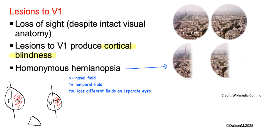
If someone has damage to V1 of the primary visual cortex, what would their clinical presentation be?
Loss of sight (despite intact visual anatomy)
Cortical blindness (complete loss of vision)
Homonymous hemianopsia (loss of vision on same side of both eyes.
What is the function of the visual association areas (V2 and higher)?
Interpretation of visual input (analysis of color, motion, and recognition of visual objects)
Where meaning is attached to incoming visual data
V2 is dependent on the acquisition of the experience
What would the clinical presentation be if someone had a lesion to V2?
Visual agnosias: inability to attach meaning to visual data/recognize objects despite intact vision.
Optic ataxia*: inability to use visual information to direct movements despite the ability to visually identify and describe objects.
Have hard time reaching for objects.
*Ataxia: lack of muscle coordination and balance
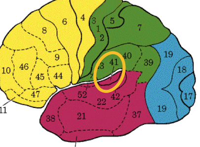
Where in the brain is the primary auditory center (A1)?
Areas 41 and 42.
A1 is located within the insula in the temporal lobe
What is the role of the primary auditory center (A1; area 41&42)?
Responsible for detecting sounds form environment
Conscious and discrimination of loudness and pitch of sounds.
What would the clinical presentation of someone by if they had a lesion to A1?
Lesion to primary auditory center (area 41 and 42):
Unilateral deafness if only 1 hemisphere is affected
Bilateral deafness if both hemispheres are involved.
Loss of localization of sounds (cannot determine where sound is coming from)
Where in the brain is the secondary auditory area (A2)?
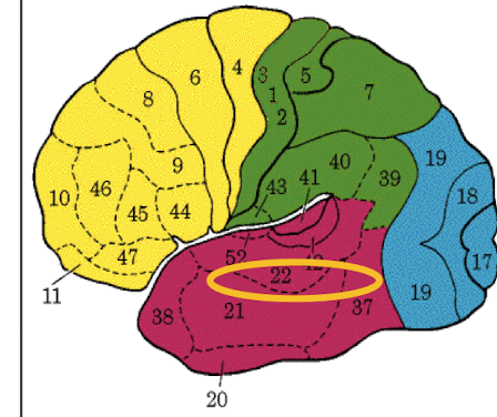
Area 22 (not as precise of visual association areas)
What is the role of secondary auditory area (A2) (area 22)?
Interpretation of auditory data
What is the clinical presentation of someone with a lesion to A2?
What if the lesion to the L or R hemisphere only?
Secondary auditory area (22):
Auditory agnosia: inability to attach meaning to specific sounds (unable to recognize sounds)
Left A2 lesion (dominant side): unable to understand speech.
Right A2 lesion: interferes with environmental sounds.
Where in the brain is Broca’s area?
Frontal lobe (area 44,45). Lies above the lateral fissure and sits within the inferior frontal gyrus.
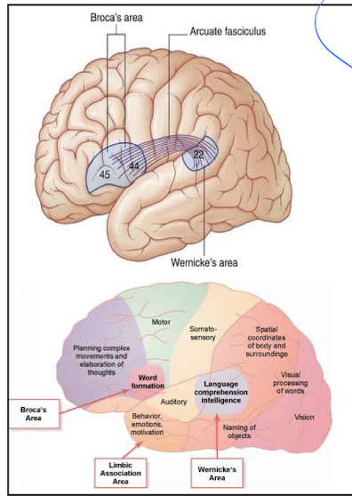
Which hemisphere is Broca’s area located?
ONLY the left hemisphere
What is the role of Broca’s area?
Contains motor programs for the verbal expression of language.
A lesion to area 44 and 45 of the brain would cause what presentation?
Expressive aphasia: pt can understand what is spoken to them, but cannot form meaningful sentences.
Where in the brain is Wernicke’s area located?
Posterior part of the superior temporal gyrus in the temporal lobe (area 22,39,40)

What is the function of Wernicke’s area?
Responsible for the comprehension of the spoken word.
A lesion to area 22 in the brain would cause what?
Receptive aphasia: pt cannot understhand what is spoken to them. They can produce sentences but the meaning does not correlate)
What joins the Broca’s and Wernicke’s areas?
A fiber bundle called the inferior arcuate fasciculus.
It runs from the temporal lobe, through the lower parietal lobe, and into the frontal lobe.

Why can damage to the temporal lobe cause language problems?
Because the inferior arcuate fasciculus runs from the temporal lobe to the frontal lobe connecting the Wernicke’s area to the Broca’s area.