18 Equine Cardiac Arrhythmias
 Call Kai
 Learn
Learn Practice Test
 Spaced Repetition
 Match
Match Flashcards
 Knowt Play
1/41

There's no tags or description
Looks like no tags are added yet.
Name | Mastery | Learn | Test | Matching | Spaced | Call with Kai | Chat |
|---|
No analytics yet
Send a link to your students to track their progress
42 Terms
The SA node is taken down as the "ruler", so multiple spots can generate electrical impulses at once, causing a "crawling atria"
Describe briefly what is happening in atrial fibrillation (just to the heart itself, not the ECG)
Atrial fibrillation
What is the most common cardiovascular cause of poor performance in horses?
Large horses with large atriums like thoroughbreds and warmbloods who regularly get their heart rates up. They can't work as hard.
Name the type of horse atrial fibrillation is typical in
Pathologic or paroxysmal
What is the etiology (cause) of atrial fibrillation in horses?
1. diastolic fill is less efficient (because remember to end off diastole, the atria contract to get the last bit of blood to the ventricles. since they are "crawling and twitching" with impulses, the atria are not contracting right)
2. the crawling and twitching impulses may cause additional arrhythmias in the heart
3. become exhausted easier because heartrate is disproportionately high compared to exercise (because the AV node is no longer controlling impulses like it normally does so the signals can slip by and cause abnormal firing)
Horses are often able to tolerate atrial fibrillation at rest. So why do horses with atrial fibrillation have poor performance during exercise?
Atrial fibrillation
A horse has a heart rate of 34 at rest. During the trot, it gets to 150 then skyrockets to 300 during the gallop and the horse is not able to keep pace for long. What do you suspect?
This horse is already in cardiac failure. If the horse had a normal heart rate at rest, we would be suspecting more A fib. But the abnormally high heart rate at rest tells us the horse is already in cardiac failure
A horse has a heart rate of 70 at rest, which shoots to 150 at the trot and 250 at the gallop, which is cannot maintain for long. What do you think?
Missing P waves, replaced by F waves (because remember P waves indicate the heart's atrial depolarization which is not in rhythm since the AV node cannot properly conduct impulses)
What wave is missing on an ECG of a horse with atrial fibrillation? What is it replaced by?
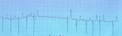
Atrial fibrillation: no p waves present. F waves are present in that long stretch of no QRS as a result of the AV node telling some of the impulses to back off. Classic Atrial fibrillation is present here, where you do not have to change the width of your fingers as you slide them over the ECG meaning QRS waves themselves are fine. No consistent spacing between QRS complexes aka the R to R interval.
Describe this horse ECG
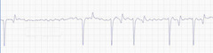
F waves
What is this called in Atrial fibrillation?
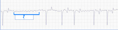
When it has been present for over 72 hours. Do a full cardiac exam to search for cause. If the horse already is in cardiac failure (because the heart rate is high at rest) control the damage the best you can.
When should you treat atrial fibrillation in horses?
1. medical quinidine sulfate by NG tube
2. transvenous electric cardioversion TVEC = shock treatment (safer but horse must be under anesthesia)
Name the treatment options for atrial fibrillation in horses
Oral ulcers (if not given by NG tube), laminitis, colic, nasal inflammation, urticaria (hives), ataxia
Quinidine sulfate is one of the treatment of choice for atrial fibrillation in horses. Name some complications it can cause
False
TRUE OR FALSE: Atrial fibrillation disappears with exercise in horses.
A "hit and run attack", where there is an abnormally shaped premature p wave causing shorter time between P and QRS complex
What happens in premature atrial conduction?
It can occur in healthy horses or those with underlying disease
What causes premature atrial conduction in horses?
Premature atrial contraction (can be called APC or PAC)
During this rhythm, a horses regular rhythm is interrupted by a premature beat, causing an irregularly irregular rhythm
Atrial premature contraction. P wave occurs prematurely and is followed by a normal QRS. There is a non compensatory pause which indicates the SA node is at rest. Normal QRS morphology with shortened P to P interval
Describe this ECG

With rest and corticosteroids
How can you correct pathologic atrial premature contraction in horses such as those which occur frequently at rest, with episodes of tachycardia, cause poor performance, or lead to fibrillation?
Atrial fibrillation and atrial premature contraction
Which abnormal equine ECG rhythms originate in the atrium?
2nd degree AV block and advanced AV blocks
Which abnormal equine ECG rhythm originate in the atrioventricular node?
Ventricular premature conduction (VPC) and ventricular tachycardia
Which abnormal equine ECG rhythms originate in the ventricle?
Physiologic: has a low HR at rest and the block disappears with exercise so there are no clinical signs
Pathologic: has an inappropriately low heart rate at rest so the block persists with exercise leads to exercise intolerance, ataxia, syncope (passing out), collapse, and unexplained trauma
What is the difference in clinical signs between physiologic and pathologic atrioventricular ECG abnormalities in horses?
2nd degree AV block
What is the most frequent physiologic arrhythmia in horses?
Regularly irregular rhythm with slow to normal HR and diastolic pause
What will a 2nd degree AV block sound like in an equine auscultation?
CAUSE: high vagal tone (parasympathetic nervous system)
ORIGIN: partial conduction at AV node
What causes 2nd degree AV block? Where does it originate?
2nd degree AV block. Normal QRS complex shape. Not every P is followed by a QRS, but every QRS is preceeded by a P. In the long pause, you can see a lonely P with no QRS
Describe what this ECG is saying?
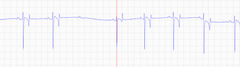
Irregularly irregular
Are VPC (ventricular premature contractions) *irregularly or regularly* irregular?
Ventricular premature conduction which happens at the level of the ventricle. Shortened QRS interval. The QRS is abnormal and not preceeded by a P wave but is followed by a compensatory pause. If you run your fingers over the ECG, you have to change the width of them to get all the complexes = classic of VPC.
What is happening in this ECG
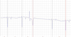
VPC: QRS is premature and abnormal, P is normal
PAC: P is premature and abnormal, QRS is normal
What is the main difference between the ways QRS and P are affected by VPC and PAC?
VPC: A compensatory pause on an ECG is the duration of the pause after a beat, where the timing of the next sinus beat is on schedule (because VPC don't interrupt the normal rhythm), meaning the sum of the pre- and post-ectopic intervals equals two normal sinus intervals=rhythm continues as if beat hadn't occurred
PAC: A non-compensatory pause is a shorter pause where the next sinus beat occurs earlier than expected, bc the PAC resets the sinus node and shortens the cycle.
We say that VPCs are followed by a "compensatory pause" and PAC are followed by "noncompensatory pauses". What does this mean?
Ventricular tachycardia
What can pathologic VPCs lead to?
Sinus arrhythmia from change in vagal tone that appears at rest when the horse is cooling down from exercise. It is transient.
Which ECG pattern is this in a horse?
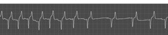
SA node
Where does sinus arrhythmia originate in horses?
Ventricular tachycardia demonstrated by more than 4 successive VPCs.
What is this ECG telling you
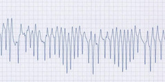
Lidocaine, quinidine, magnesium sulfate, phenytoin, and propanolol
What is the treatment like for ventricular tachycardia?
Ventricular tachycardia
Which abnormal equine ECG rhythm has the treatment of:
Lidocaine, quinidine, magnesium sulfate, phenytoin, or propanolol?
Healthy exercise ECG (even tho no P wave)
Describe this ECG during exercise
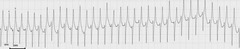
AV block during exercise. 2 P waves with no QRS.
Describe this ECG during exercise
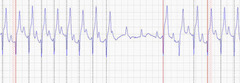
Atrial fibrillation. Irregular QRS spacing and F waves present
Describe this ECG during exercise
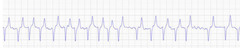
2 VPCs (notice the two taller waves followed by a compensatory pause)
Describe this ECG during exercise
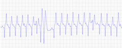
Atrial fibrillation
This picture shows which arrhythmia?