Cerebrum, Ventricular System, and Vasculature
1/47
There's no tags or description
Looks like no tags are added yet.
Name | Mastery | Learn | Test | Matching | Spaced | Call with Kai |
|---|
No analytics yet
Send a link to your students to track their progress
48 Terms
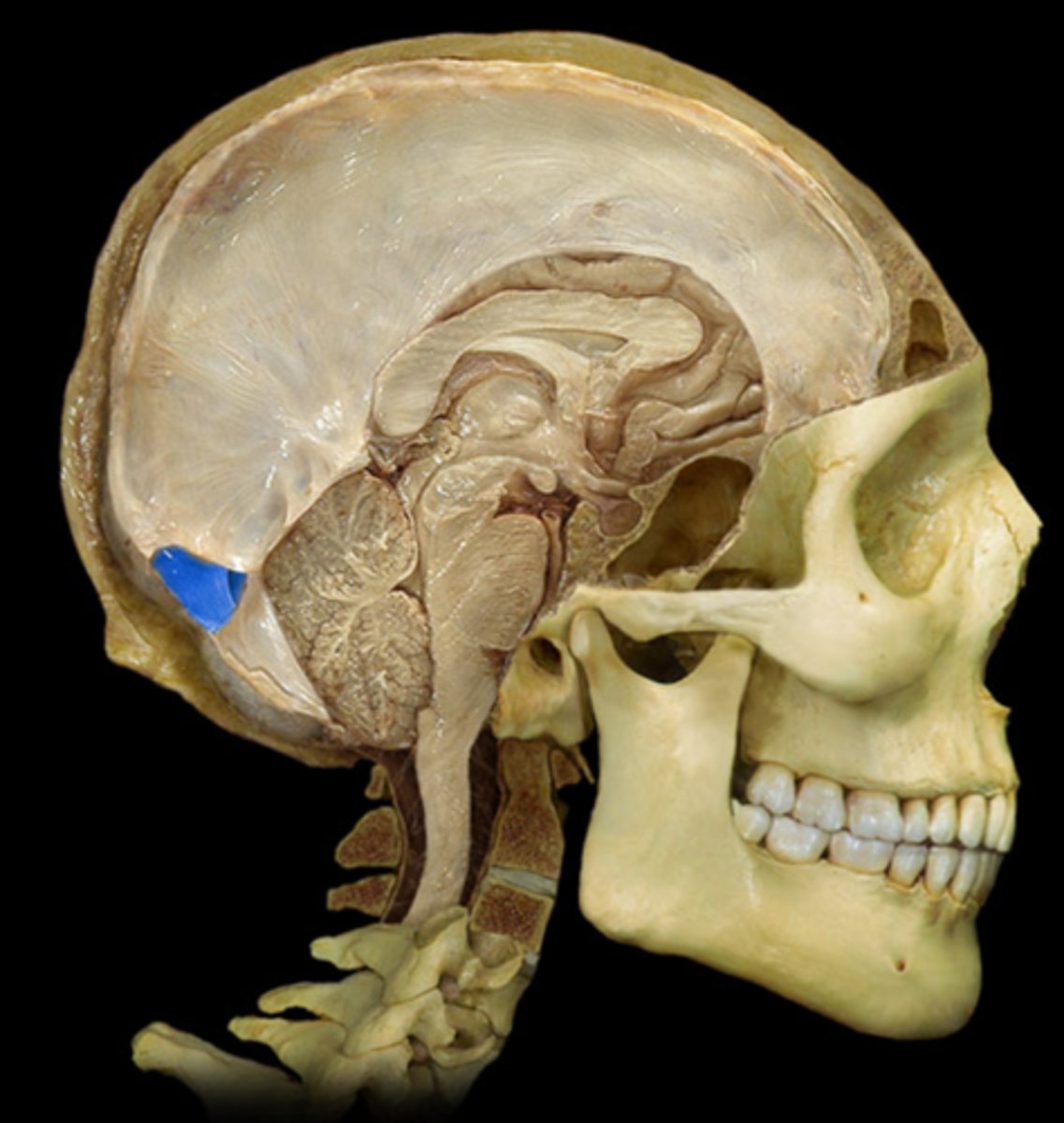
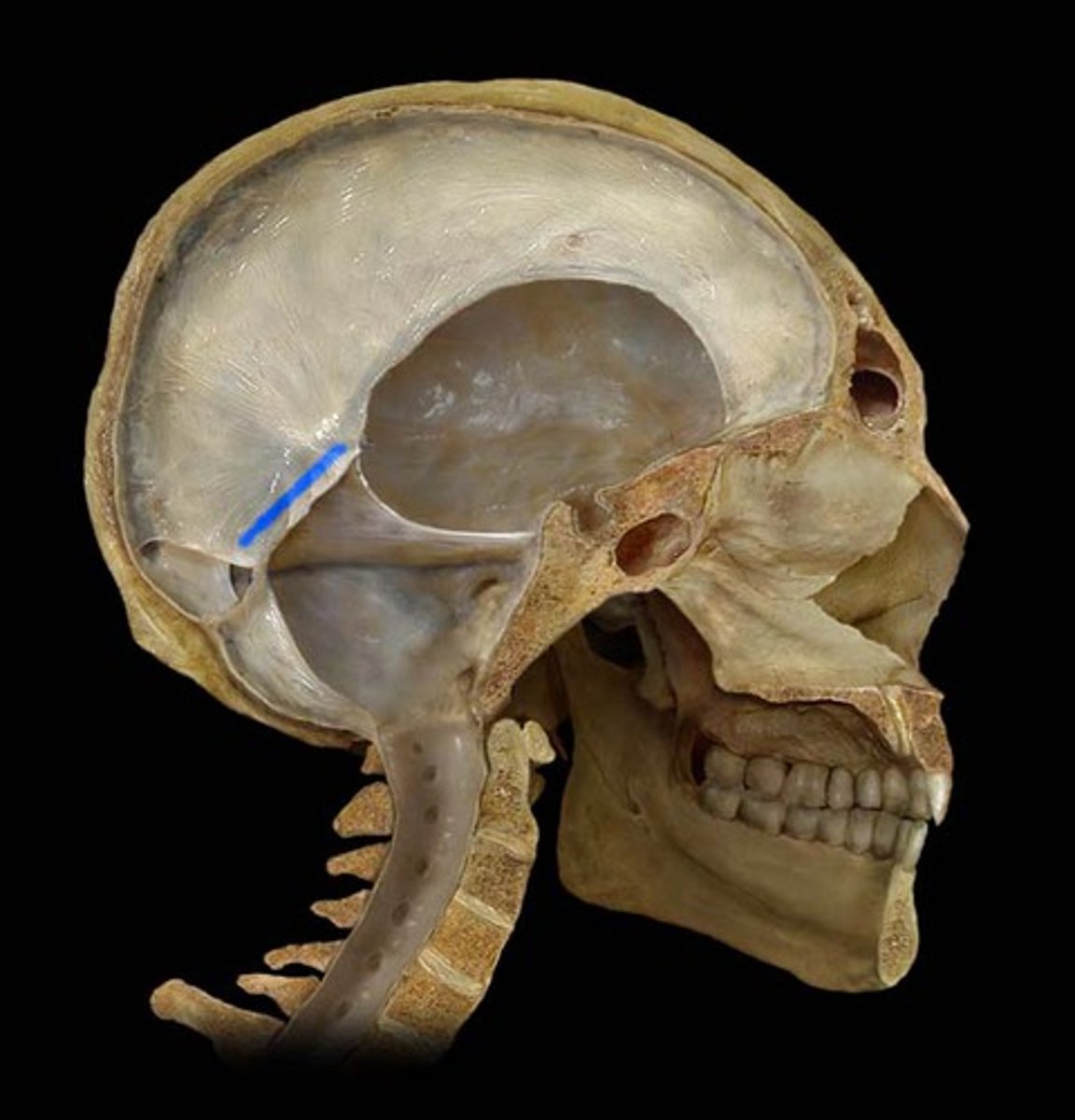
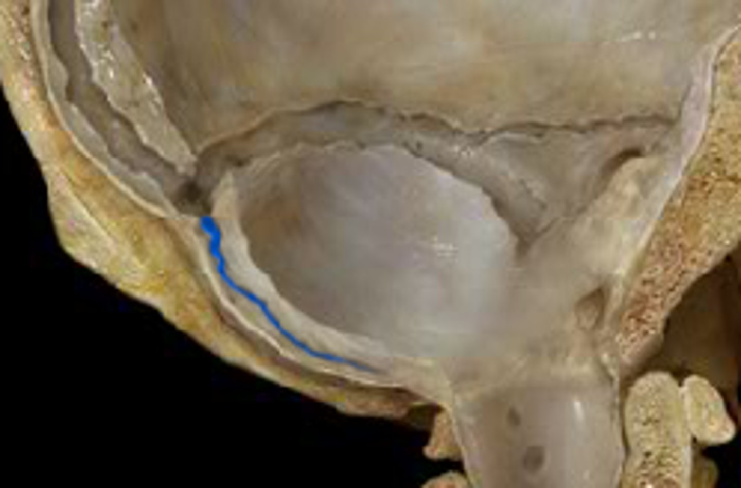
Arachnoid Granulations
Groups of arachnoid villi; protrude through the meningeal dura into dural venous sinus
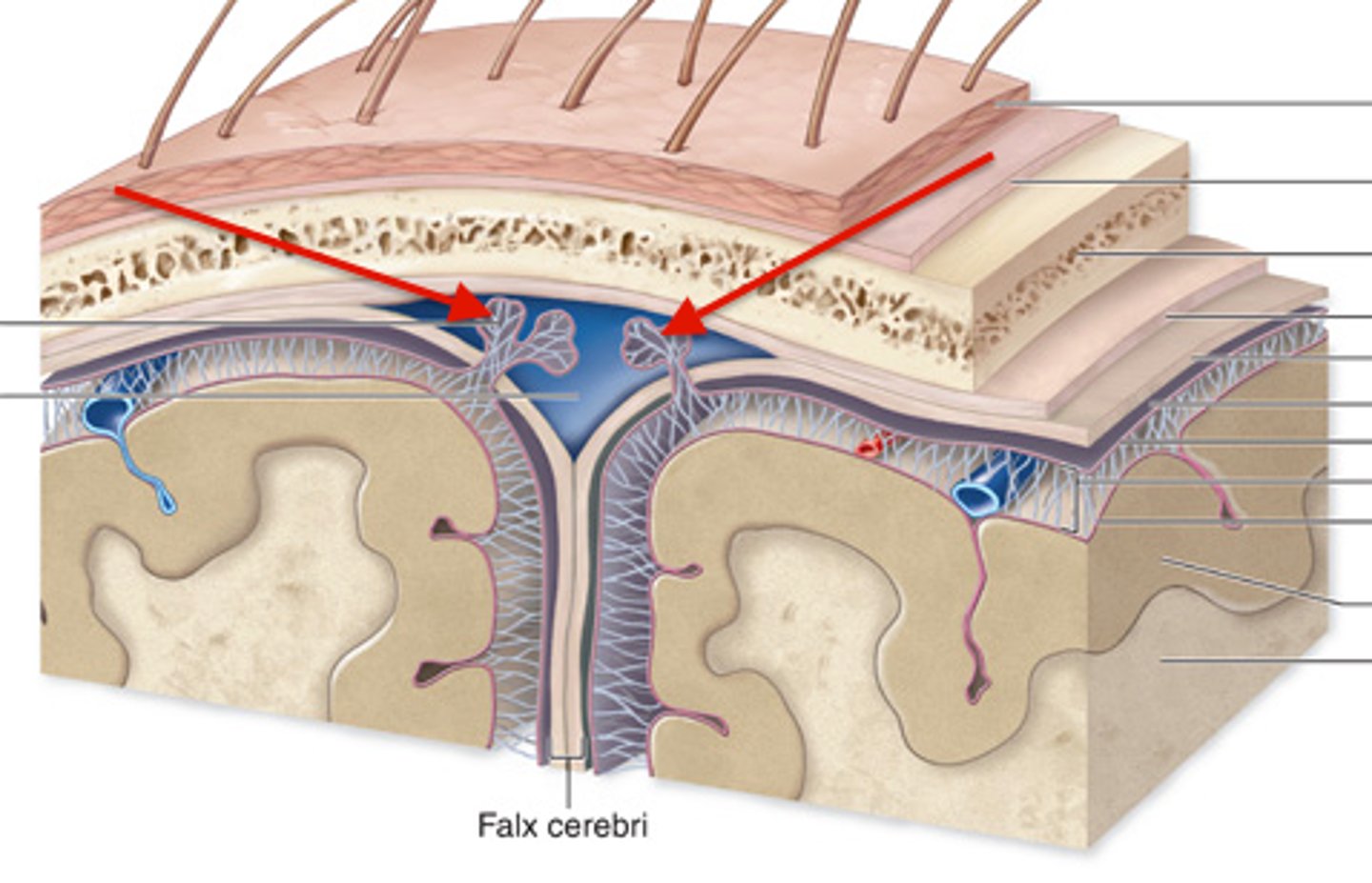
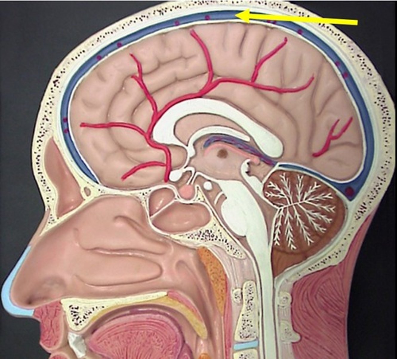
Periosteal, meningeal
Two layers of the dura mater
Dural septae infoldings, venous sinuses
Areas where dura mater layers are not continuous with one another
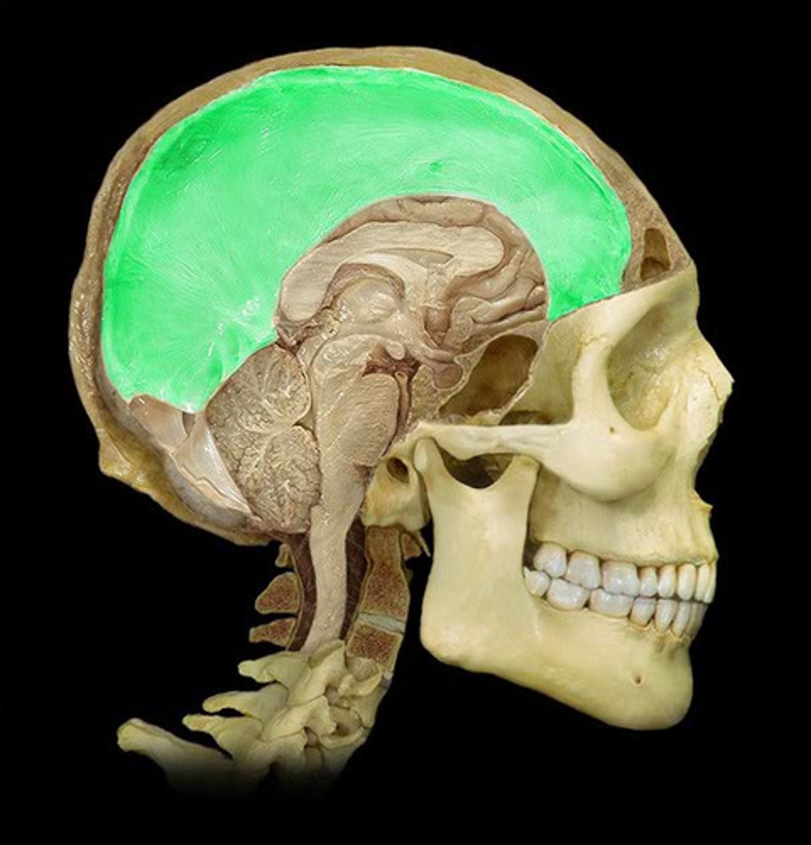
Falx Cerebri
Separates two cerebral hemispheres
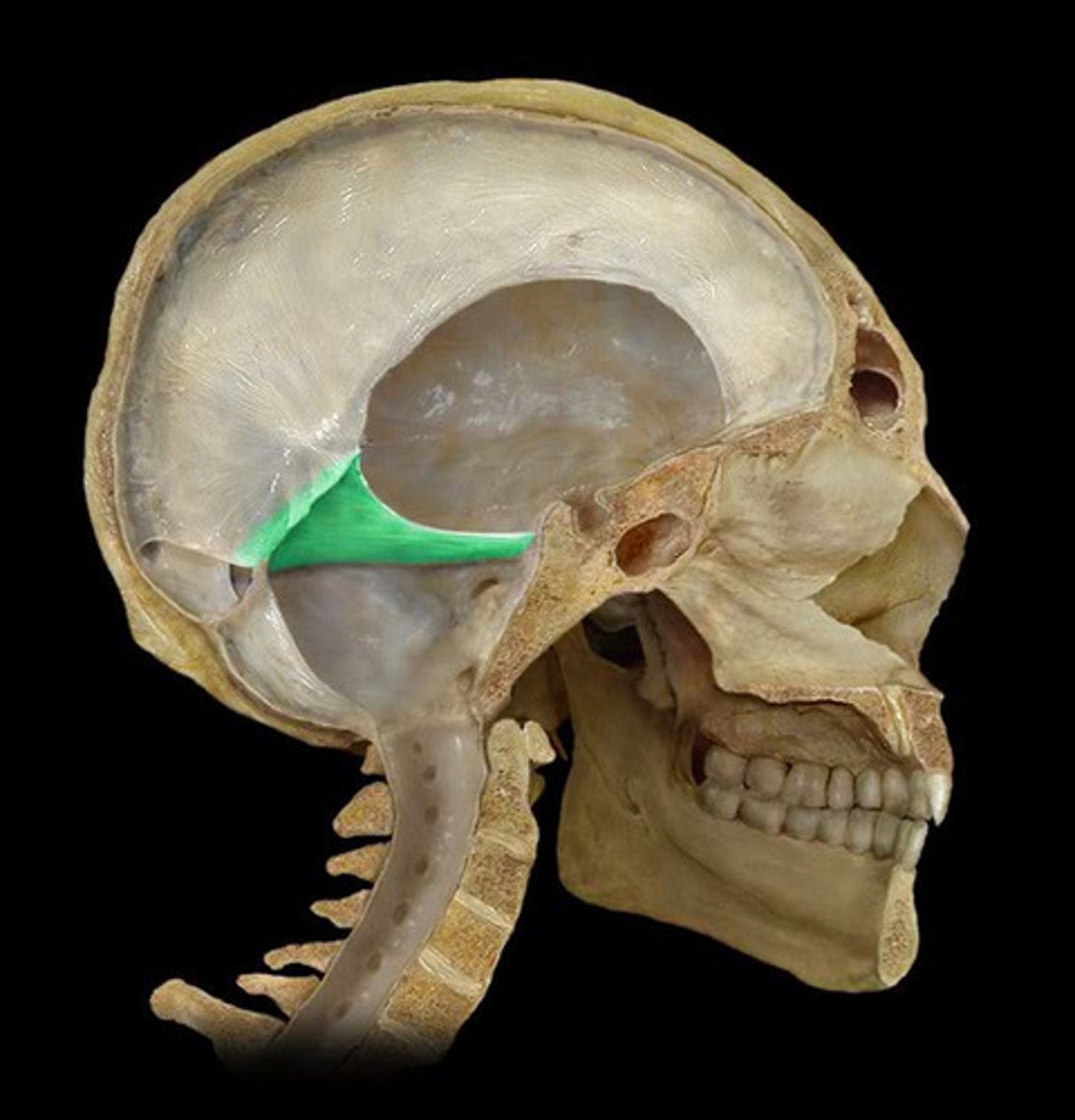
Tentorium Cerebelli
Separates cerebellum and posterior parts of cerebrum
Falx Cerebelli
Separates two cerebellar hemispheres
Dural Venous Sinuses
large veins in the dura mater that drain the cranium
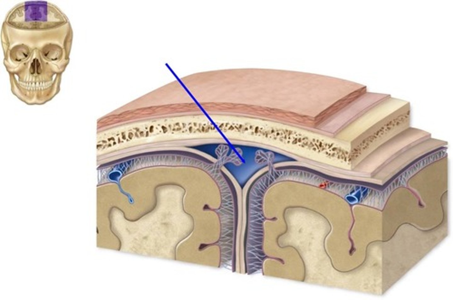
Diploic Veins
Veins that exist in the bones of the skull
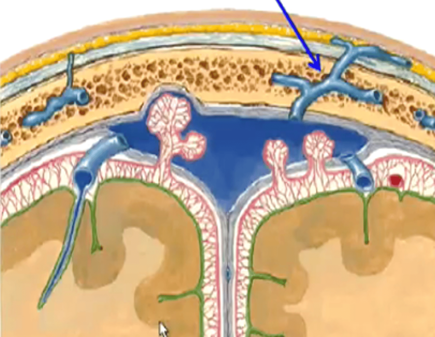
Emissary Veins
Veins that drain from the loose connective tissue layer of scalp into the cranial cavity
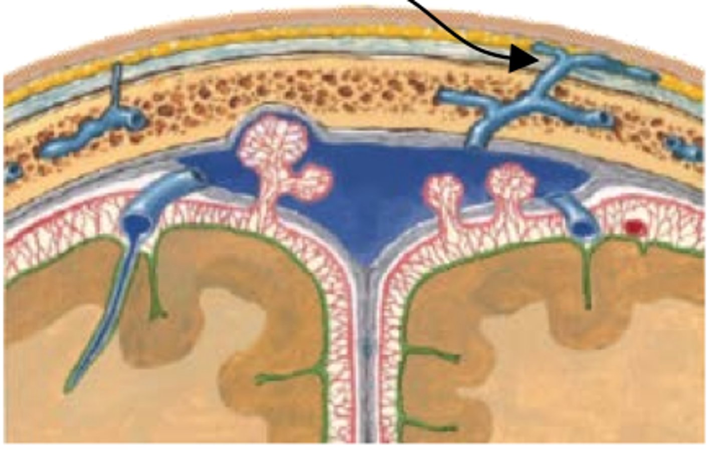
Cerebral bridge veins
Connect veins from cerebral cortex to the dural venous sinus; drain brain tissue
Internal Jugular veins
Where do the dural venous sinuses drain?
Confluence of sinuses
Meeting place for superior, straight, occipital, and transverse sinuses
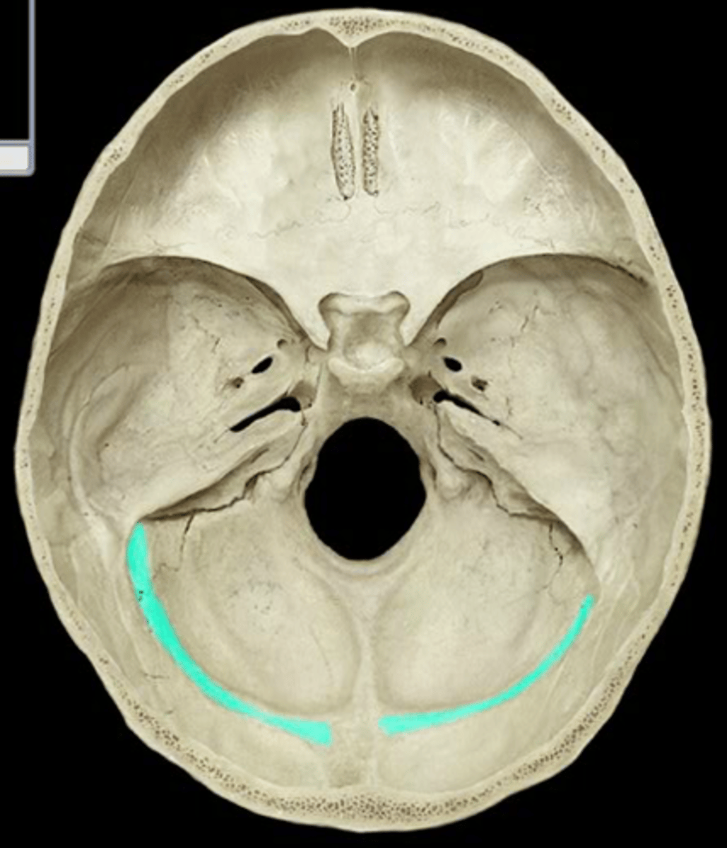
Straight sinus
Where does the inferior sagittal sinus drain into?
Transverse sinus
Where does the sigmoid sinus drain into?
Superior sagittal sinus
Straight sinus
Occipital Sinus
Transverse sinus
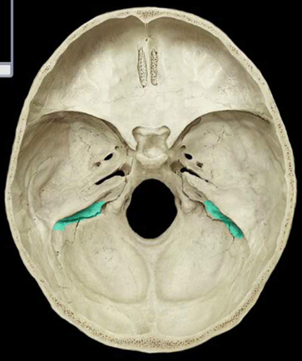
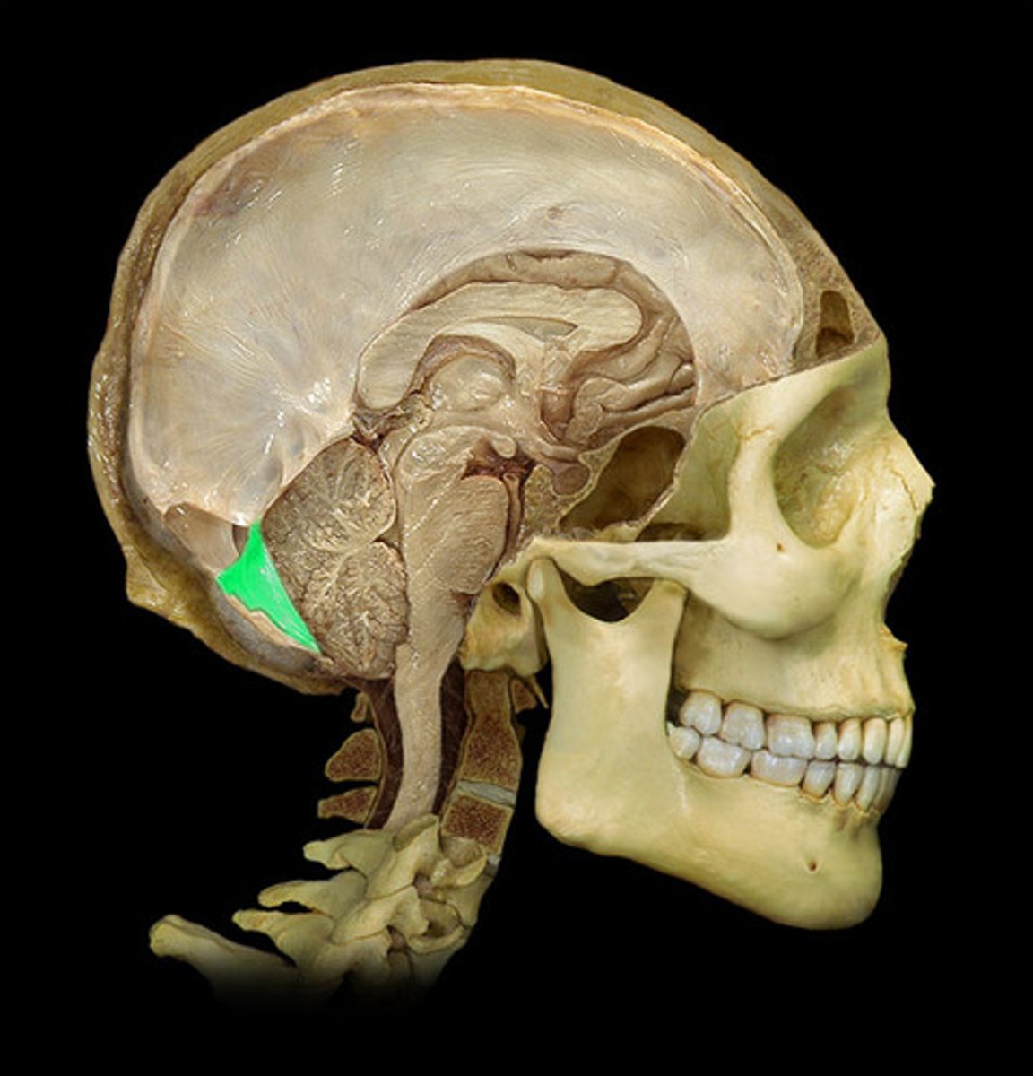
Cavernous sinus
Located on either side of sella turcica
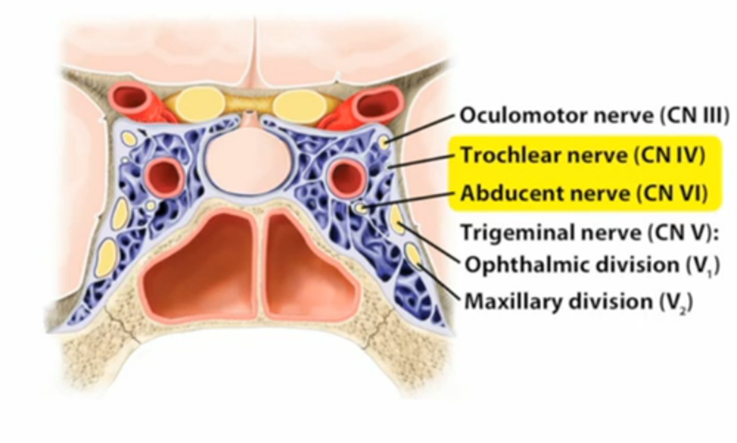
CN III, CN IV, CNV1, CNV2, CN VI (Carotid plexus of sympathetic nerves)
Nerves that run through the Cavernous sinus
Internal Carotid artery
What artery run through the cavernous sinus?
Pituitary Tumors
Tumors that can compress the cavernous sinus causing paralysis of the extraocular muscles and sensory loss in the forehead and maxillary regions
Choroid Plexus (ventricles)
What secretes CSF?
fringes of vascular pia mater covered by epithelial cells
What is the choroid plexus?
Cerebrospinal Fluid
Protects brain and keeps brain buoyant
Monro-Kellie Doctrine
Any increase in volume of one component in cranial cavity must be compensated by the decrease in volume of another
Change in intracranial blood can only occur through the displacement or replacement of CSF
What is an example of the Monro-Kellie Docterine?
Lateral Ventricles
A set of paired ventricles lying within the cerebral hemispheres.
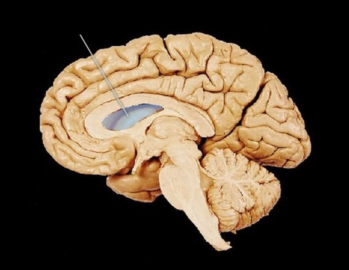
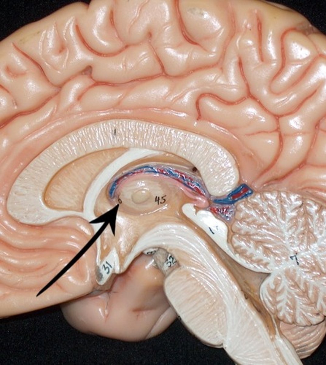
3rd ventricle
found in the diencephalon and communicates with lateral ventricles via intraventricular foramen
4th ventricle
between pons and cerebellum
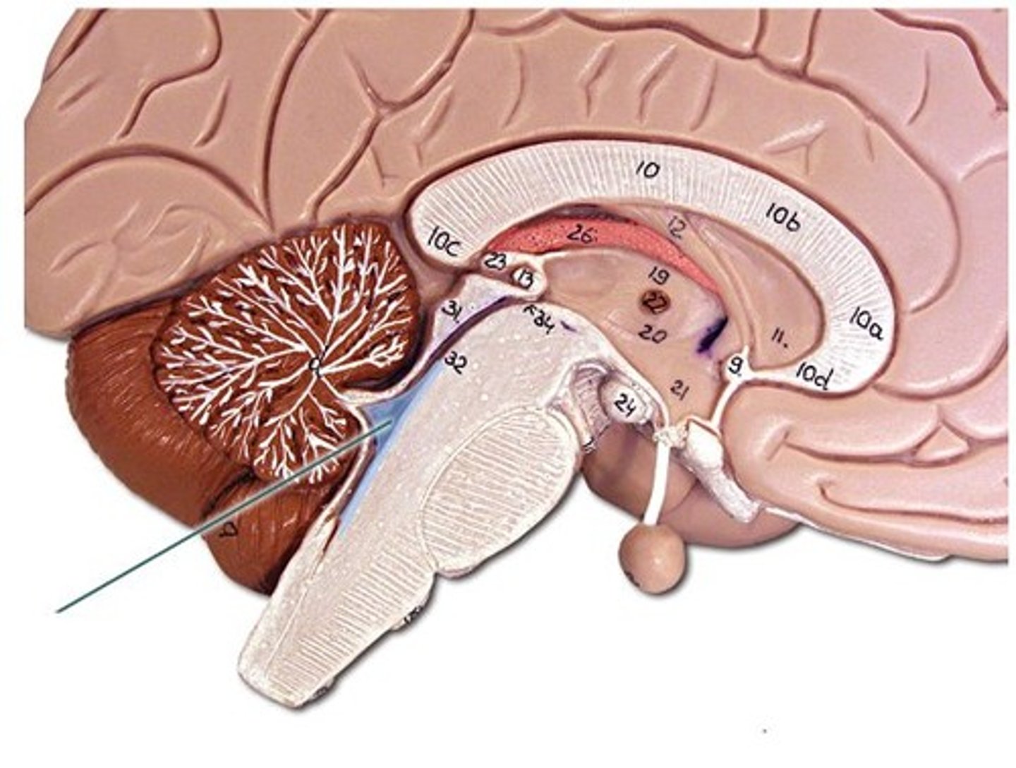
Lateral Ventricle → Interventricular Foramen → 3rd Ventricle (diancelphalon) → Cerebral aquaducts → 4th ventricle → Subarachnoid space
CSF Flow
In the dural venous sinus through the arachnoid granulations
Where is CSF reaborbed?
Hydrocephalus
Excessive CSF in ventricular system; can be caused by obstructed flow, overproduction, or failure to reabsorb; treated through VP shunt placement
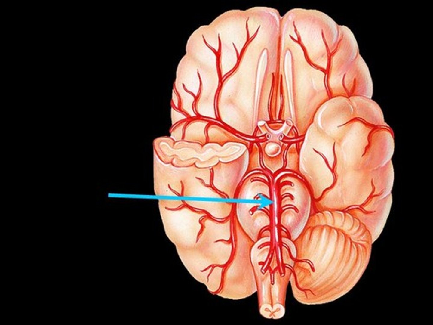
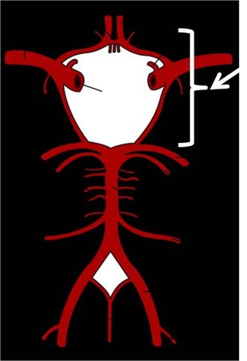
Internal Carotid, Vertebral Arteries
Blood supply to the brain
Carotid Canal
Where does the ICA enter the cranium through?
Subclavian artery
What gives rise to the vertebral artery?
Transverse foramen in C-spine
Where does the vertebral artery pass through?
Foramen Magnum
Where does the vertebral artery enter the crainum?
Basilar Artery
Artery formed by right and left vertebral artery; divides into posterior cerebral arteries
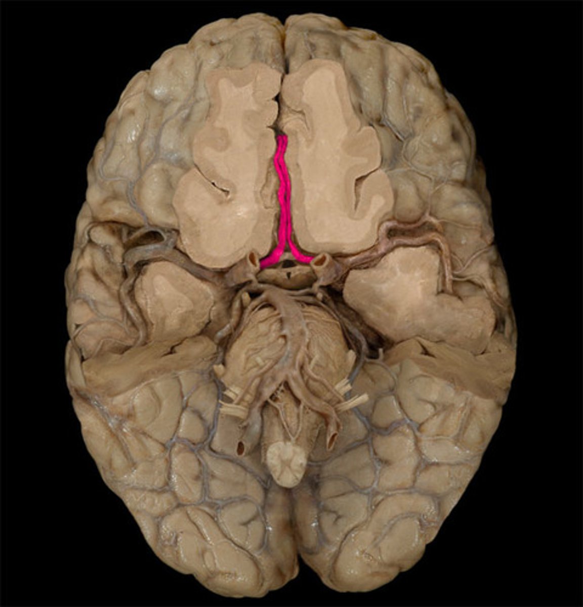
Anterior cerebral artery
Supplies medial and superior surfaces and frontal pole
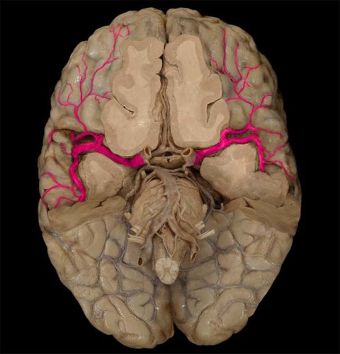
Middle cerebral artery
Supplies lateral surface and temporal pole
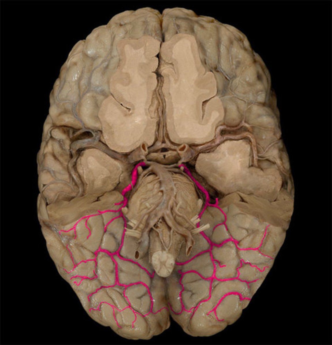
Posterior cerebral arteries
Supplies inferior surface and occipital pole
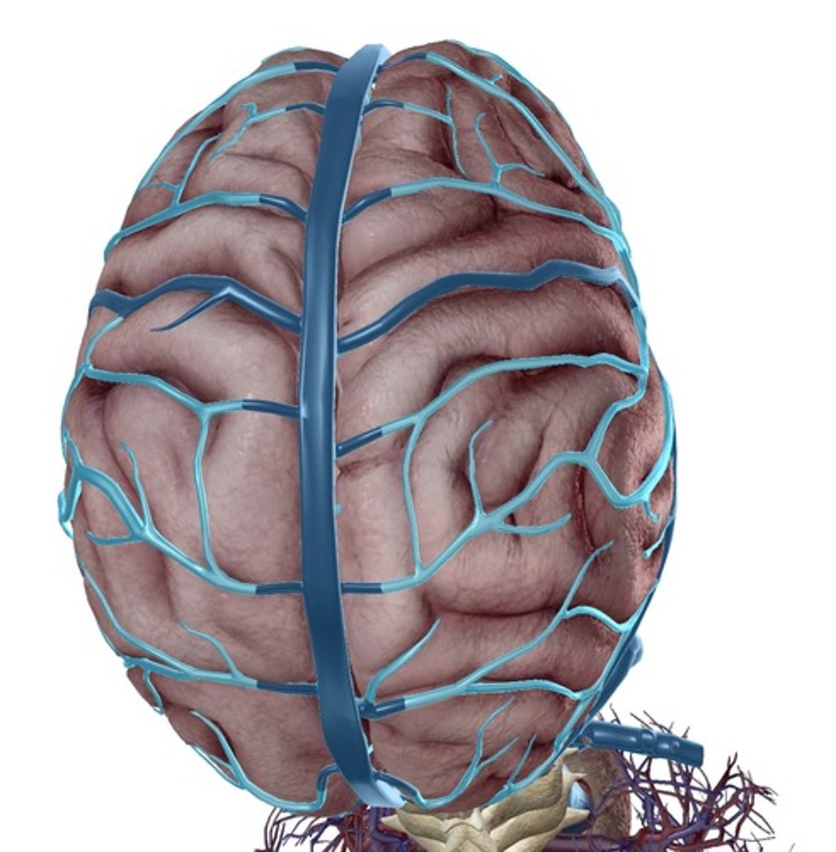
Cerebral veins
Drainage of the brain
MCA
Supplies region associated with face and upper extremity function; stroke here leads to more contralateral face and upper extremity deficits
ACA
Supplies regions associated with trunk and lower limb function; stroke here leads to more contralateral trunk and lower extremity involvement
Circle of Willis (cerebral arterial circle)
Anastomosis between branches of the ICA and vertebral arteries at base of brain
Posterior and Anterior Cerebral artery, Posterior and Anterior communicating artery, Internal Carotid Artery
Which arteries are part of the circle of Willis?
Sigmoid Sinus
continuation of the transverse sinus