Connective Tissue Identifiaction
 Call Kai
 Learn
Learn Practice Test
 Spaced Repetition
 Match
Match Flashcards
 Knowt Play
1/10

There's no tags or description
Looks like no tags are added yet.
Name | Mastery | Learn | Test | Matching | Spaced | Call with Kai | Chat |
|---|
No analytics yet
Send a link to your students to track their progress
11 Terms

Areolar Tissue
Appearance: Loose network of fibers (collagen and elastic), scattered fibroblasts
Functions: Nourish and remove waste for epithelial Tissues; binds tissues together
Locations: Underlies epithelial tissue, surrounds nerves and blood vessels, the body’s “filler material”
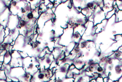
Reticular Tissue
Appearance: Loose meshwork of reticular fibers and fibroblasts, infiltrated with leukocytes
Functions: Forms supportive stroma (framework) for lymphatic organs
Locations: Lymph nodes, spleen, bone marrow

Adipose Tissue
Appearance: Empty-looking cells with thin margins, nucleus pressed against the cell membrane, vascular
Functions: Energy storage, insulation, cushioning
Locations: Subcutaneous (under skin) breast, surrounds internal organs

Dense Regular Connective Tissue
Appearance: Densely packed, parallel collagen fibers, often wavy, compressed fibroblast nuclei
Functions: Withstands predictable stresses
Locations: Tendons attach muscle to bone and ligaments attach bone to bone
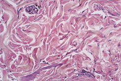
Dense Irregular Connective Tissue
Appearance: Densely packed collagen fibers running in random directions, few visible cells
Functions: Withstands unpredictable stresses; durable
Locations: Dermis of skin, capsules around organs
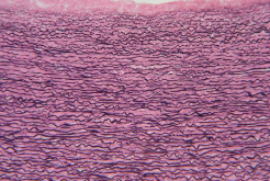
Elastic Connective Tissue
Appearance: Densely packed collagen fibers and elastic fibers
Functions: Expansion and recoil of tissue/organs
Locations: Walls of large arteries (aorta), vocal cords

Elastic Cartilage
Appearance: Abundance of elastic fibers in web-like mesh, chondrocytes in lacunae
Functions: Provides flexible, elastic support
Locations: External ear and epiglottis

Fibrocartilage
Appearance: Coarse bundles of parallel collagen fibers; rows of chondrocytes in lacunae between collagen fibers
Functions: Resists compression and absorbs shock
Locations: Pubic symphysis, meniscus of knee, intervertebral discs

Hyaline Cartilage
Appearance: Smooth/glassy matrix; chondrocytes in lacunae, often in small clusters
Functions: Eases joint movement, holds airway open, moves vocal cords, growth of long bones in children, precursor to fetal skeleton
Locations: Articular/joint cartilage on ends of long bones, costal (rib) cartilage, trachea, larynx, fetal skeleton
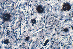
Osseous Tissue (Bone)
Appearance: Hard, calcified matrix arranged in concentric lamellae around a central canal; osteocytes in lacunae; osteons are the structural units of compact bone
Functions: Movement, mineral storage (calcium and phosphorus), framework of body, protection of viscera (internal organs)
Locations: Bones of the skeleton

Blood
Appearance: RBCs, WBCs, and platelets; fluid matrix (plasma)
Functions: Transports substances, contributes to immune defense
Locations: Inside blood vessels and heart