Neurophysiology Revision
1/96
There's no tags or description
Looks like no tags are added yet.
Name | Mastery | Learn | Test | Matching | Spaced |
|---|
No study sessions yet.
97 Terms
What is the main mechanism by which neurons work
Transmitting electrical signals
What is the defining characteristic of neurons
The cell process
Information arrives at the cell body via
dendrites (and cell bodies) where it is assimilated and processed
What happens to the processed information that arrives at the cell body
Processed information is then digitised into APs which are transmitted along the axon
What happens to the processed information at the end of the axon
The information is passed to the target (muscle or neuron) at boutons
What is a bouton?
axon terminal
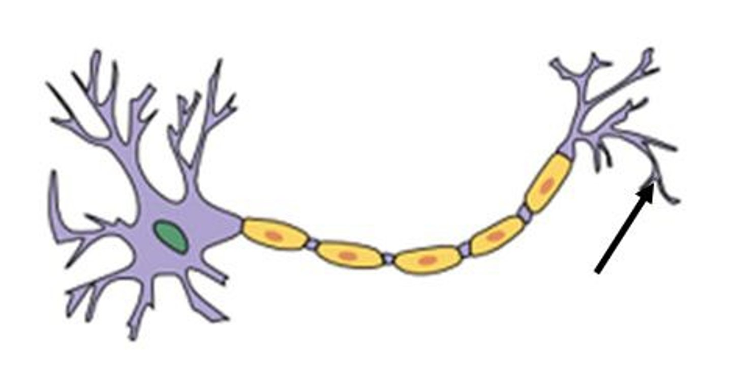
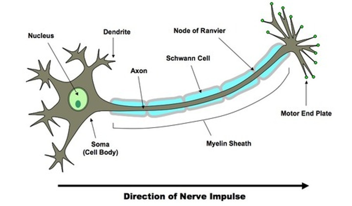
Describe the structure of neuron
Dendrites send and receive messages to other cells. These messages get passed to and from the cell body, which is the cell's life-support center, and then pass through the axon (covered by the myelin sheath) to the terminal branches of the axon. There are also gaps in the myelin sheath called nodes and the sections covered by myelin are called internodes
Where do action potentials occur?
axon
Where do action potentials start in the axon?
Cell body
How are action potentials graded?
By frequency
What is the only way an AP can increase in intensity?
To increase its frequency
Does the inside of a neuron (cell) have a negative or positive resting potential
Negative
Is there a physical limit to how big the resting potential can be and why?
Yes - otherwise you would get sparking inside the cell
What maintains a negative resting potential inside the neuron
Membrane ion (Sodium) pumps
What is the resting potential of a neuron?
-70mV
How can the resting membrane potential be measured?
Using a glass electrode
What causes depolarisation?
Opening of Na+ channels allowing Na+ into the cell. AP is started by initial stimulus which pushes voltage towards threshold causing depolarisation.
What happens during depolarisation?
The membrane potential is depolarised towards positive values.
What is repolarisation
The change from a positive action potential back to a negative resting potential (caused by opening of potassium channels)
What is the refractory period?
A period in the hyper polarised state when the neuromembrane is unresponsive.
What are channels in the neuron membrane sensitive to?
Voltage - meaning they open when the threshold is met
What ion flows into the cell once threshold has been reached
Sodium
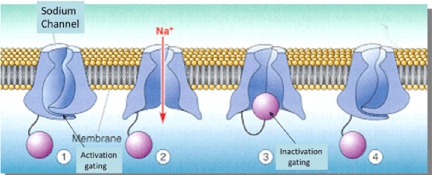
Describe the mechanism of voltage gated sodium channels
At resting potential Na+ channels are closed - the activation gate is closed
Depolarization opens the activation gate and Na+ flows into the cell along it's electrochemical gradient
A delayed component of voltage dependent activation is the blocking of the channel by the inactivation gate (after about 0.5ms)
Repolarization of the cell re-sets the two gates to their equilibrium positions.
There is a time dependent conformational change which will eventually end up in the inactivation gate closing the channel resulting in the refractory period.
Further conformational change causes the inactivation gate to relax and the membrane potential returns to the resting potential.
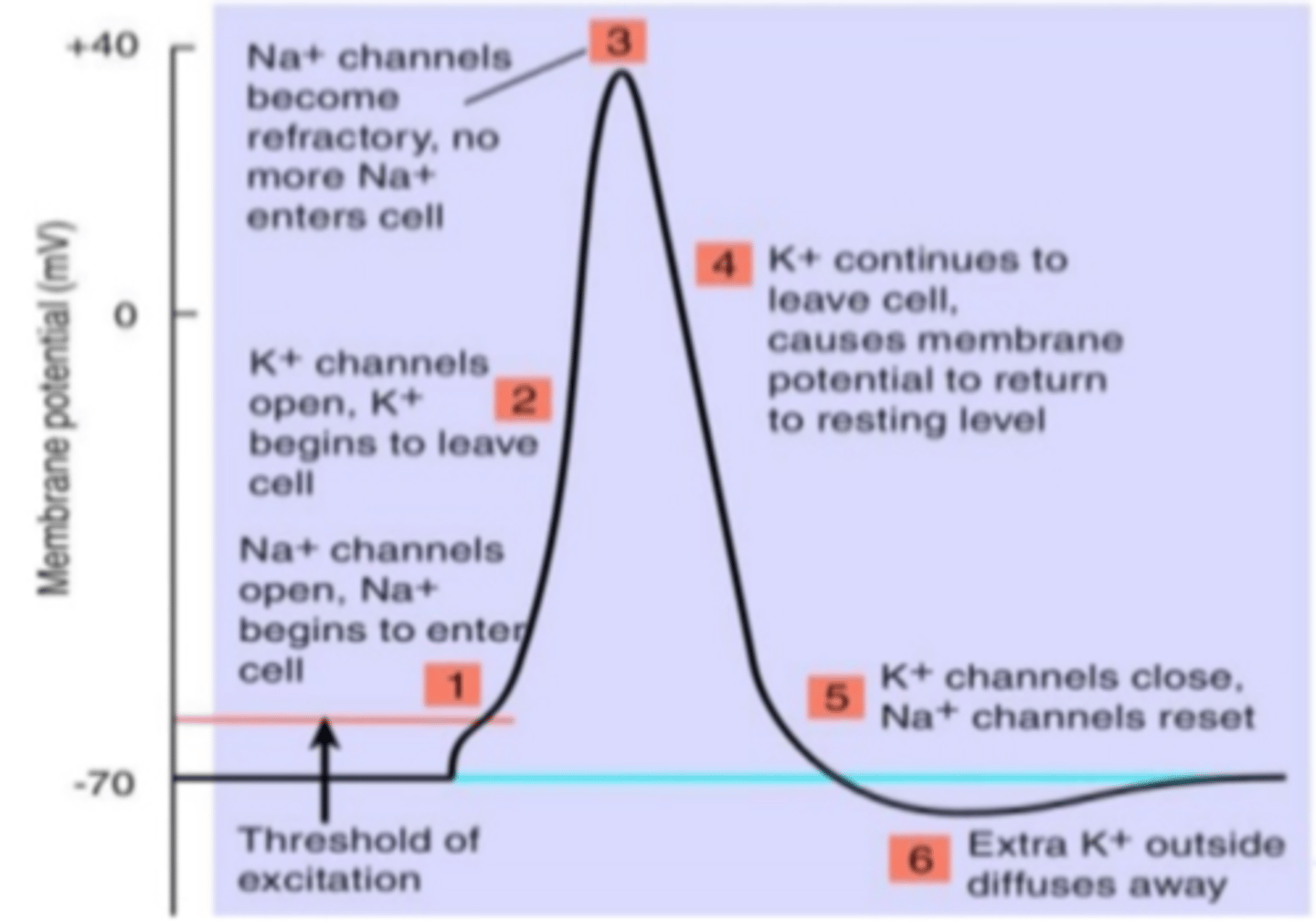
Describe the evolution of an action potential in terms of ions
Na channels open, Na begins to enter the cell.
K+ channels open, K+ begins to leave the cell.
Na+ channels become refractory, no more Na+ enters the cell.
K+ continues to leave the cell causing the membrane potential to return to resting level.
K+ channels close, Na+ channels reset.
Extra K+ outside diffuses away.
What mops up the excess K+ that diffuses away
Glial cells called astrocytes
What is the absolute refractory period?
Time from the opening of the Na+ activation gates until the closing of inactivation gates. Prevents the neuron from generating an action potential. Ensures that each action potential is separate. Enforces one way transmission of nerve impulses.

What is the relative refractory period?
During relative refractory period a stronger stimulus than normal could induce an action potential - some Na+ ready but more K+ channels are open than usual - cell still hyperpolarised
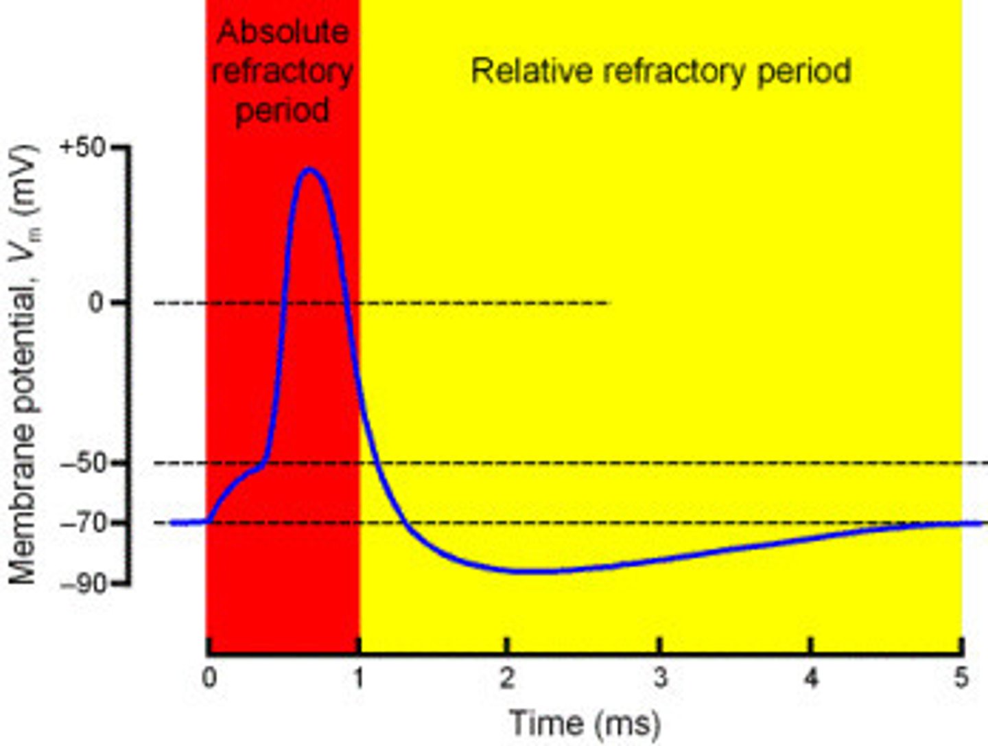
Why during the absolute refractory period can the cell not be stimulated to its threshold potential
As all Na channels are closed
What is myelination?
the process of forming a fatty sheath known as a myelin sheath around a nerve to allow nerve impulses to move more quickly and to reduce size
What cells are responsible for myelination in the PNS
Schwann cells
What cells are responsible for myelination in the CNS
Oligodendrocytes
The bigger the fatty sheath...
The better the protection
Where on the axon are APs generated?
Nodes of ranvier
What is saltatory conduction?
Saltatory conduction is the "jumping" of an action potential between the unmyelinated nodes of ranvier. In this way the conduction of the nerve impulse flows rapidly along the inside of the axon to the node, where it slows and ionic depolarisation (action potential) takes place. Note only a few ions are needed to do this thus energy is saved. And then the fast conduction along the inside of the axon resumes afresh
What are internodes?
Stretches of myelin in-between nodes
What conducts nerve impulses faster, small myelinated fibres or large unmyelinated ones
Small myelinated ones - these help keep the NS compact
What is normal conduction velocity?
50-60 meters/second
What is the conduction velocity of fastest alpha motor neurons?
120 m/s
Any slowing of the speed of conduction is consistent with...
Demyelination
Where in the neuron are chemical signals secreted?
Axon terminal
What is a synapse?
Specialised regions of close approach between axon and another cell (target is mostly a neuron but can be a muscle cell)
What is the synaptic cleft?
gap between adjacent neurons
What is a presynaptic cell?
a neuron that delivers a signal to a synapse
What is a postsynaptic cell?
the cell that receives the signal
The terminal of the presynaptic cell forms a swelling called a
bouton
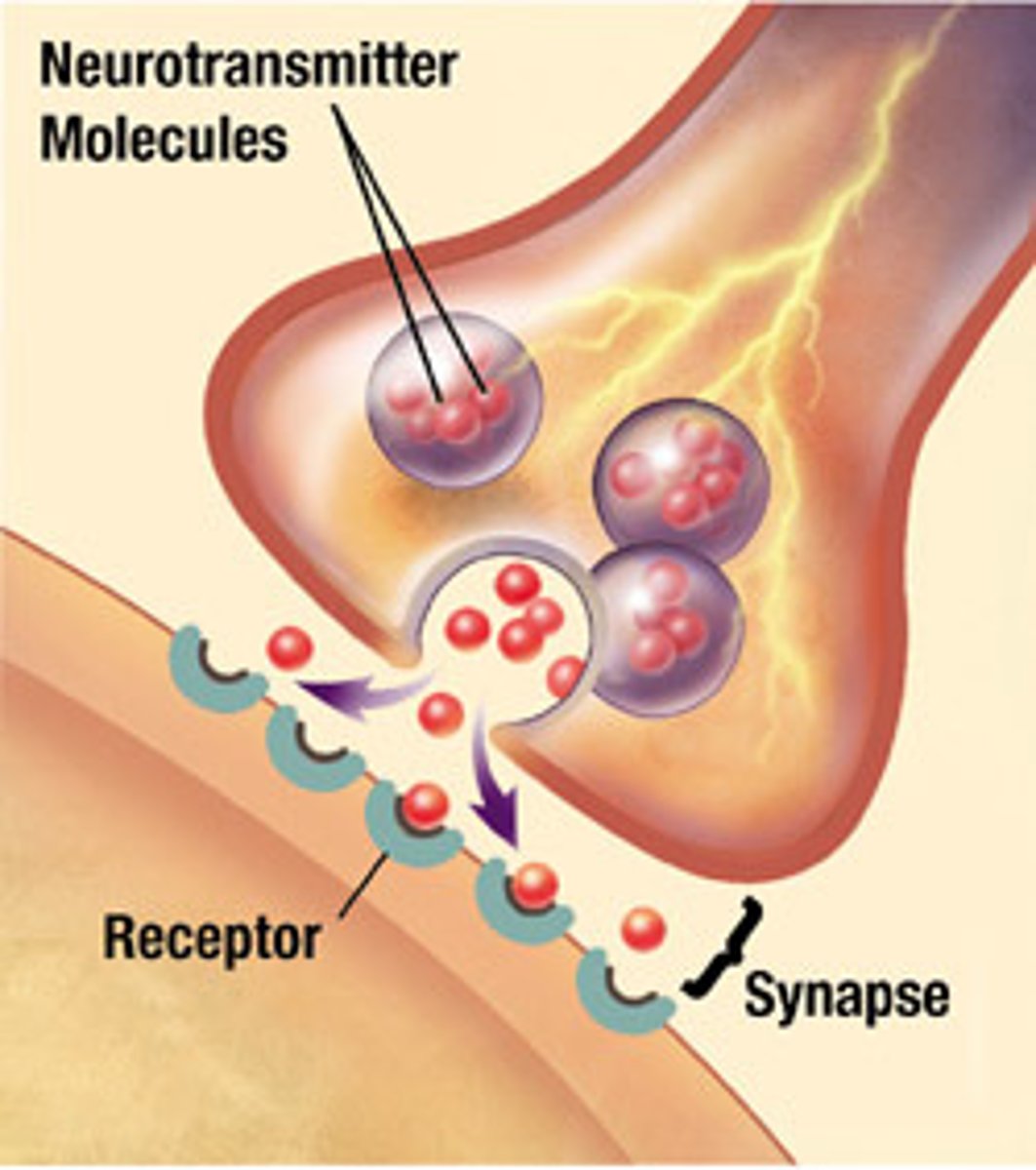
The vesicles contain molecules of
neurotransmitter
How big is the synaptic cleft?
20nm
What happens once the neurotransmitter has diffused across the synaptic cleft
It binds to receptors on the postsynaptic cell which can convert it into a further electrical signal on the postsynaptic membrane
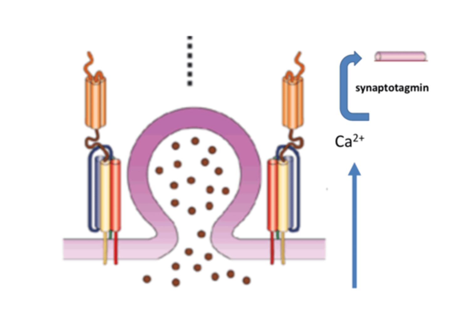
What are SNARES
Pairs of molecules on a vesicle which allow it to dock at a target to prepare for exocytosis
What are the 2 types of SNARES
v-SNARES and t-SNARES
What is the function of SNARES
v-SNARES and t-SNARES pair together and this pairing draws the vesicle through the zippering mechanism allowing it to dock
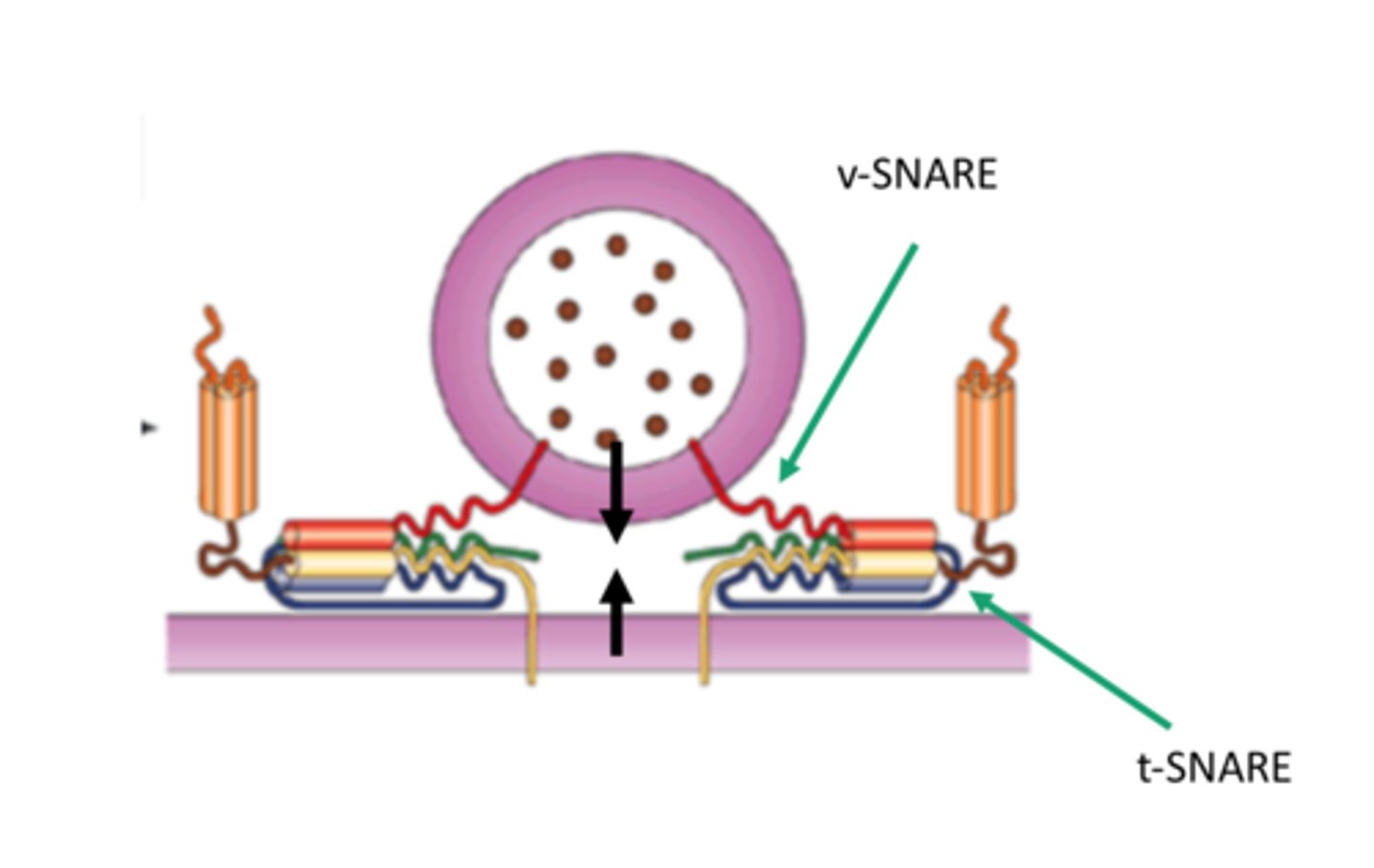
What happens once a vesicle is docked on the membrane
Action potential triggers calcium influx at end-bulb. Calcium induces synaptotagmin to displace complexin and exocytosis proceeds
What can bind to SNARES and inactive them
Botulinium toxin - these people become paralysed
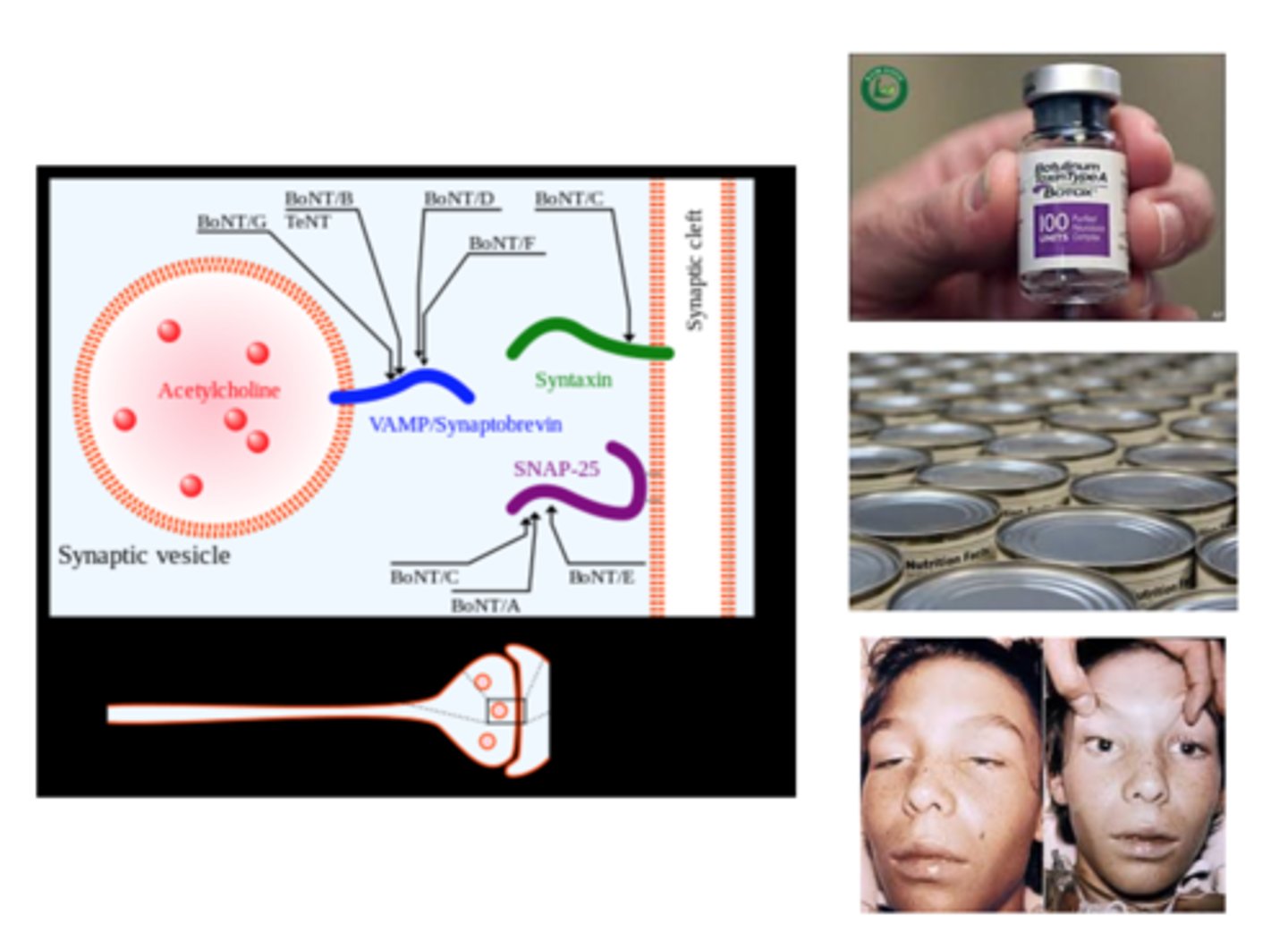
Neurotransmitters are released by
Exocytosis and diffuse to the post-synaptic membrane where they bind to receptors
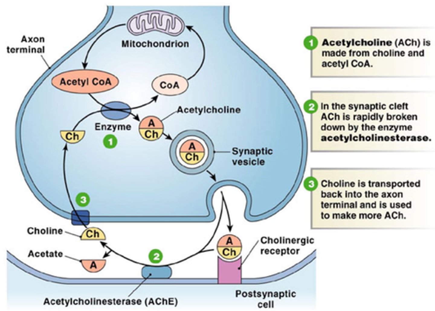
Neurotransmitters are always...
Inactivated
How are neurotransmitters inactivated?
Diffusion, re-uptake or enzymal inactivation
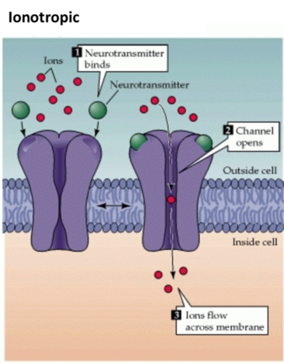
What 2 different types of receptors are present on the post-synaptic cleft?
ionotropic (directly gate ion flow)
metabotropic (indirectly gate ion flow or activate other pathways)
What is an ionotropic receptor?
a receptor protein that includes an ion channel that is opened when the receptor is bound by an agonist
Why are metabotropic receptors indirect
As there is a receptor but this receptor is not a channel. The receptor sends a signal via a G protein to allow the channel to open
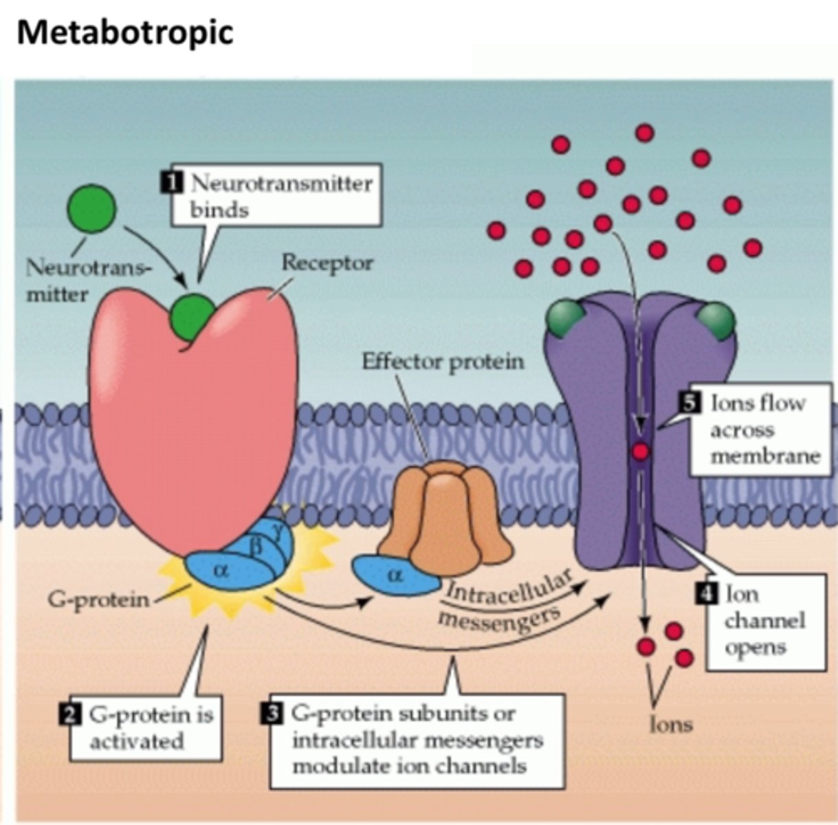
Which is faster, ionotropic or metabotropic
Ionotropic
Receptors can be...
excitatory or inhibitory depending on the ions they let into/out of the cell
Opening of pores was visualised by
Cryo-electron microscopy
Post synaptic potentials or PSPs are caused by
The passage of ions through ion channels which have been opened following receptor/neurotransmitter interactions
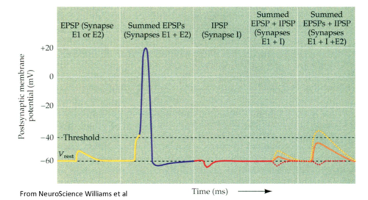
What is an excitatory PSP (EPSP)
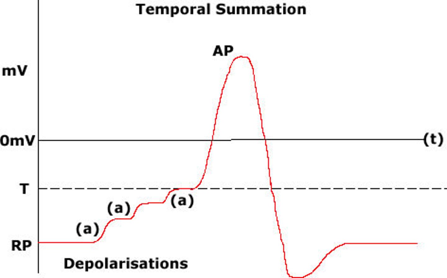
A net flow of positive ions into the cell depolarises the membrane (brings it closer to the threshold) and is thus termed EXCITATORY (EPSP). Single EPSPs rarely result in an action potential, they can be additive
What is an inhibitory PSP (IPSP)
The net flow of negative ions causes a further depolarisation of the membrane making it resistant to generating an action potential
For both inhibitory and excitatory PSPs the amplitude of the signal decreases with...
Distance as well as time
What is spatial summation?
postsynaptic neuron is stimulated by a large number of terminals at the same time
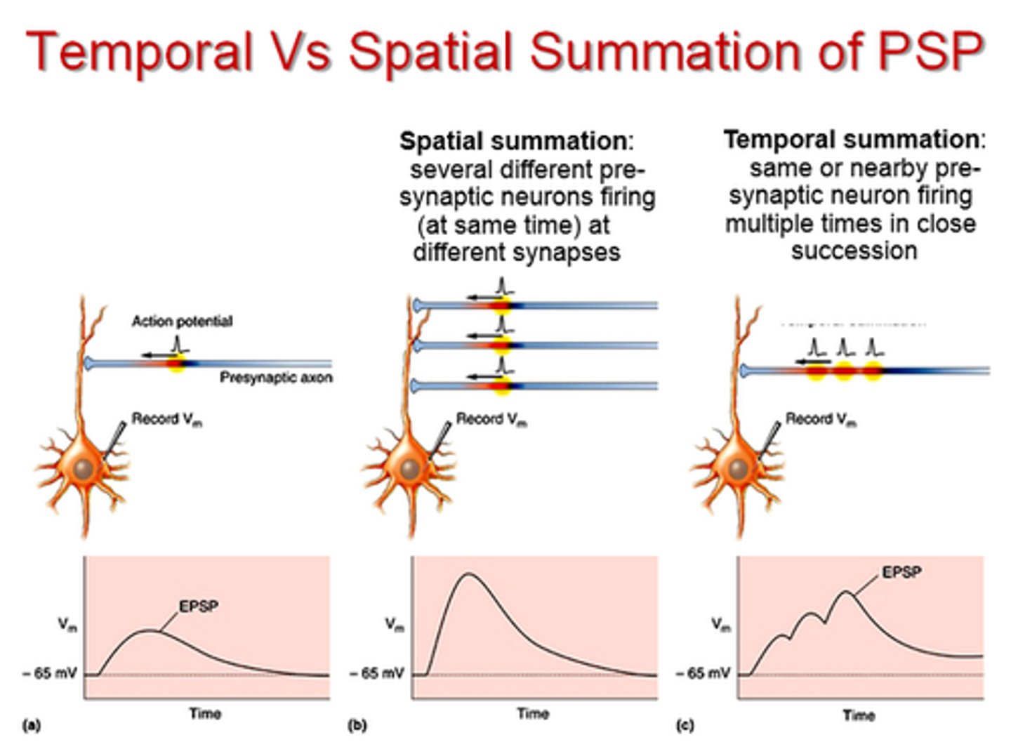
What is temporal summation?
summing several EPSPs from one presynaptic neuron
What determines whether or not an action potential occurs at a post-synaptic neuron
Summation of IPSPs and EPSPS
An increase in what ion drives the SNARE process
Ca
Stronger external stimuli result in...
A higher frequency of APs in the axon
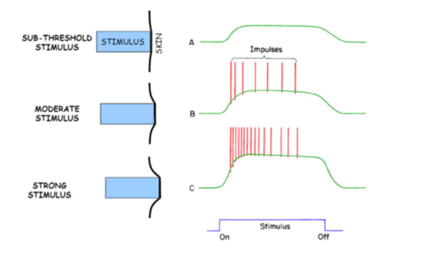
Why do stronger external signals result in a higher frequency of APs in the axon?
Each receptor type has a modality and feeds information about modality to the CNS
Most receptors code duration and magnitude of external signals as a generator potential
What is generator potential
The change in potential difference due to a stimulus
Generator potentials are produced as...
Sodium and potassium flow down their electrochemical gradients to depolarize the terminal ending
In most cases, the magnitude and duration of the generator potentials are related to the...
Applied mechanical force: the greater the mechanical force, the greater is the depolarization, and the longer the mechanical force is applied, the longer the terminal remains depolarized
What are rapidly adapting generator potentials
Terminals that do not sustain the depolarization for the duration of the mechanical distortion
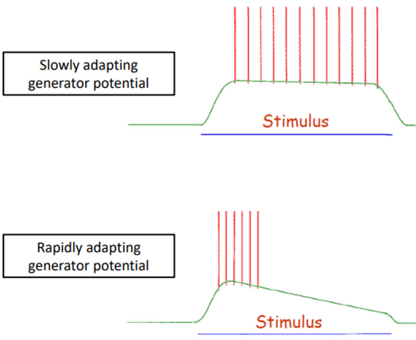
What are slowly adapting generator potentials
Terminals that sustain the depolarization with minimal decrease in amplitude for the duration of a stimulus

Where in the body can slowly and rapidly adapting generator potentials be found?
Muscle spindles
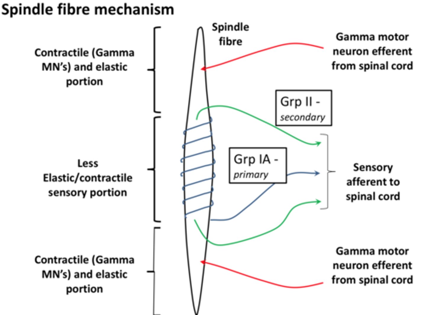
What are muscle spindles?
Assemblies of a number of sensory specialised fibres found in skeletal muscle - measures active stretch and the extent of stretch
What is the function of a muscle spindle
The muscle spindle is a proprioceptive receptor in the muscle which senses muscle stretch and sends excitatory inputs to the lower motor neurons to regulate muscle length. Prevents muscles from being overstretched
Describe the structure of a muscle spindle
Either end is a contractile and elastic portion which measures active stretch and the centre is less elastic/contractile sensory portion which measures the extent of the stretch, preventing overstretch
How is nerve conduction measured?
We measure the sum of all the action potentials in a nerve or the resulting stimulus inside a target such as a skeletal muscle
Why is it important to measure nerve conduction
As clinically we cannot measure individual action potentials
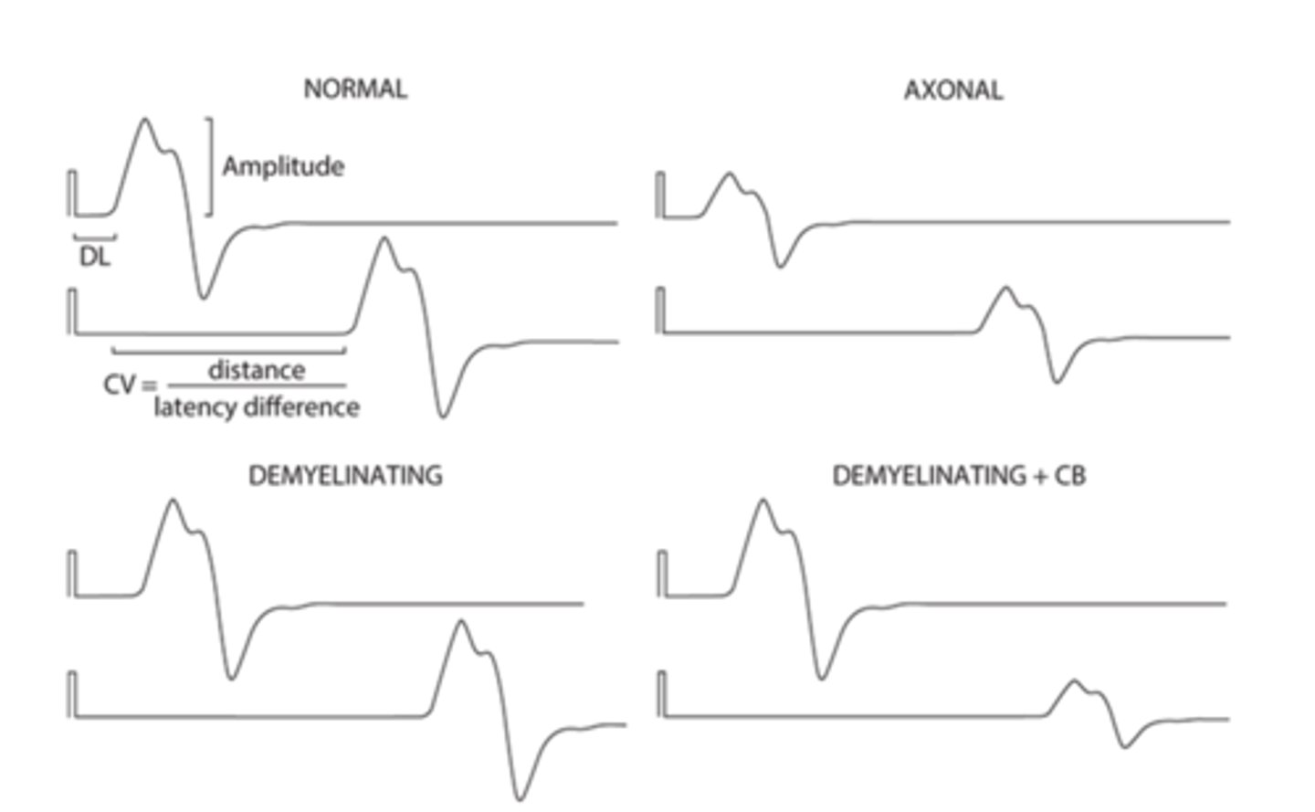
In a nerve conduction study what does a reduced amplitude suggest
Less axons
In a nerve conduction study what does a slowed conduction velocity suggest?
Less myelin
What are the 2 different ways of measuring nerve conduction
SNAP and CMAP
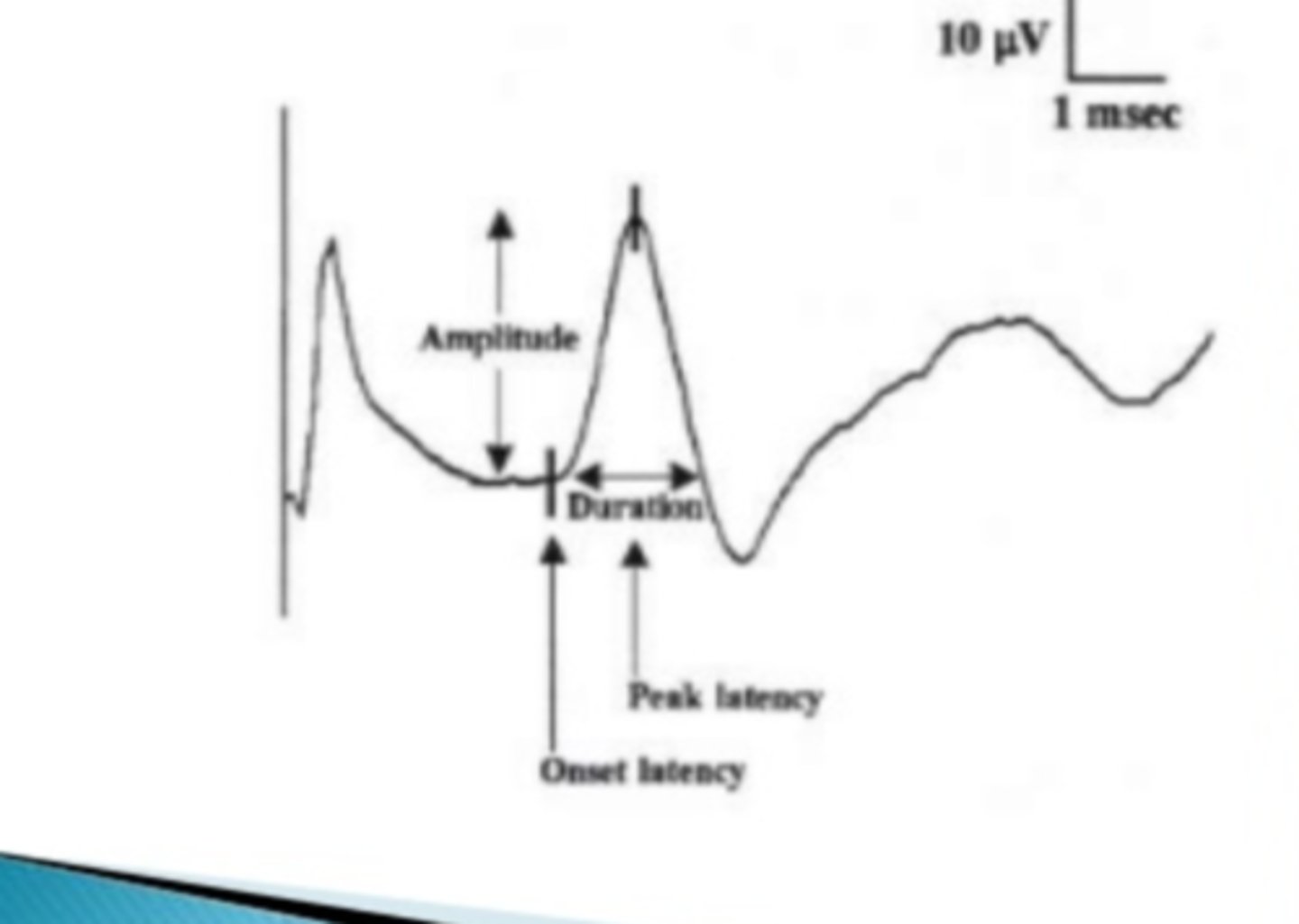
What is SNAP
sensory nerve action potential
What is CMAP
compound muscle action potential
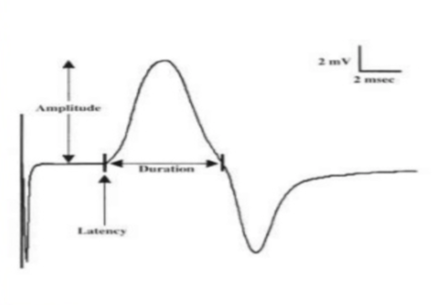
Where would CMAP be measured?
Thenar eminence
What can nerve conduction studies be used to differentiate?
Neuropathies e.g. carpal tunnel syndrome
What would the nerve conduction study of someone with carpal tunnel syndrome show?
A decrease in the function of the axons themselves - not demyelinating
What is a channelopathy?
Disease from a dysfunction of an ion channel.
What are the 2 types of channelopathy
Inherited and autoimmune
What ions are involved in inherited channelopathies
Calcium, chloride potassium, sodium, glycine, GABA, Ach
What parts of the body can be affected by inherited channelopathies
Brain and heart
Give examples of autoimmune channelopathies
Myasthenia gravis
Lambert-Eaton myasthenic syndrome (associated with small cell lung Ca)
Limbic encephalitis (memory loss; associated with underlying neoplasm
What is myasthenia gravis?
- An autoimmune neuromuscular disease leading to fluctuating muscle weakness and fatigue.
- Antibodies bind to the neuromuscular acetylcholine receptor and diminish its function
- Improved with rest