Immmunology - Unit 3: The Adaptive Immune Response
 Call Kai
 Learn
Learn Practice Test
 Spaced Repetition
 Match
Match Flashcards
 Knowt Play
1/83

There's no tags or description
Looks like no tags are added yet.
Name | Mastery | Learn | Test | Matching | Spaced | Call with Kai | Chat |
|---|
No analytics yet
Send a link to your students to track their progress
84 Terms
what are the branches of the adaptive immune response?
adaptive → active, passive (passed on to you, ex. babies get IgG from mother), and adoptive (getting cells from someone else to produce immune product. ex. bone marrow transplant)
active→ humoral immunity (B-cells producing Abs) and cell-mediated immunity (T-cells use cytokines to tell cells what to do→ B-cells produce Ab after signaled by T-cells)

Active immunity
building your own defense
immune response produced by an immunocompetent individual following exposure to a challenge
Immune products produced → may take time (weeks) to develop; lifelong protection
Can be naturally acquired (ex. recovery from infection) or artificially acquired (vaccination)
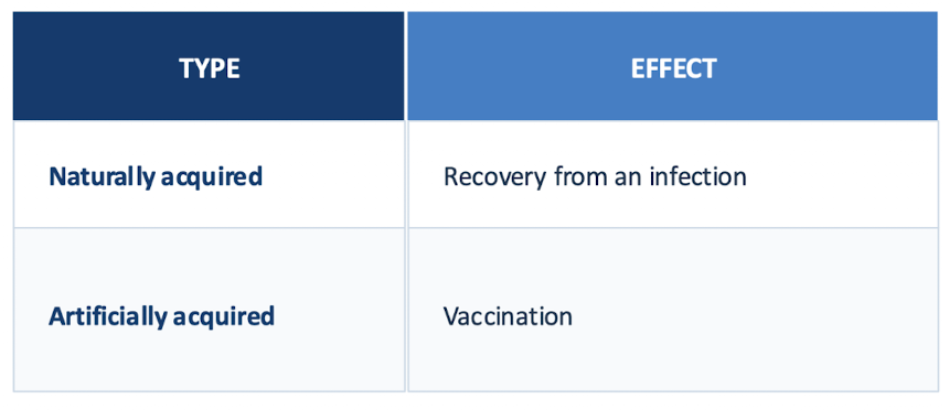
what is an example of naturally acquired active immunity?
recovery from an infection
what is an example of artificially acquired active immunity?
vaccination
passive immunity
borrowing pre-made protection
given pre-formed immune products → transient protection acquired when preformed immune products are administered to an individual
confers immediate protection
Naturally acquired (ex. IgG crossing mother’s placenta, IgA in breast milk) or artificially acquired (Intravenous Ig, mAb therapy)
what is an example of artificially acquired passive immunity?
intravenous Ig, mAb therapy
what is an example of naturally acquired passive immunity?
IgG crossing mother’s placenta, IgA in breast milk
Adoptive immunity
transferring immune cells
transfer of immunocompetent cells from one individual to a second individual to establish immunocompetence in the second individual
Reconstitute the immune system→ ex. BMT, CAR T-cell therapy (cells recognize certain antigens and attack it)
what are examples of adoptive immunity?
BMT
CAR T-cell therapy
how are blood cells produced?
through hematopoiesis in the bone marrow
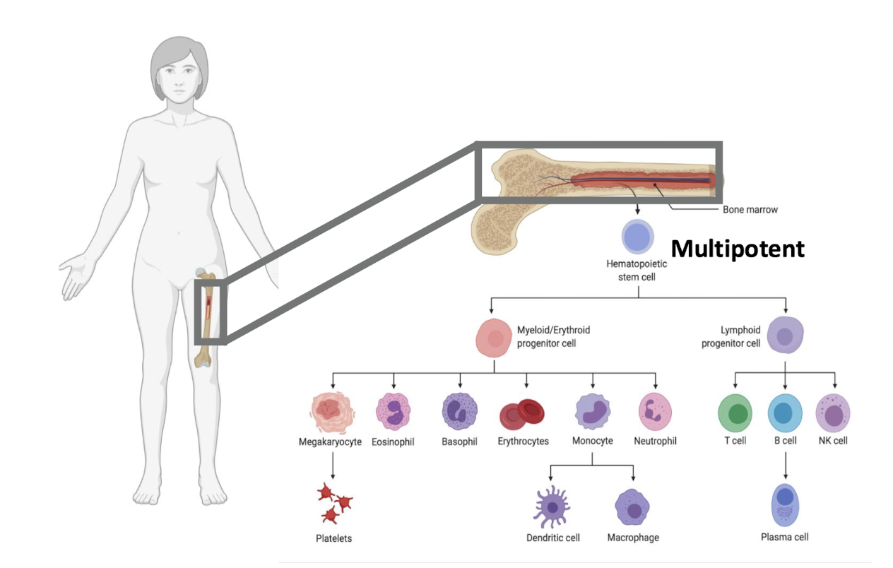
What are the lymphocytes (like B and T cells) formed from?
hematopoietic stem cell (bone marrow) → Lymphoid progenitor cell → T cell, B cell, or NK cell
B cell can further develop into plasma cell
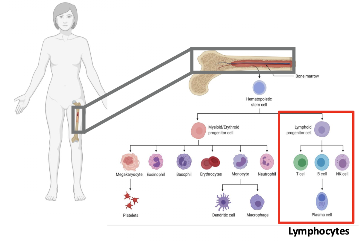
where do lymphocytes develop?
primary lymphoid organs
development and maturation of lymphocytes
selection process→ self vs. non-self
Locations:
Bone marrow → B cells
Thymus → T cell progenitor migrates to thymus
where do B cells develop?
primary lymphoid organs → in the bone marrow
where do T-cells develop?
primary lymphoid organ→ progenitor migrates to the thymus
where are lymphocytes activated?
secondary lymphoid organs
after maturation, lymphocytes enter circulation
may hang out in specific organs → encounter Ags → activated
Ex. lymph nodes, spleen, mucosal-associated lymphoid tissue (MALT)
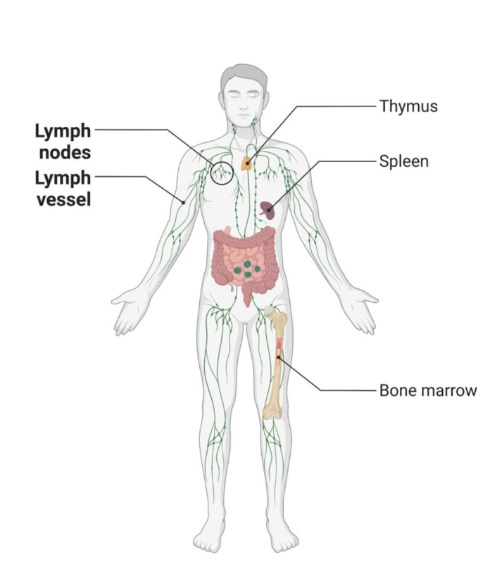
what are examples of secondary lymphoid organs
Lymph nodes, spleen, mucosal-associated lymphoid tissue (MALT)
Secondary lymphoid organs: lymph nodes
filter lymph fluid from draining tissue
B cells concentrated in follicles and cortex
form germinal centers after B cells activated
T cells concentrated in paracortex
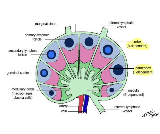
where are B cells in the lymph nodes?
follicles and cortex
form germinal centers after ___ cells activated
Where are T-cells concentrated in the lymph nodes?
paracortex
secondary lymphoid organs: Spleen
filters blood
similar to lymph node
Secondary lymphoid organs: MALT
non-encapsulated
gut associated lymphoid tissue (GALT)
Peyer’s patches, tonsils, adenoids
M cells deliver Ags from gut to adaptive immune cells
Bronchial-associated lymphoid tissue (BALT)
respiratory epithelium
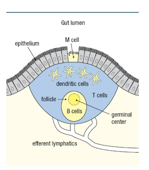
what is GALT?
Peyer’s patches, tonsils, adenoids
M cells deliver Ags from gut to adaptive immune cells
what is BALT
respiratory epithelium
flowchart of how lymph nodes organize B- and T- cell encounters
Antigen found by APC → APCs enter draining lymph node → APC scan for cognate naive T-cell → T cell activated → clonal expansion and B cell interaction

what control is immune response under?
genetic control
linked to MHC → Major Histocompatability Complex/ HLA
found on all nucleated cells → brings small peptide antigen to cell surface→ recognized by T-cells
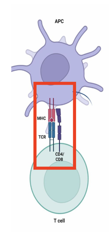
where are MHC genes found?
short arm of chromosome 6
closely linked
inherited as a haplotype→ one chromosome from each parent, co-dominant
>1.7 billion combinations gives diversity
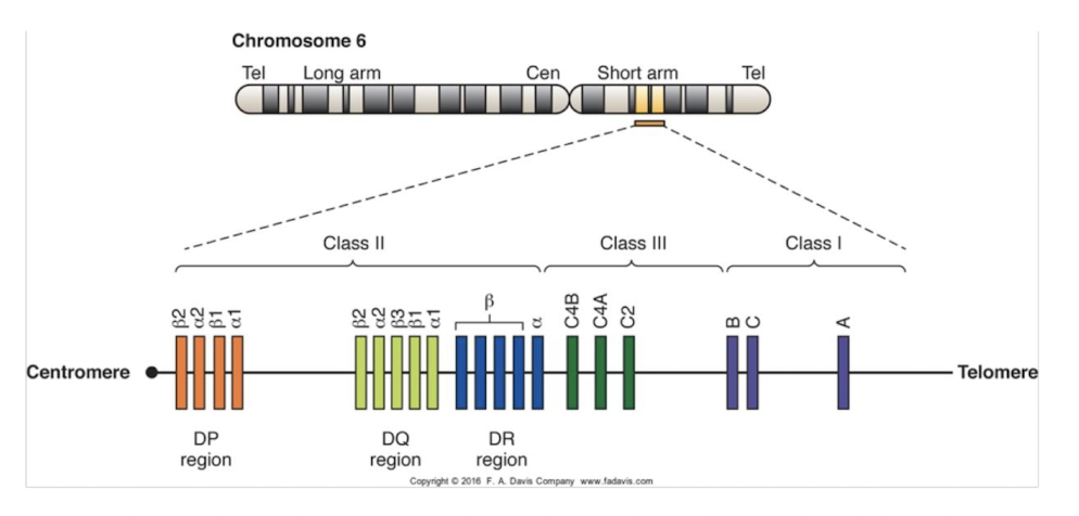
what are the 3 classes of MHA/MLA
Class I → all nucleated cells; 6 gene loci (Major: HLA-A, -B, -C; Minor: MLA-E, -F, -G)
Class II→ Antigen Presenting Cells (APC); 5 Isotypes (HLA-DM, -DO, -DP, -DQ, -DR)
Class III → not found on cells; complement proteins

what are features of MHCI (expression and genes)
expressed on all nucleated cells
six gene loci:
Major: HLA-A, -B, -C
Minor: MLA-E, -F, -G
G: part of the placenta → produces inhibitory signals so that mother’s immune cells don’t attack baby
what are features of MHCII (expression and genes)
on Antigen Presenting Cells (APC)
5 Isotypes
HLA-DM, -DO, -DP, -DQ, -DR
what are features of MHCIII (expression and genes)
not found on cells
complement proteins
Clinical relevance of MHC/HLA involvement
Transfusion reactions
Platelet → HLA-A
Transplant and graft rejections
Autoimmune disease
Prevention → HLA matching
what is the structure of MHCI
glycoprotein dimer with 2 non-covalently linked polypeptide chains
Alpha chain → 3 domains → a1 and 2 create a cleft for Ag; a3 binds CD8 and is inserted in membrane by cytoplasmic tail
Beta chain
encoded by a single gene on chromosome 15
does not penetrate cell membrane
essential for alpha chain folding

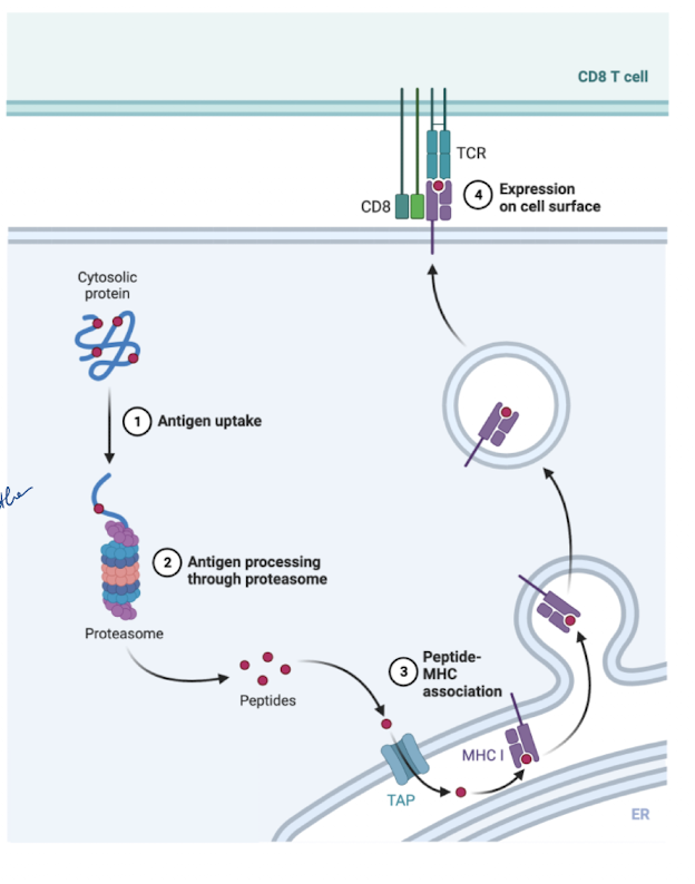
what is the function of MHCI and the steps to that process
detect endogenous antigens
intracellular pathogens: viruses, tumor antigens → degraded by proteasome → peptides produced
peptides transported to rough ER → binds to MHCI
Peptide-MHCI complex transported to golgi → then cell surface
TCR binds antigen → MHCI binds co-receptor: CD8
CD8 + T cell lyses cell
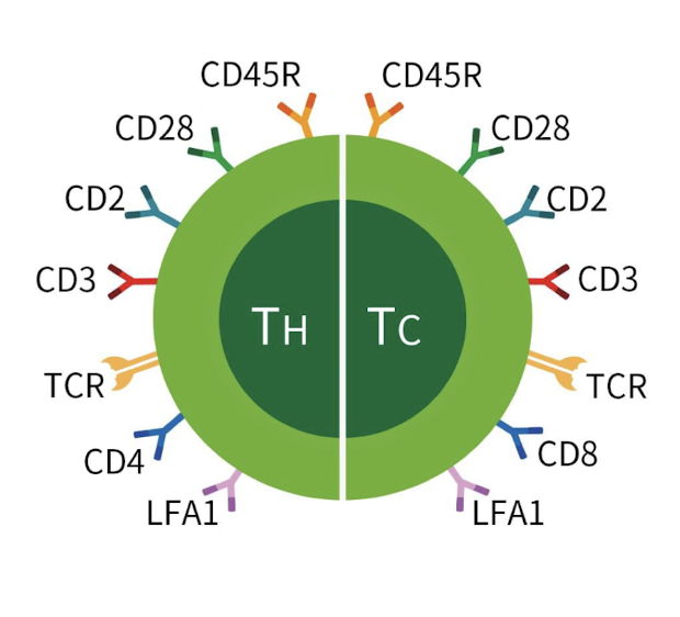
How are immune cells classified?
CD markers
Cell surface proteins that can show immature vs. mature, B cell vs. T-cell
More than 1 used typically
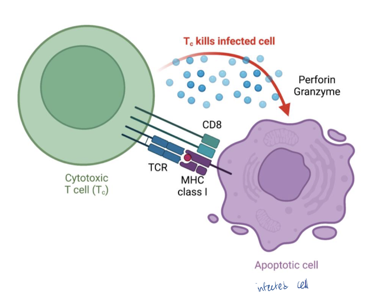
what is the main role of MHCI
bind viral/tumor peptides
Present to cytotoxic (CD8) T-cell
10-100 identical Ag-MHCI complexes needed to induce response
after contact, CD8 T-cell produces granzyme and perforin to lyse cell
what type of cells is MHCII expressed on?
Antigen presenting cells (APCs)
Monocyte
Macrophage
Kuppfer cells, microglia, alveolar, etc.
Dendritic cell → most effective
B cell
Present exogenous antigens


why are dendritic cells the most effective as APCs?
multiple arms that can bind/grab many Ags/ have a higher chance of grabbing Ags
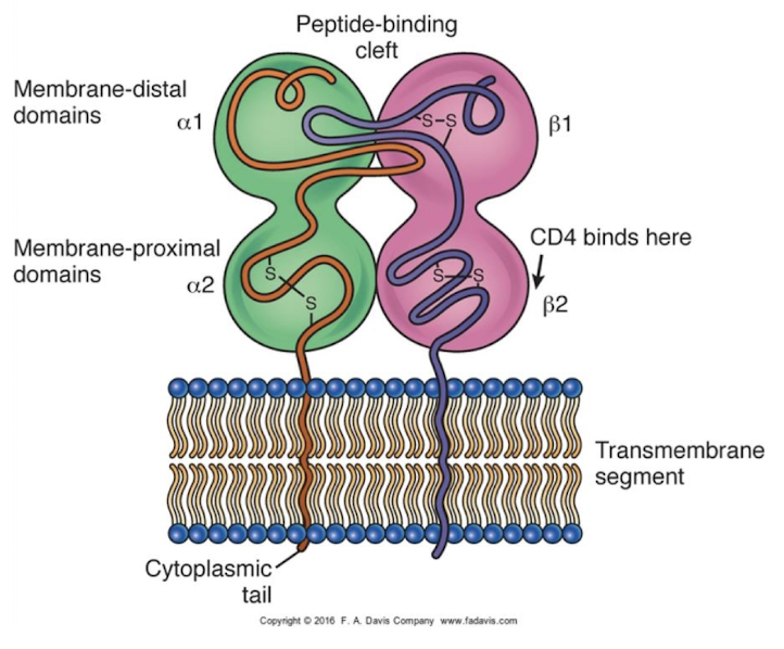
what is the structure of MHCII
two non-covalently bound polypeptides
Alpha and beta chains
both inserted into membrane by cytoplasmic tails
a1 and b1 regions form peptide binding site → deeper than Class I, longer peptides
what is the function of MHCII and the steps to the process
present exogenous antigens
exogenous protein taken up → endocytosed
Ag processed into peptides in endosome
MHCII exists in rough ER
bound by invariant chain→ binding to other peptides now blocked
MHCII leaves rough ER thru Golgi
fuses w/ endosome
endosome cleaves invariant chain
peptide-MHCII complex can bind
Peptide-MHCII complex transported to cell surface
TCR binds Ag and MHCII binds co-receptor (CD4)
Results in clonal expansion of T-cells → proliferation and activation

what is the main role of MHCII
Clonal expansion of T-cells
Proliferation
Activation → T cells able to do their job

what is the coreceptor/ T-cell type for MHCII when the T-cell binds
CD4
what is the coreceptor/ T-cell type for MHCI when the T-cell binds
CD8
what kind of immunity are T-cells a part of?
Cell-mediated immunity
main features of T-cells (size, amount, markers)
Small lymphocyte (7-10 µm)
75% of circulating lymphocytes
Markers:
TCR: T cell receptor
CD2: sheep RBC receptor
CD3: part of TCR → normally how T cells classified
CD4: co-receptor for MHCII
CD8: co-receptor for MHCI
list the main markers found on T-cells
TCR: T cell receptor → every T cell has unique one
CD2: sheep RBC receptor → historically used to identify T-cells→ rosettes of sheep blood around cells
CD3: part of TCR → normally how T cells classified
CD4: co-receptor for MHCII
CD8: co-receptor for MHCI
what gives T-cells their specificity?
TCR → recognizes peptide-MHC complex
can’t bind Ag w/o MHC
Every T cell has a unique TCR
Recognizes unique Ag
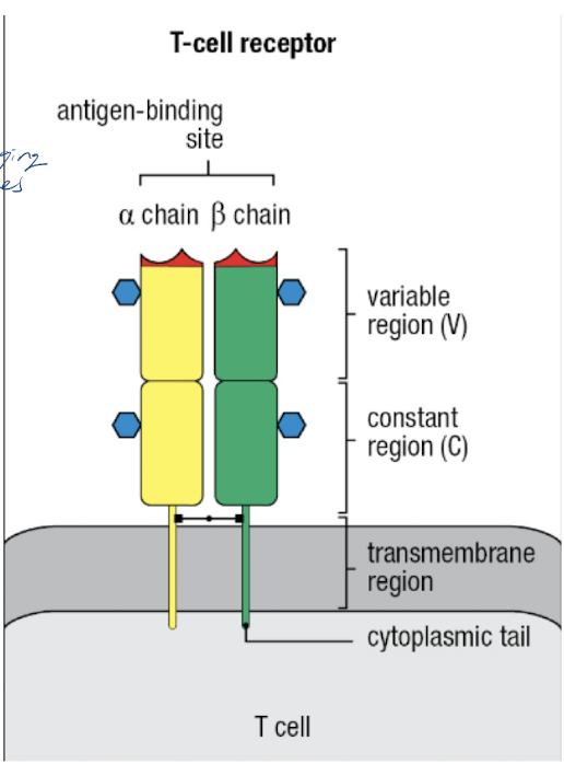
what are the two parts of the TCR?
Antigen receptor
Alpha and beta chains
variable and constant region
similar to Ab
CD3 glycoprotein
intracellular signaling → tell nucleus to turn on gene expression when T-cell activated
what proteins give uniqueness to TCR and how?
Recombination activating genes (RAG) 1 and 2
rearrange gene segments of TCR → V(D)J recombination
RAG1/2 deficiencies prevent normal TCR formation → cause forms of SCID (Severe Combined Immunodeficiency)
what are the steps to T-cell maturation in the thymus in order?
T cell precursor migrates from bone marrow to thymus
Double negative T-cell (No expression of CD4 or CD8)
Double Positive T-cell (both CD4 and CD8 expressed on membrane)
Positive selection → binds to an Ag to make sure T-cell works (binding shouldn’t be too strong→ autoimmune or too weak→ non functional)
If T-cell survives, becomes singly positive (expresses either CD4 or CD8, but not both)
Negative selection → self-antigen presented to T-cell→ if it binds, apoptosis of T-cell
Mature CD4+ and CD8+ (SP) cells exit thymus → naive cells (have not encountered an Ag yet)
-takes ~3 wks, only 1% survive (positive/negative selection remove most)

What are the two subsets of T-cells
CD4+ Helper T cells
CD8+ Cytotoxic T cells
what are features of CD4+ Helper T cells?
exogenous Ags presented by APCs using MHCII
Coordinate immune response by releasing cytokines to activate and regulate other cells
Called helper T cells (TH) → B cell help, etc.
what are features of CD8+ Cytotoxic T-cells
endogenous Ags presented by any nucleated cell using MHCI
Kill virally infected cells and tumor cells → use perforin and granzyme
Called cytotoxic T lymphocytes (CTLs or TC)
how are CD4+ helper T-cells activated?
Requires 2 signals:
TCR interaction w/ MHCII-peptide
Co-stimulatory molecules
T cells differentiate and effector T helper (TH) cell or memory T (TM) cell
Effector T cell differentiation is based on environmental cytokines

T-helper cell 1 (Th1) - CD4+
typically pro-inflammatory
responsible for cell-mediated immunity thru pro-inflammatory cytokines
Interferon-gamma (IFN-g)
stimulate macrophages
boosts tumoricidal activity
stimulates Ag presentation by MCHI and MHCII
Interleukin-2 (IL-2)
Drives proliferation of both T and B cells
what does Interferon-gamma (IFN-g) produced by Th1 cells do?
stimulate macrophages
boosts tumoricidal activity
stimulates Ag presentation by MCHI and MHCII
what does Interleukin-2 (IL-2) produced by Th1 cells do?
drives proliferation of both T and B cells
T-helper cell 2 (Th2) - CD4+
helps w/ antibody response
responsible for antibody-mediated immunity thru IL-4 production to help B cells turn into plasma cells
Interleukin-4 (IL-4)
Interacts w/ IL-6 to drive B cells into plasma cells
Regulates other immune responses (ex. allergies, autoimmune responses, parasitic infections)
what does Interleukin-4 (IL-4) produced by Th2 do?
Interacts w/ IL-6 to drive B cells into plasma cells
Regulates other immune responses (ex. allergies, autoimmune responses, parasitic infections)
T regulatory (Treg) cells - CD4+
suppress inflammation
regulate immune cell activity thru anti-inflammatory cytokines
establish peripheral tolerance (in bloodstream→ tolerating own cells so they don’t attack self)
Interleukin-10 (IL-10)
antagonist to IFN-g
inhibits Ag presentation by APCs
Transforming Growth Factor-Beta (TGF-B)
regulates cell growth, differentiation, apoptosis, migration
what does Interleukin-10 (IL-10) produced by Treg cells do?
antagonist to IFN-g
inhibits Ag presentation by APCs
what does Transforming Growth Factor-Beta (TGF-B) produced by Treg cells do?
regulates cell growth, differentiation, apoptosis, migration
what is key for proliferation of T-cells?
IL-2 → autocrine and endocrine function → T-cells release it during activation

anergy
T cells unresponsiveness after Ag recognition thru TCR
W/o co-stimulation (signal 2), T cells remain alive, but:
reduced proliferation
reduced cytokine production
how are CD8+ T cells (adaptive immunity) and NK cells (innate immunity) similar?
both are cytotoxic → use perforin and granzyme to kill
how are CD8+ T cells (adaptive immunity) and NK cells (innate immunity) different?
CD8 T cells are MCHI restricted thru TCR
NK cells don’t have TCR
If cells have low or missing MHCI, that is a sign for NK cells to kill
Use receptors as “do not kill” signal
Also use ADCC (kill signal, Ab dependent cell-mediated cytolysis)

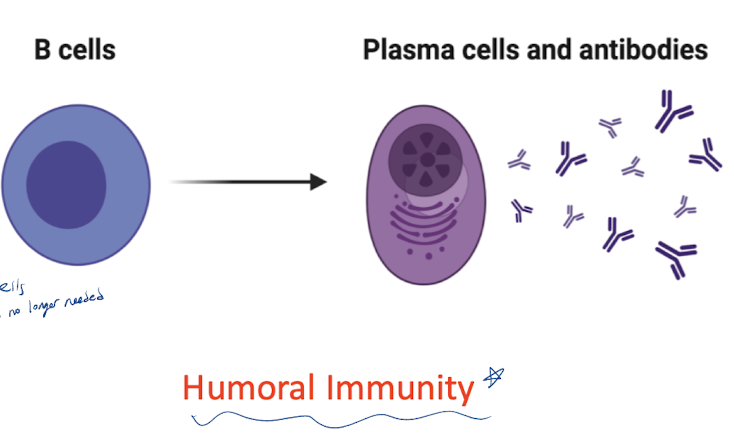
what are the characteristics of B cells (amount, type, markers)?
10-20% of circulating lymphocytes
APC
Part of HUMORAL IMMUNITY
Markers:
Surface Ig: IgM and IgD
IgG Fc receptor
C3b receptor
CD21 (EVB receptor)→ mono affects these cells b/c of this receptor
MHCII
Others: CD19, CD20, CD40 → markers to indicate it is specifically a ___ cell
what are the surface Ig’s on the surface of B cells?
IgM and IgD
List the markers on B-cells
Surface Ig: IgM and IgD
IgG Fc receptor → inhibitory signal to these cells to tell them Ab is no longer needed
C3b receptor
CD21 (EVB receptor)→ mono affects these cells b/c of this receptor
MHCII
Others: CD19, CD20, CD40 → markers to indicate it is specifically a ___ cell
what are the markers on B-cells that identify them as B-cells specifically
CD19, CD20, CD40
where does maturation of B cells occur?
bone marrow
List the steps/stages to the development of B cells
Develop in the bone marrow starting from a stem cell
stem cell → Pro-B cell
Pre-B cell - mu chains present in cytoplasm (will form into IgM)
Immature B cell - IgM expressed on the cell surface
Negative selection→ self-reactive B cells culled
IgM and IgD are expressed on surviving B cells
Naive B cells enter circulation

what protein makes every B cell specific to a unique Ag
BCR → the IgM and IgD on the cell surface

what proteins give uniqueness to BCR
RAG proteins → V(D)J recombination
what are the steps to B cell activation?
B cell binds to unique Ag thru BCR (IgM/IgD)
Leads to IgM capping
all bound BCRs move to one “pole” on the cell → two Abs w/ 1 Ag bound btwn them → cross linking → activation
B cell now has two options:
differentiate into plasma cell to produce Ab
differentiate into memory cell to wait for later exposure
what happens when a B cell is activated by a thymus dependent (TD) antigen?
Ag is recognized by B cell → B cell processes it and presents it to a T cell
T cell is activated → releases cytokines to signal B-cell to activate/differentiate
B cell is activated and transforms into a plasma cell
can produce all types of Igs (IgM, IgG, etc.)

what happens when a B cell is activated by a thymus independent (TI) antigen?
These antigens are polysaccharides that are recognized by B cell → long polysaccharide can cause cross-linking between Abs on B cell surface → activation
B cells only make IgM (will not class switch)
ABO antigens

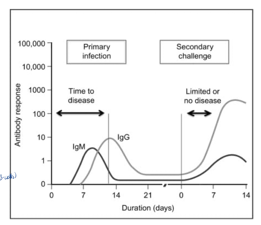
What happens during the primary antibody response to an Ag
first exposure to Ag
Latent phase:
no detectable Ab
~14 days to activate cells and produce Abs
First Ig appear: IgM
IgG follows (class switching in B-cells)
memory cells are formed
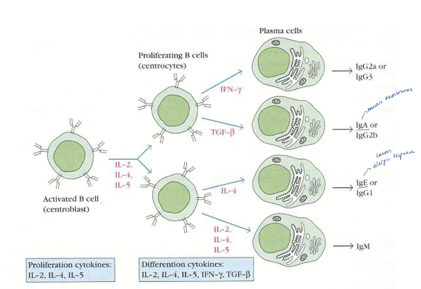
Class switching → Changeover from IgM to IgG
same plasma cell producing IgM changes to produce IgG
Ag specificity remains the same
class switching is stimulated to T cell cytokines
Ig class switching - proliferation cytokines and differentiation cytokines
IL-2, IL-4, IL-5 → proliferation
IL-2, IL-4, IL-5, IFN-gamma, TGF-B → differentiation
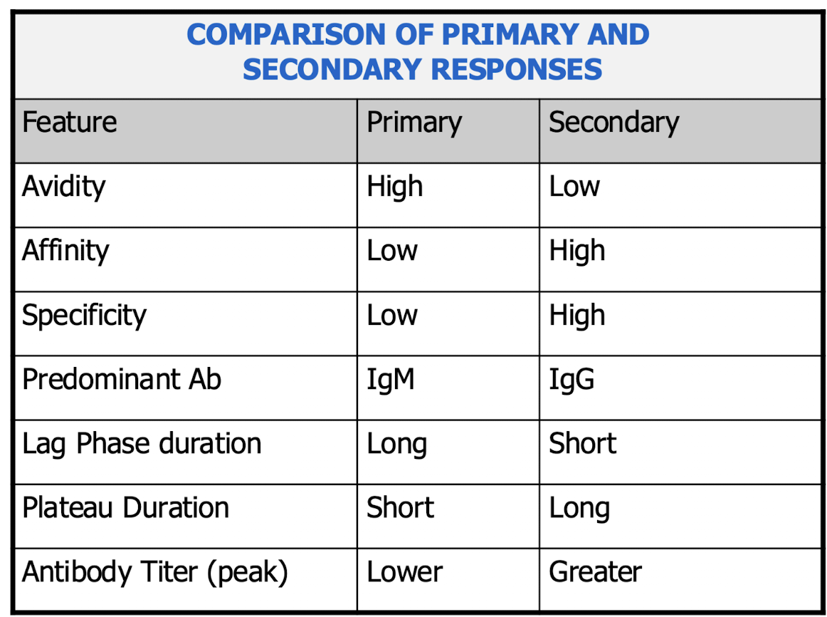
what happens during secondary antibody response to an Ag
stronger than the 1st response
evokes anamnestic response
rapid and intense
shorter latent phase
higher titer of Ig
Predominant Ig: IgG
persists longer

affinity
strength of one Ag-binding site interacting w/ one epitope
depends on molecular fit and noncovalent interactions
avidity
overall functional strength of multivalent binding
depends on affinity, valency, geometry, and Ag arrangement
Comparison of primary vs. secondary response$
list the roles of Abs in infection
protection against infection
opsonization
complement fixation
neutralization of pathogens
cell lysis via ADCC w/ NK cells

role of Abs in diagnosis
IgM vs. IgG
mother/baby → to measure baby’s immune response: IgM; for mother’s: IgG
Measure amt of antibody via titer
Patient serum (Ab) reacts w/ Ag
serial dilution → last dilution to react= titer