BIOL 224 Lab 13: Digestive System
1/100
There's no tags or description
Looks like no tags are added yet.
Name | Mastery | Learn | Test | Matching | Spaced |
|---|
No study sessions yet.
101 Terms
ingestion
entrance of materials from mouth into digestive tract
mechanical processing
breaking down of materials physically (chewing or churning)
digestion
chemical breakdown of foods into fragments small enough for absorption
secretion
release of water, enzymes, acids, buffers, and salts into digestive tract via digestive tract epithelium or glandular organs
absorption
passage of organic substrates, vitamins, electrolytes, and water into interstitial fluid of digestive tract across digestive epithelium
excretion
eliminate of wastes from body fluids
mastication
act of chewing initial mechanical breakdown of food
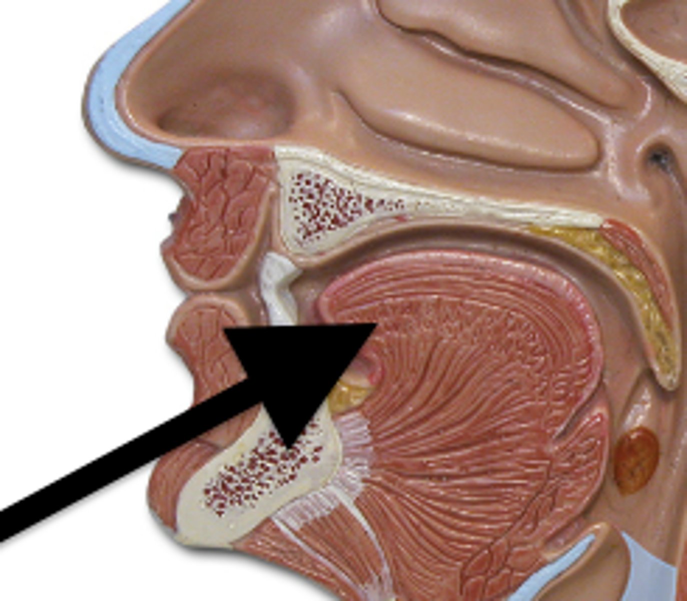
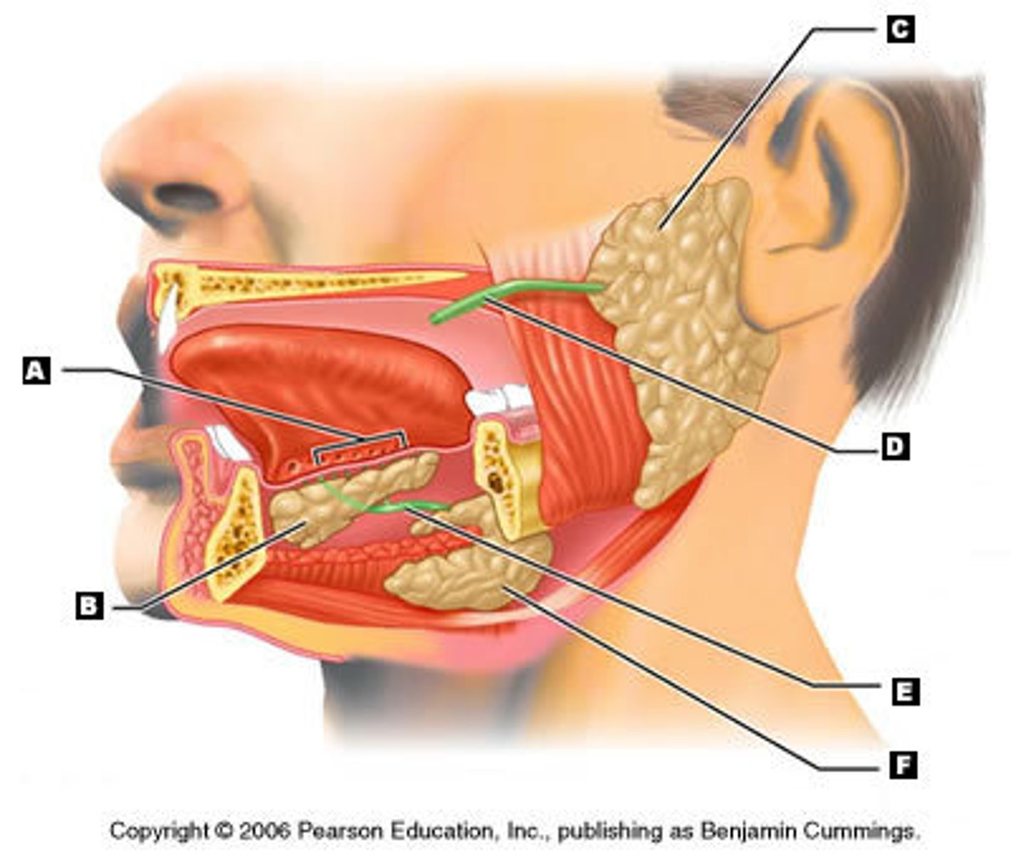
tongue
gingivae
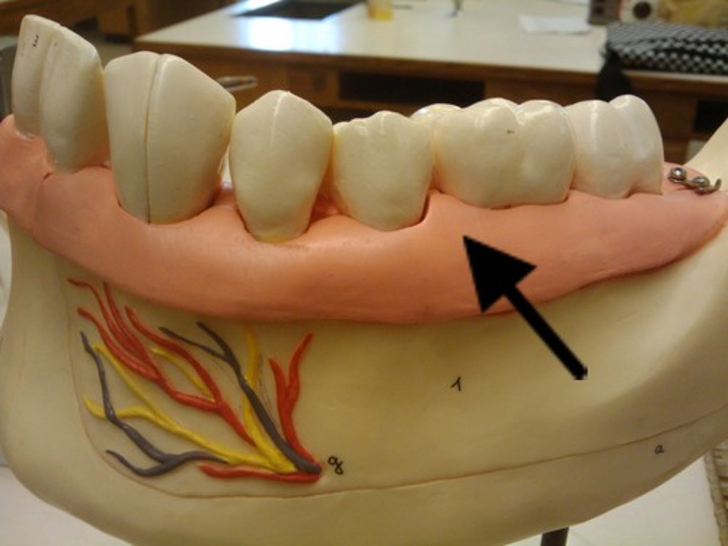
teeth
hard palate
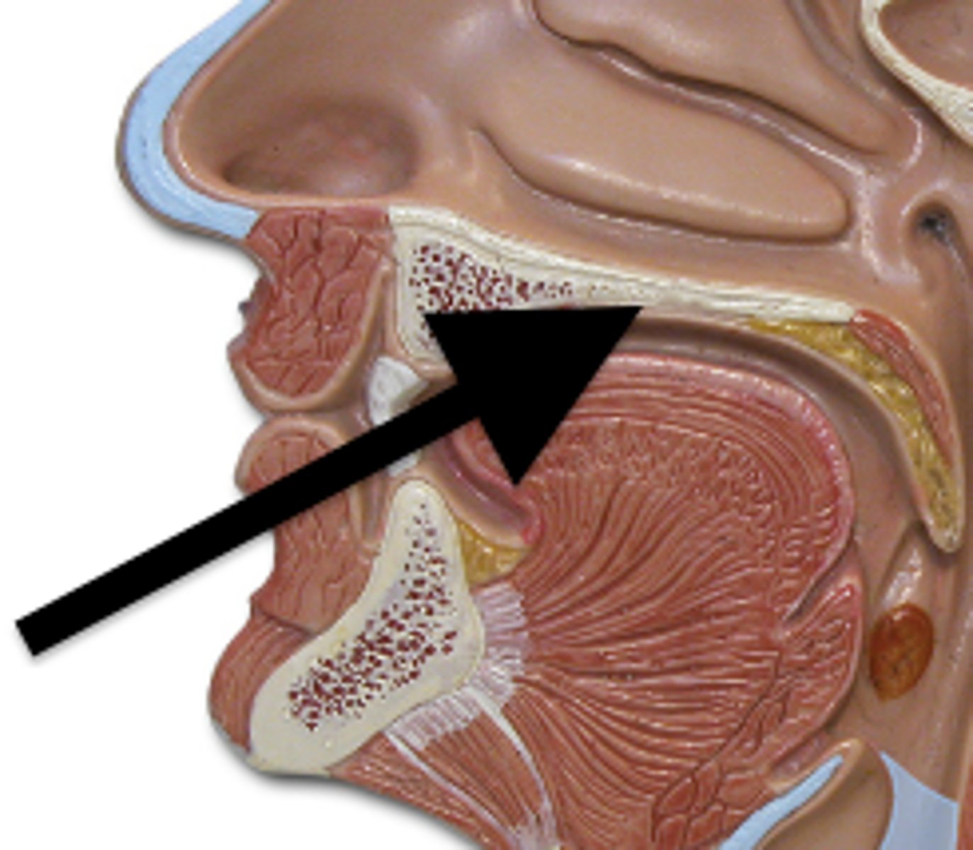
soft palate
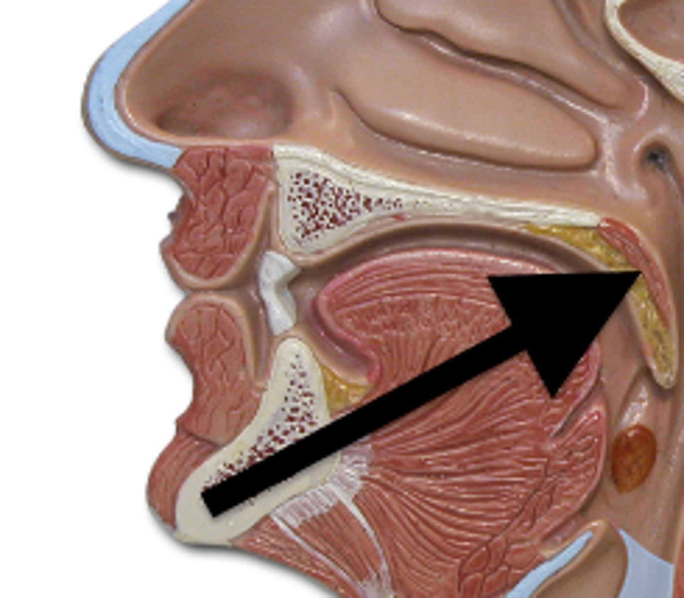
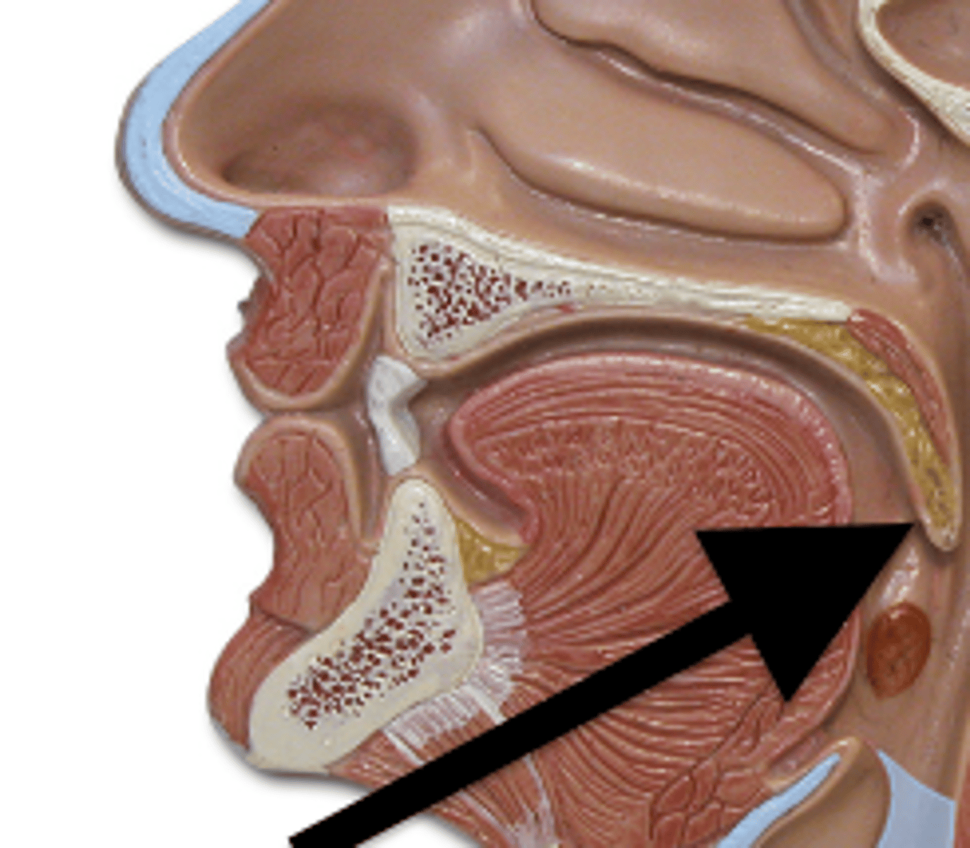
uvula
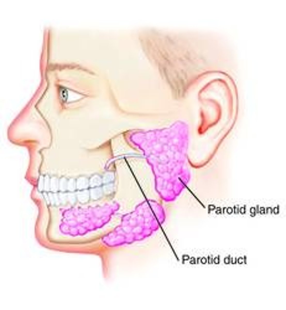
parotid salivary glands
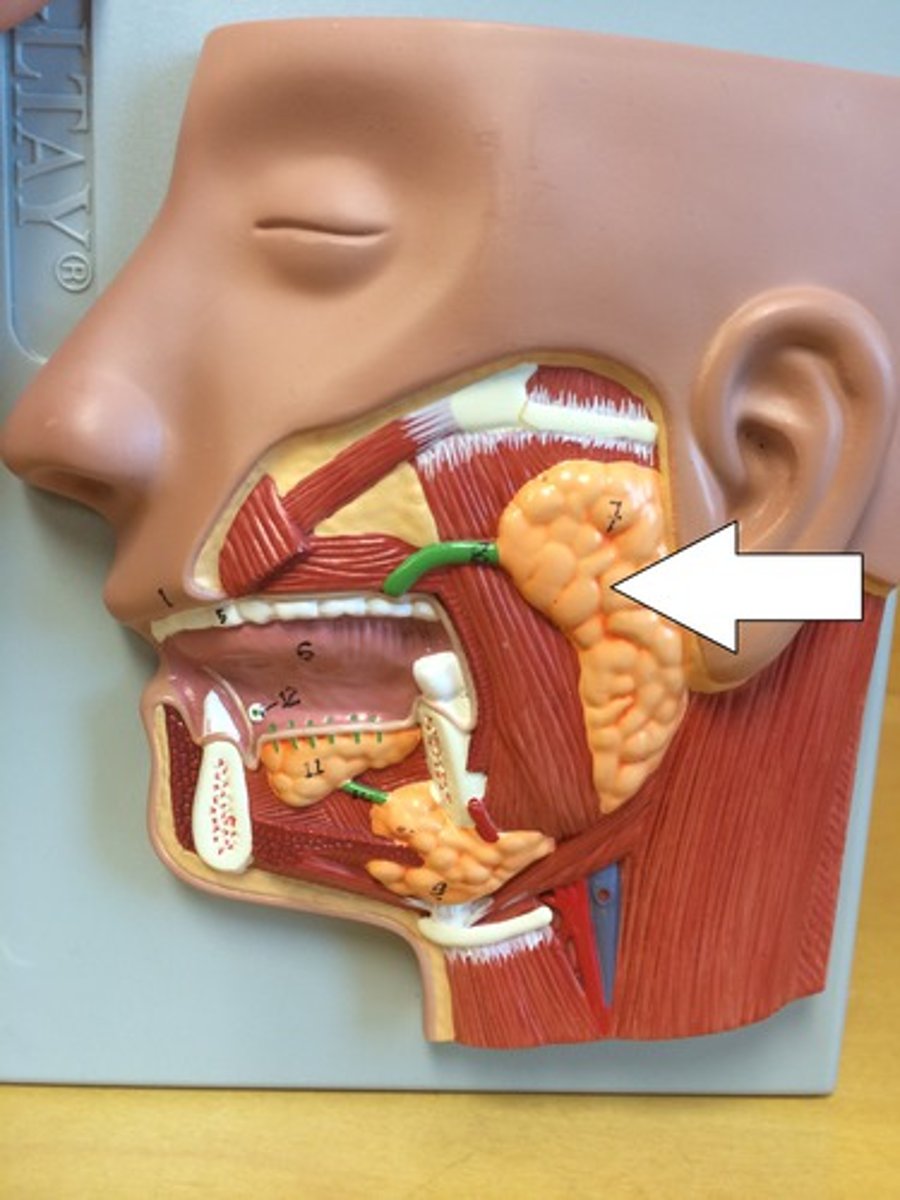
parotid ducts
duct
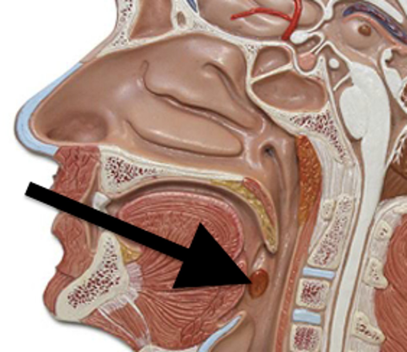
palatine tonsils
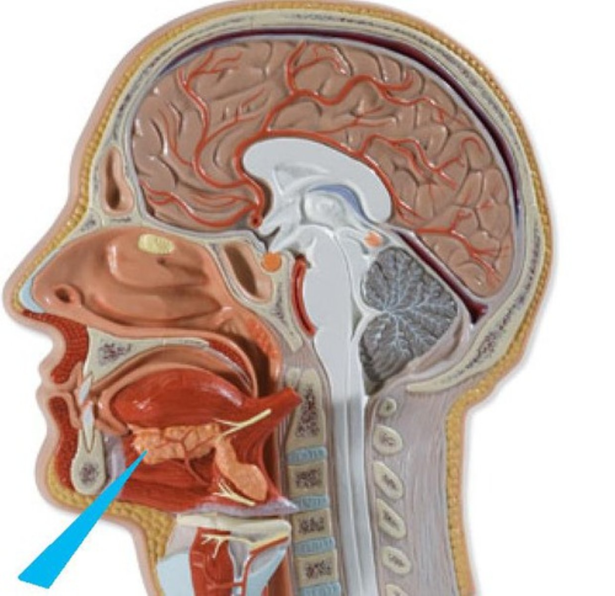
sublingual salivary glands
submandibular salivary glands
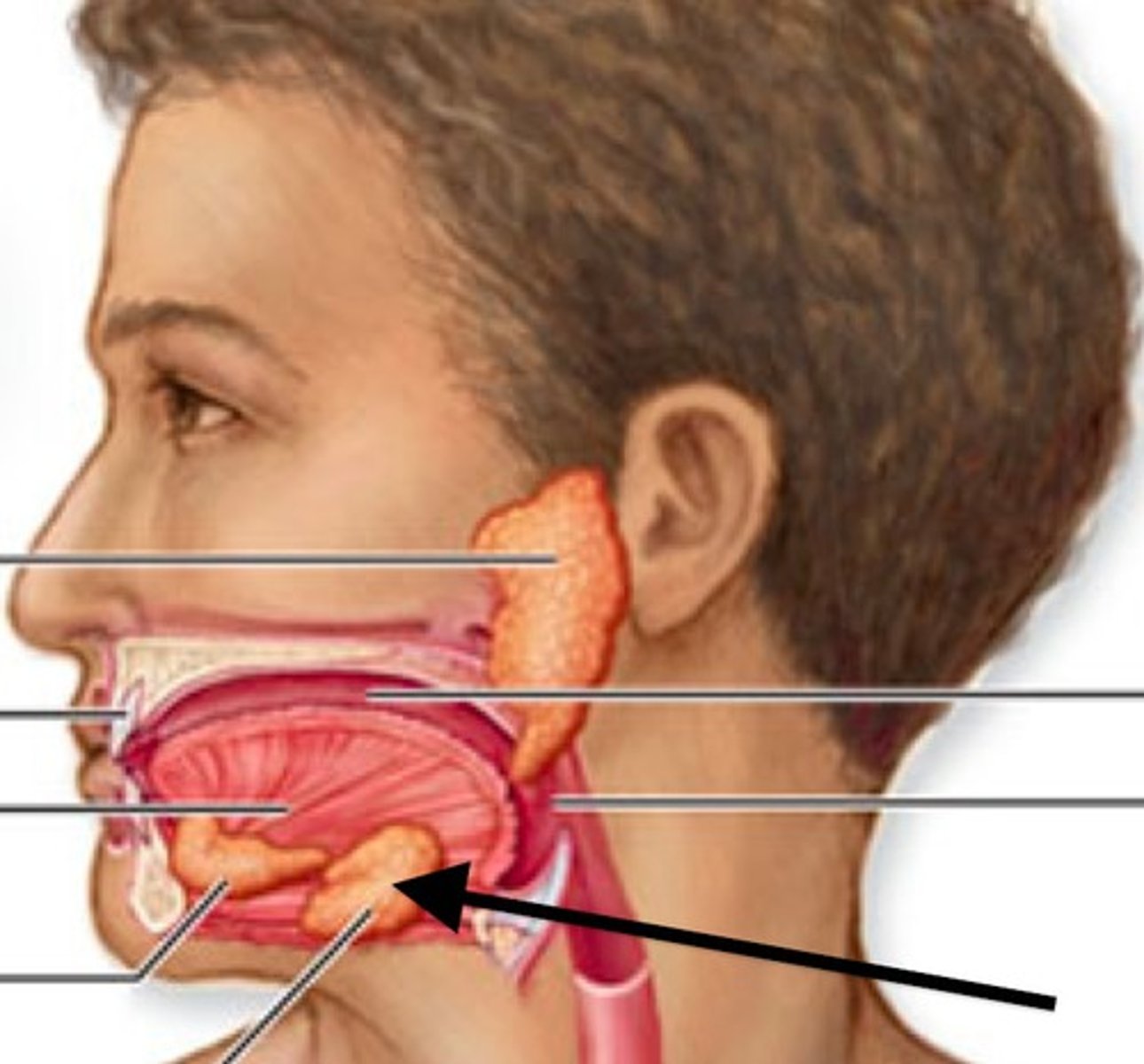
submandibular duct
lower duct
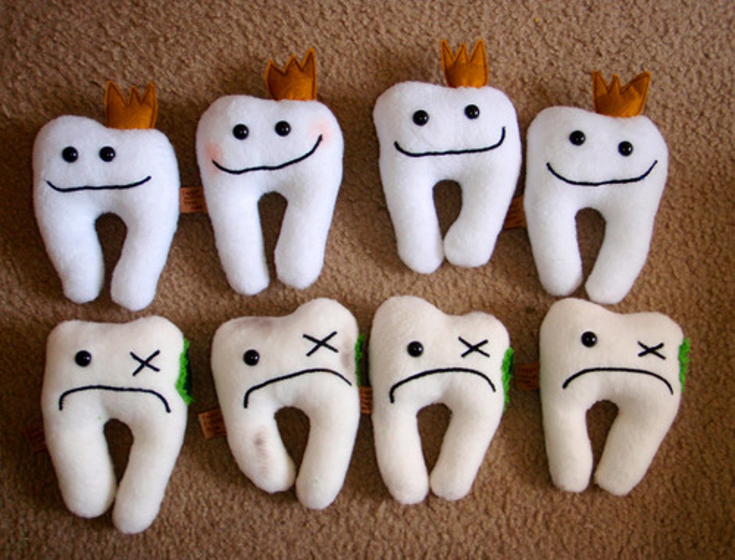
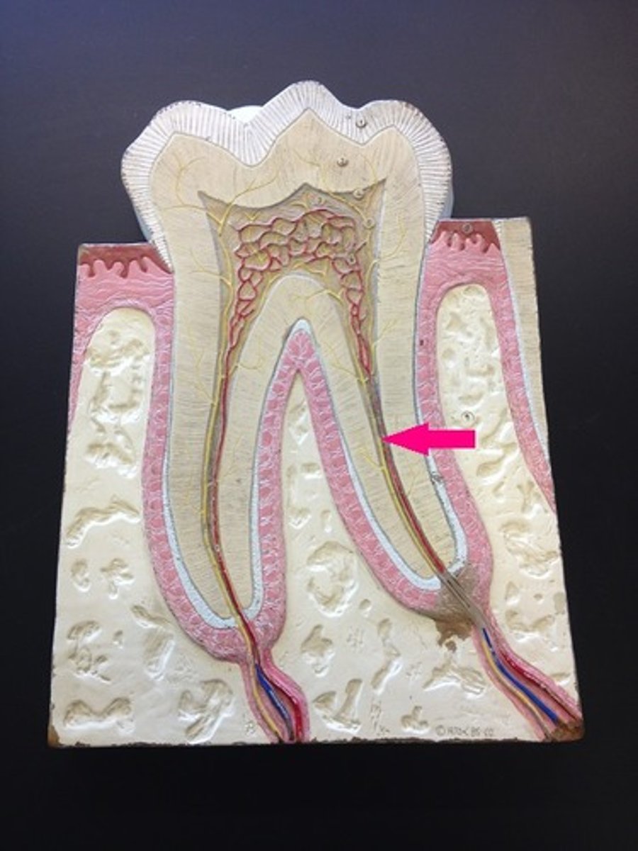
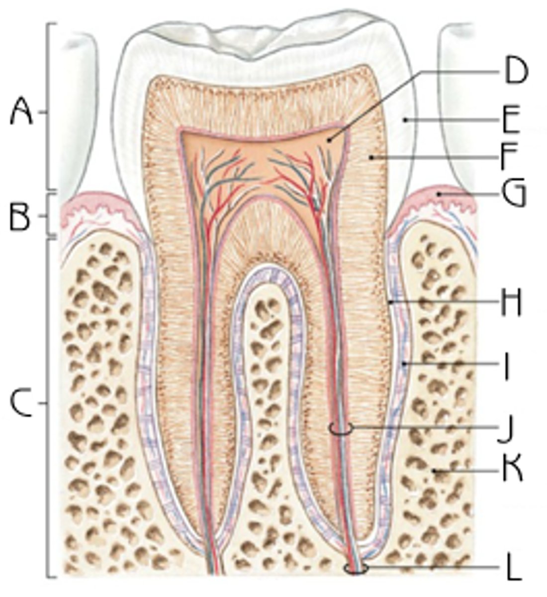
crown
portion that you can see of tooth

neck of the tooth
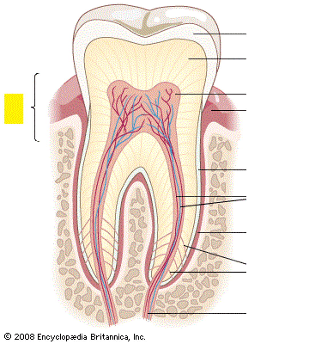
root
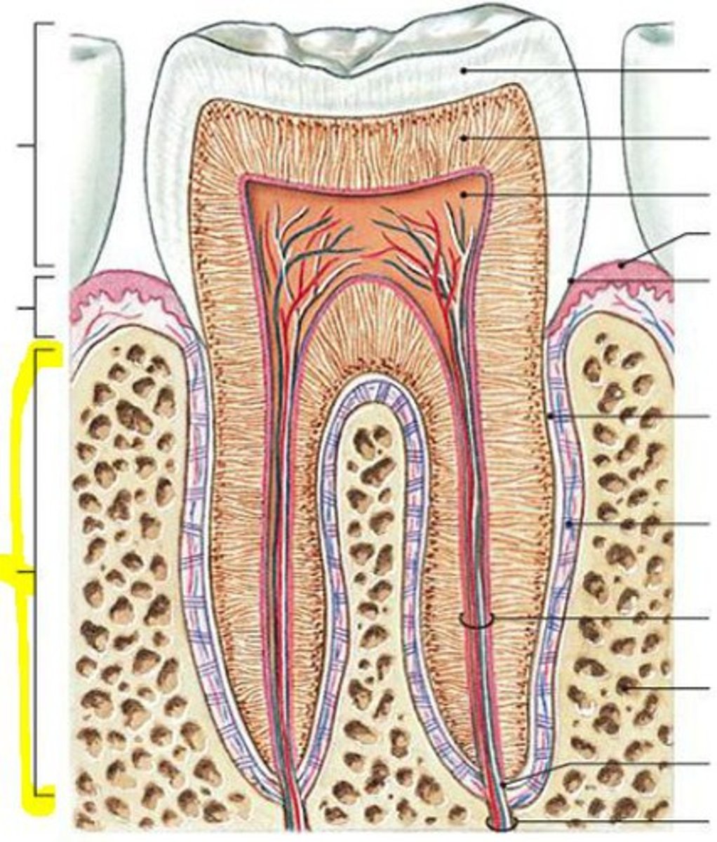
root canal
apical foramen
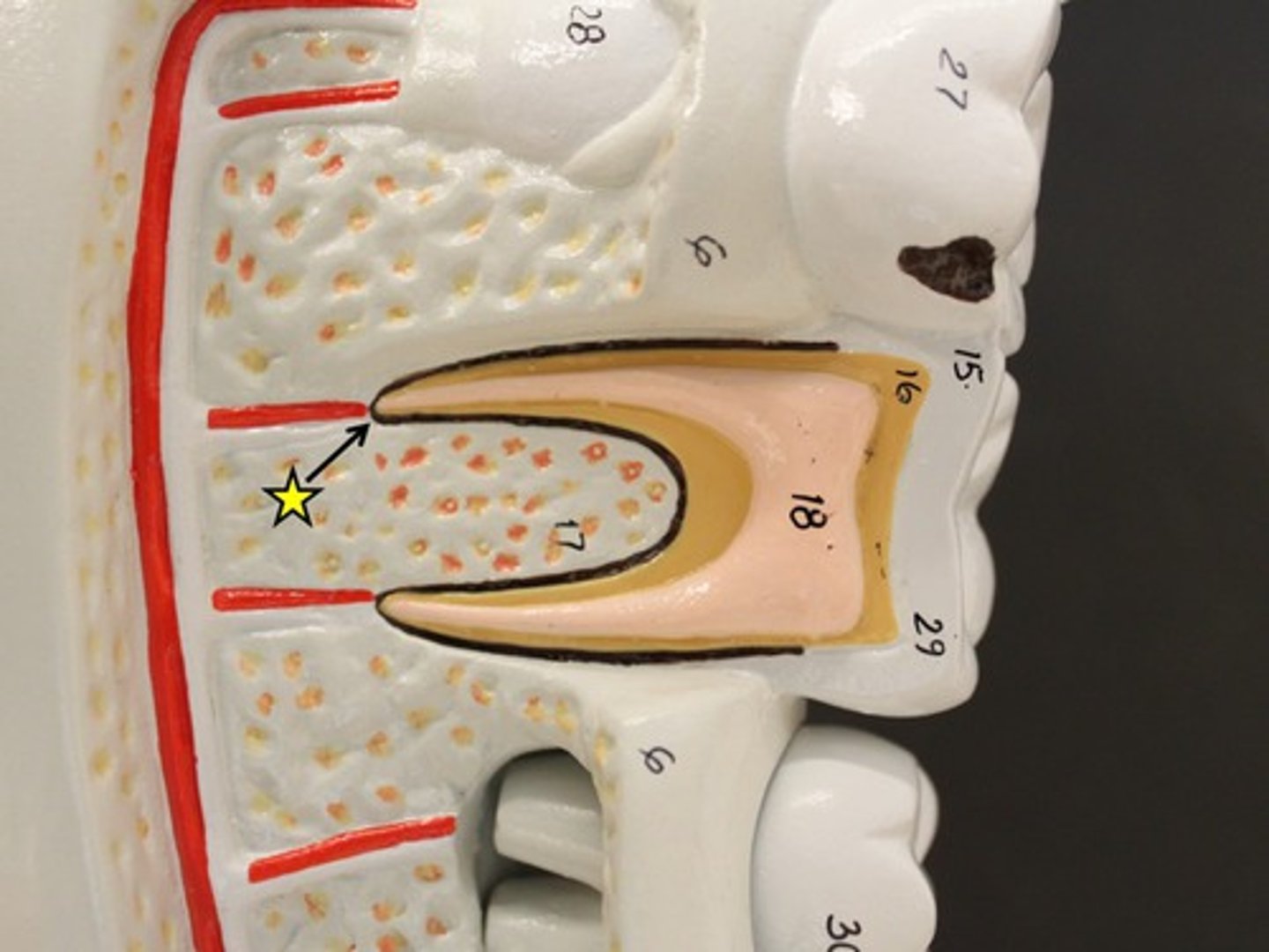
enamel
A
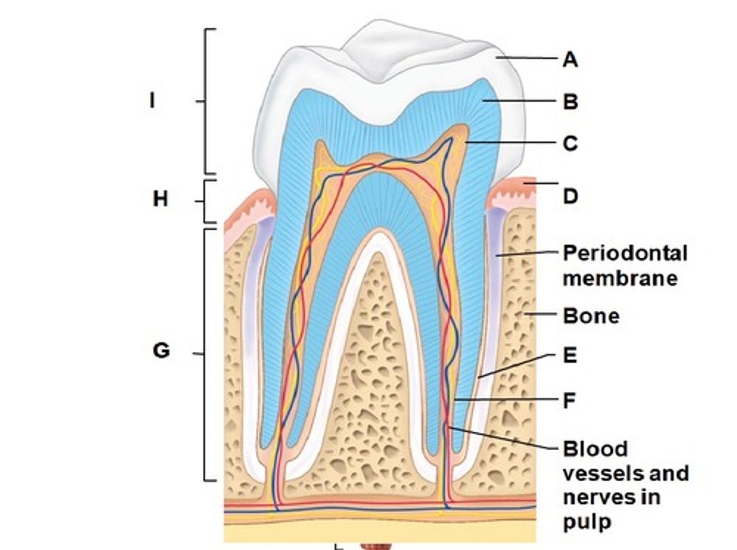
dentin
B

pulp cavity
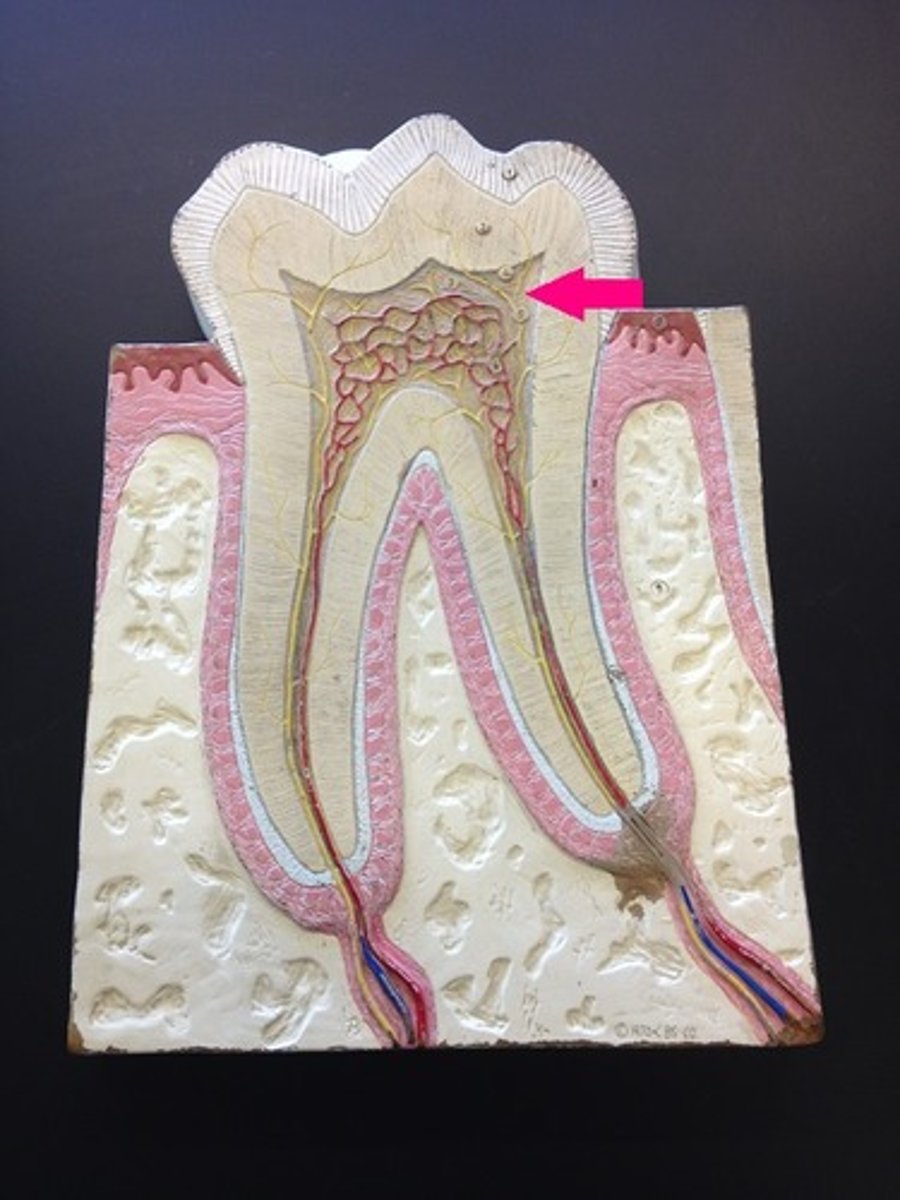
periodontal ligament
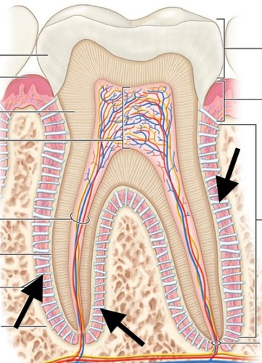
cementum
outermost layer (blue layer) outside of root
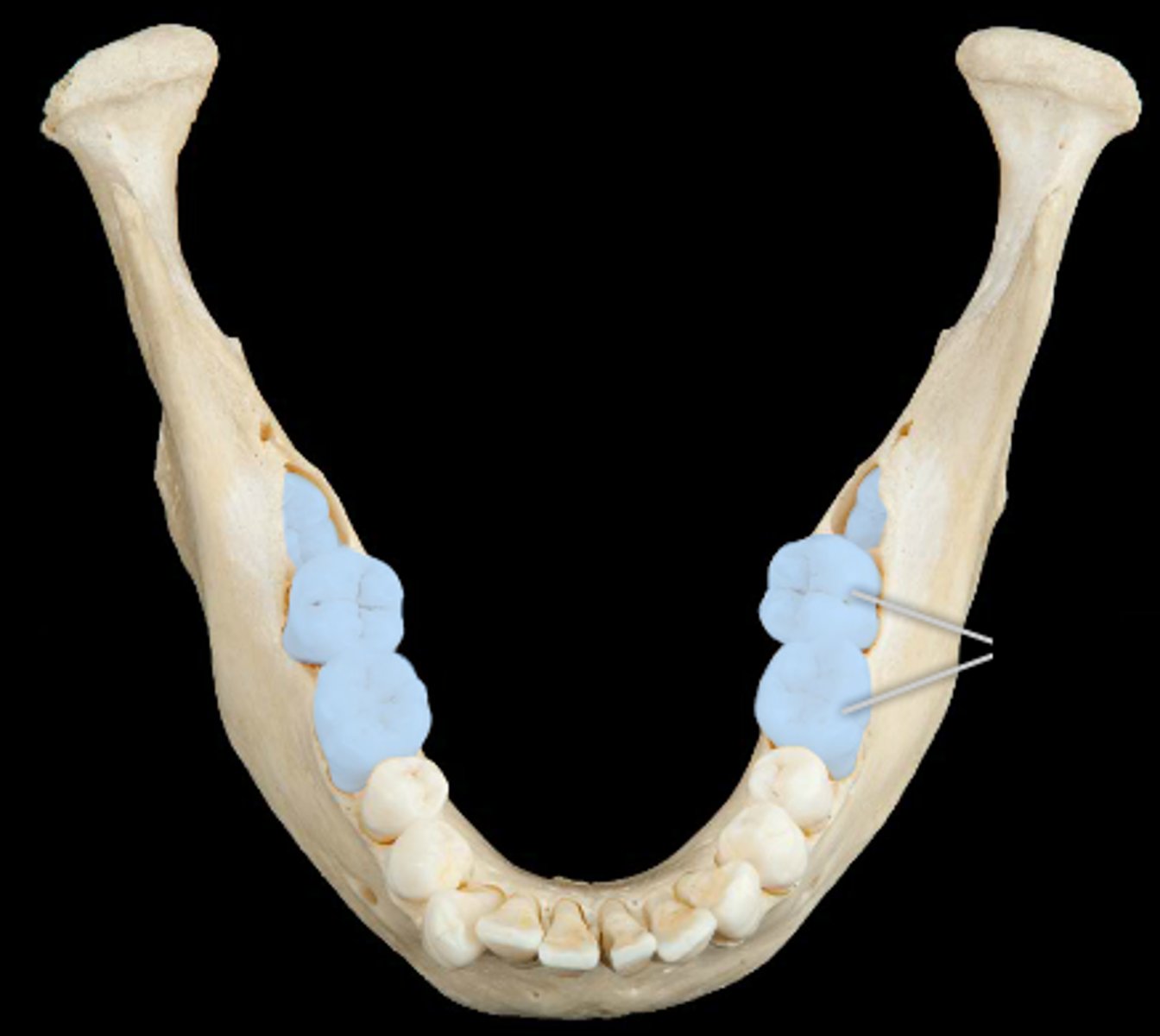
incisors
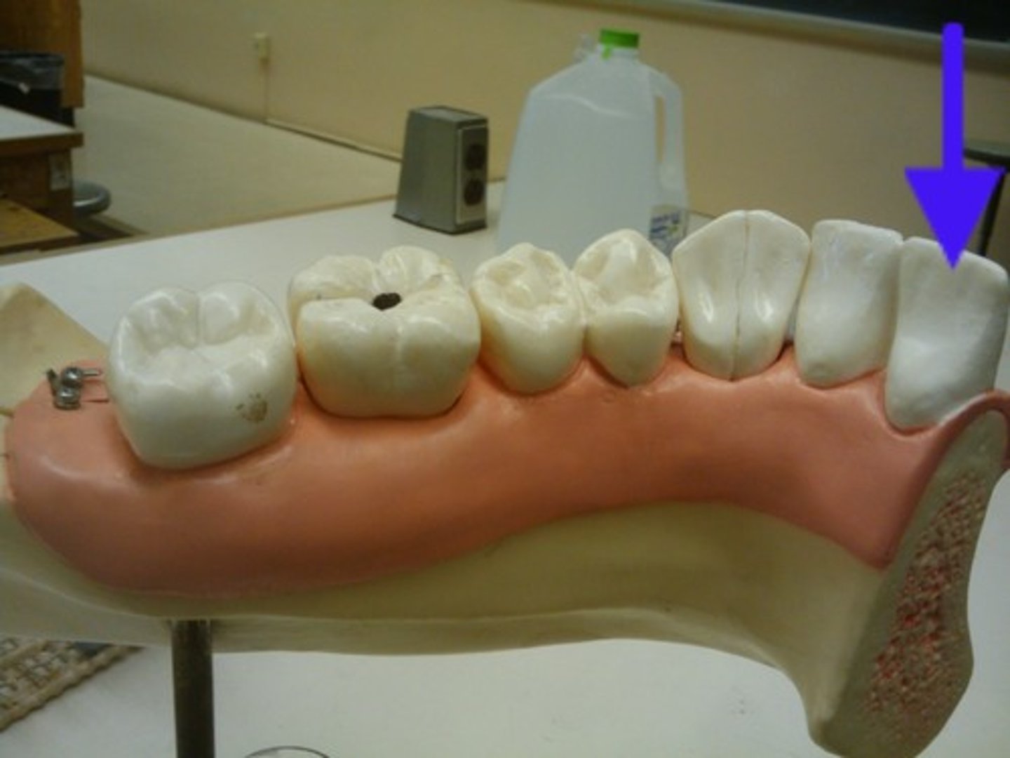
cuspids
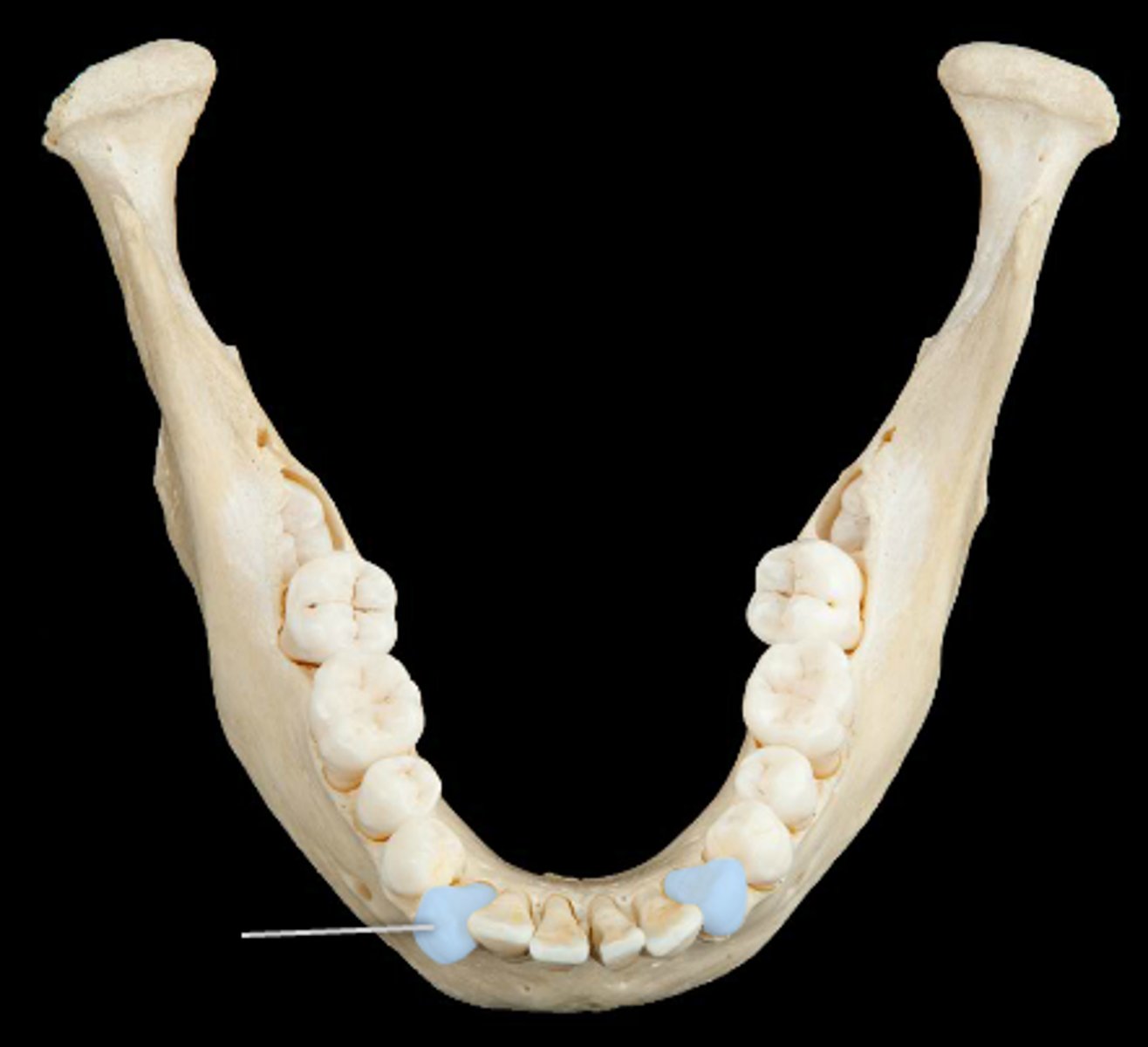
premolars
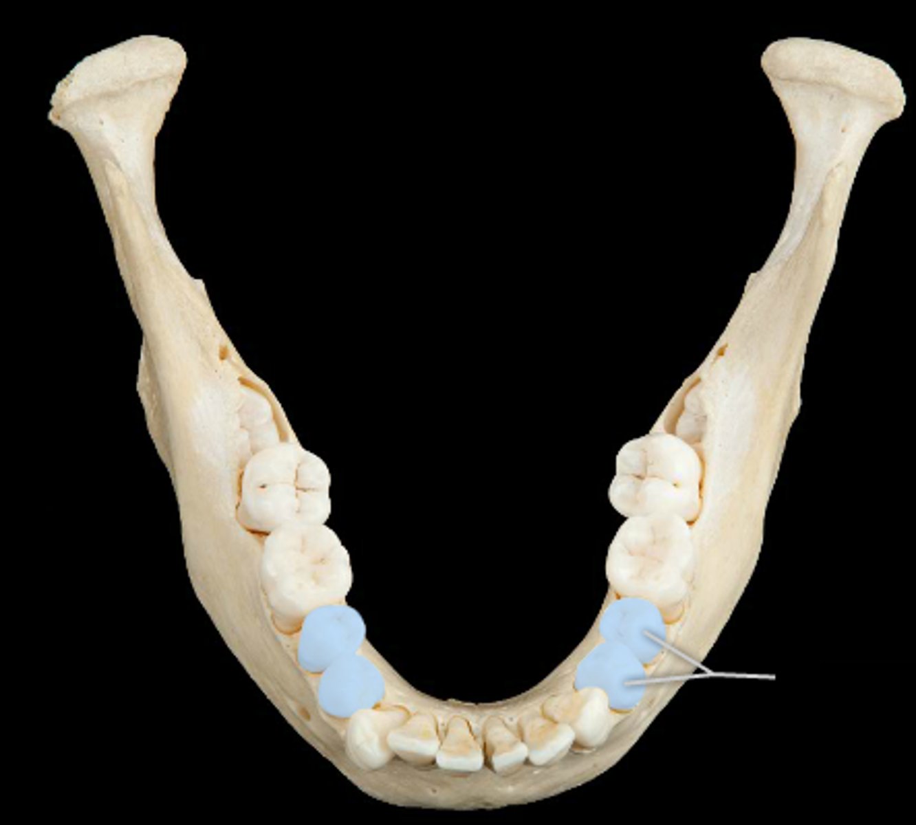
molars
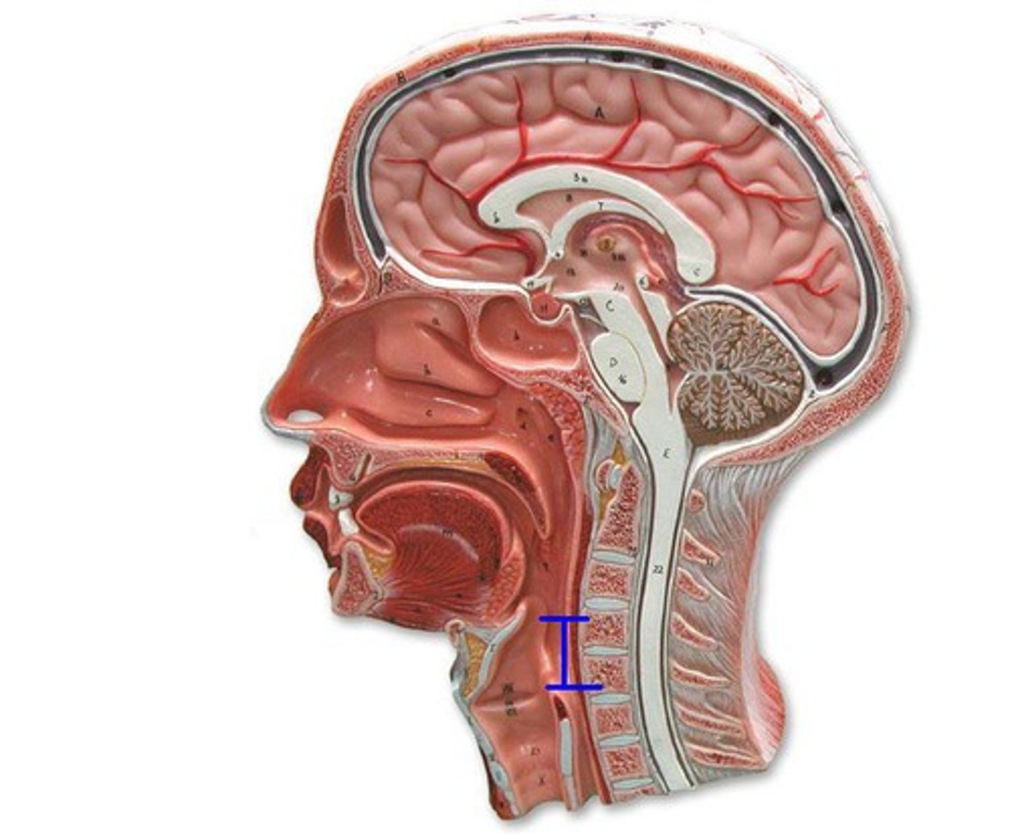
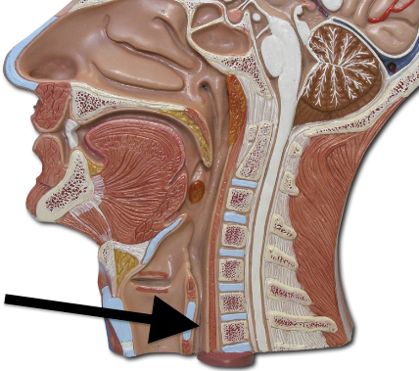
nasopharynx
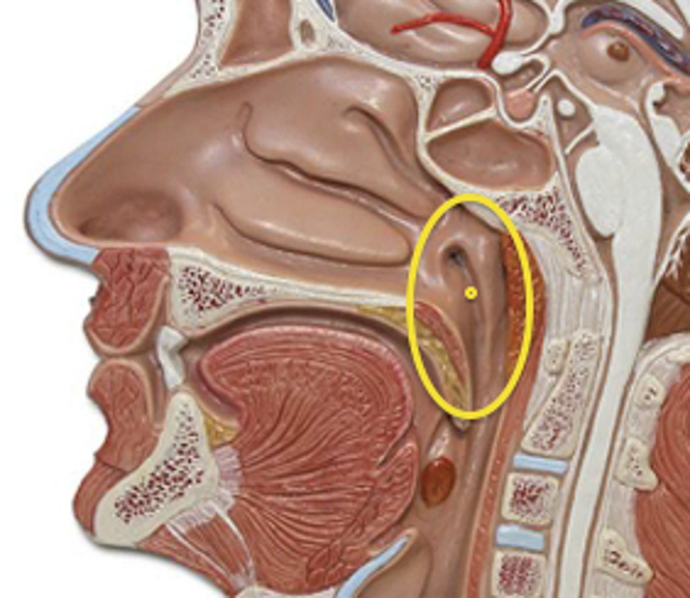
oropharynx
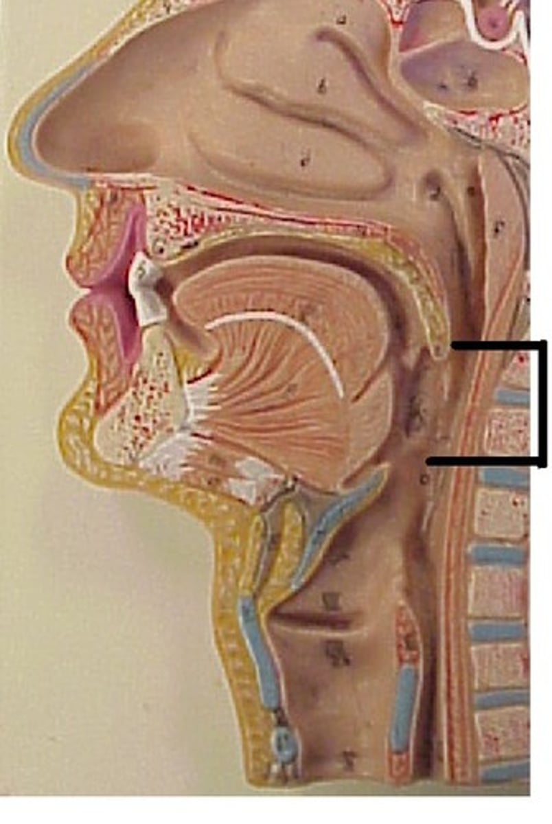
laryngopharynx
esophagus
lower esophageal sphincter
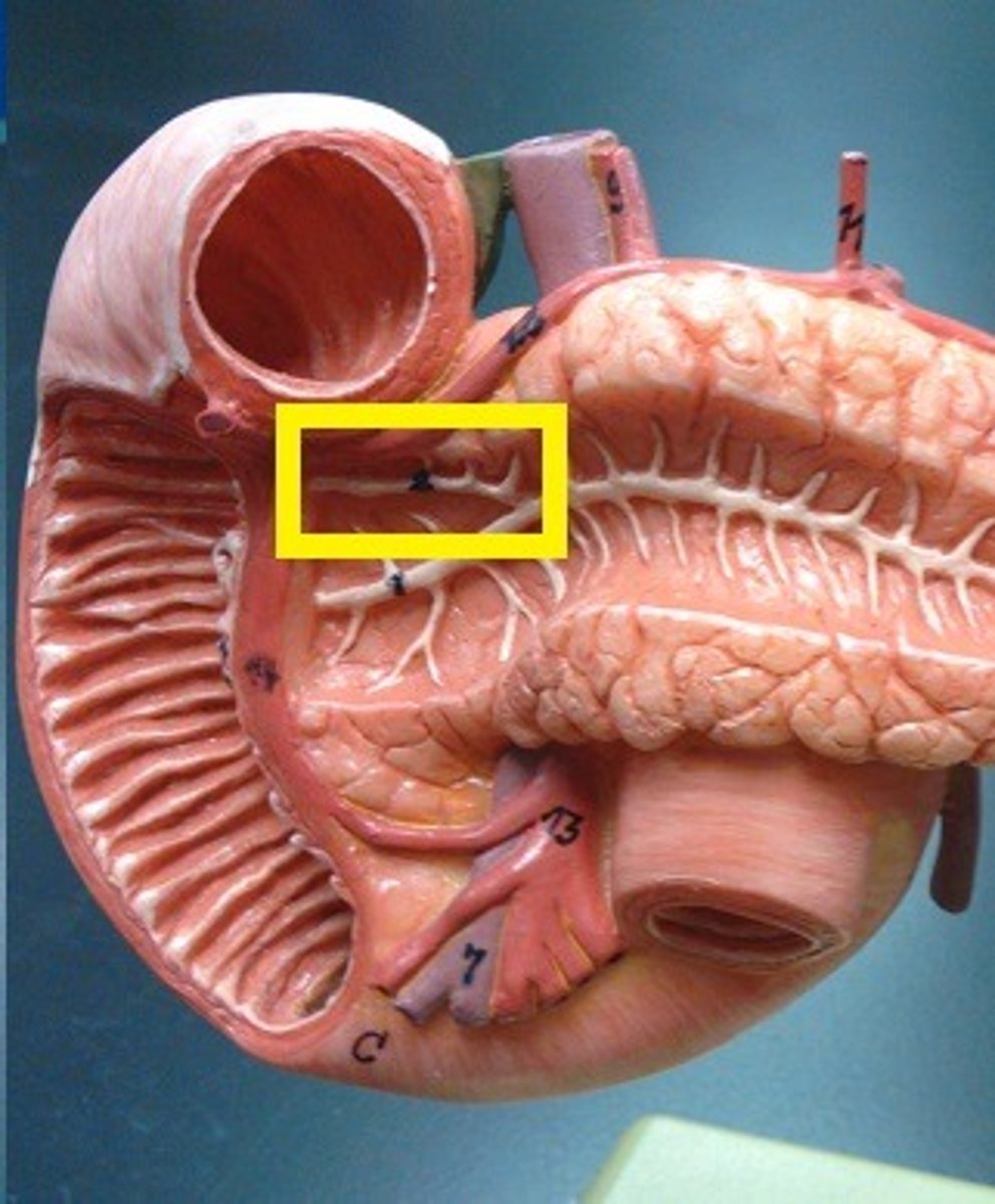
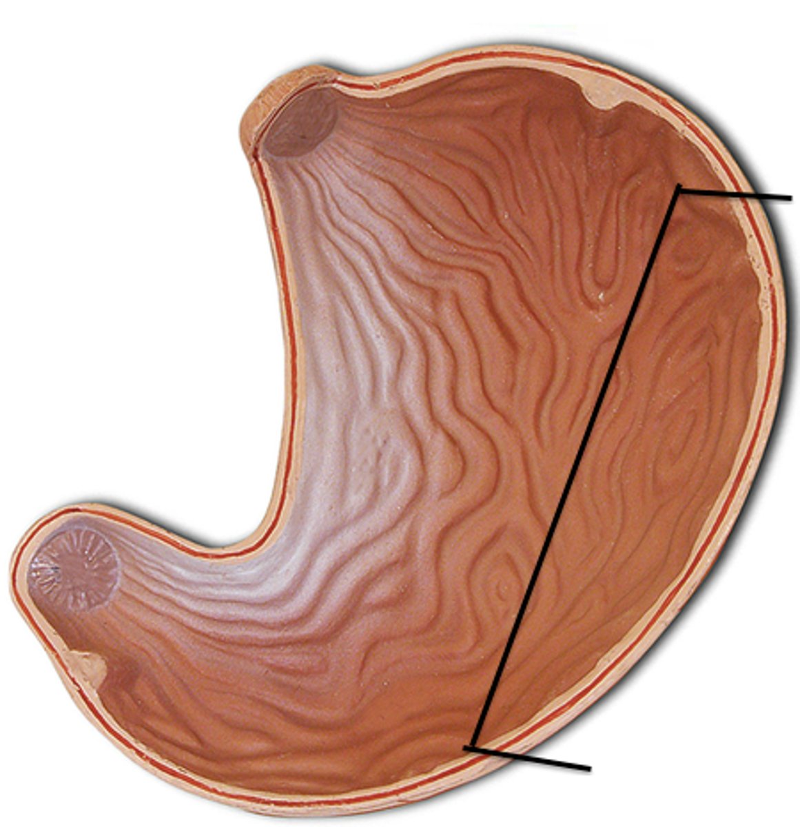
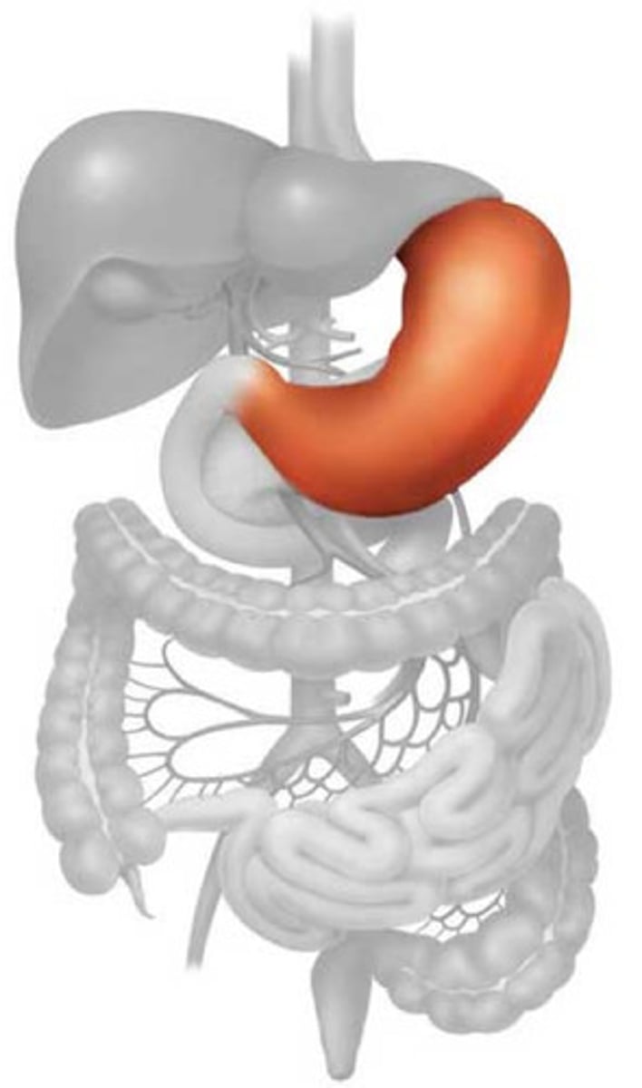
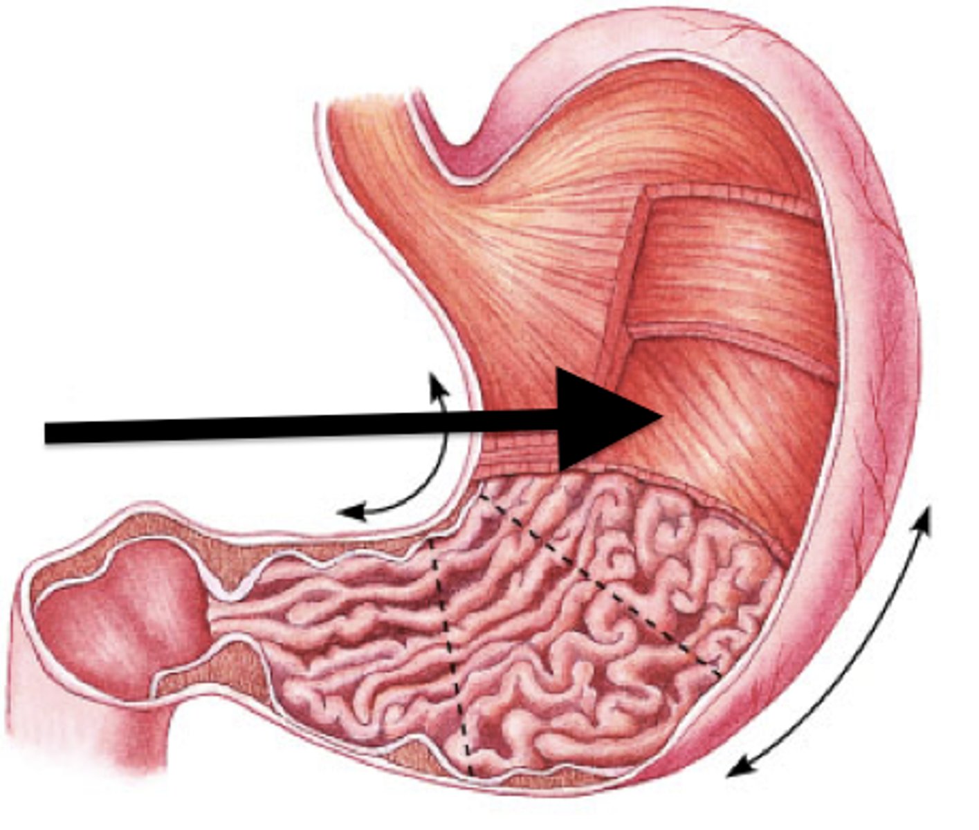
lesser curvature
greater curvature
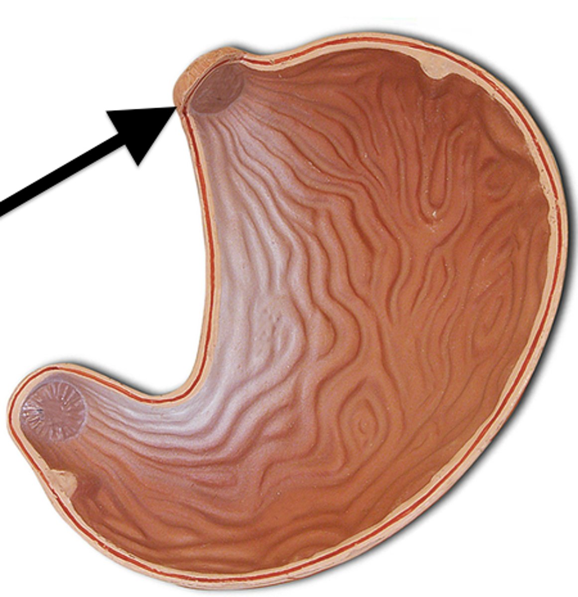
cardia of the stomach
entrance area (not a sphincter)
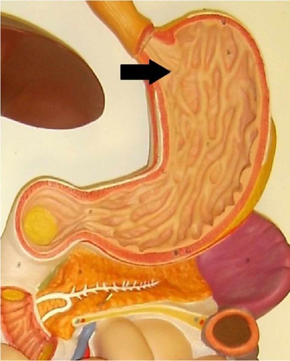
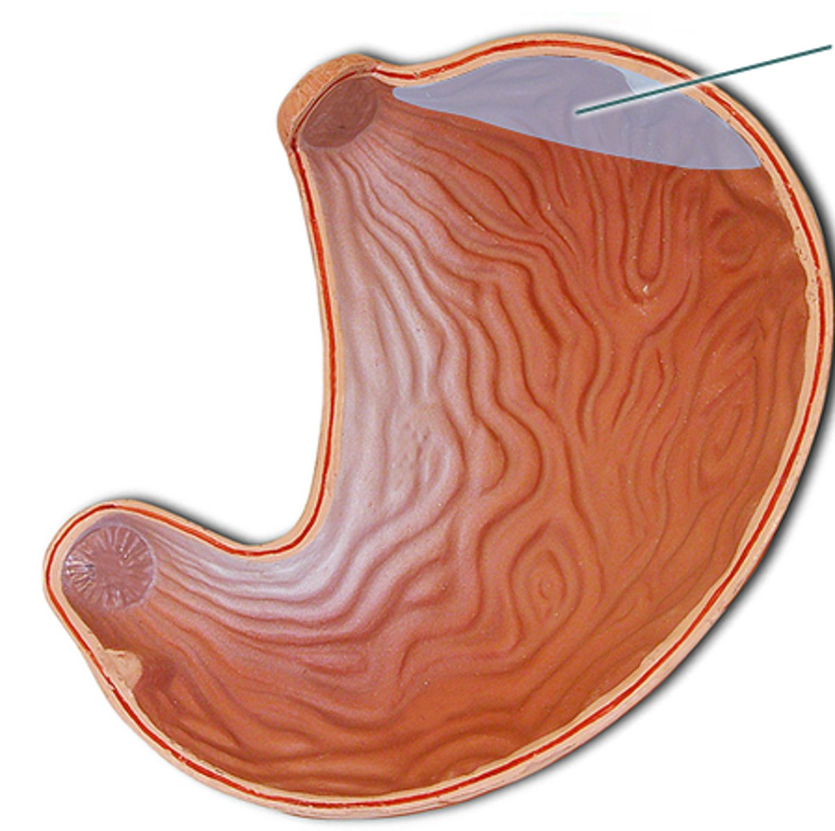
fundus of the stomach
bulge
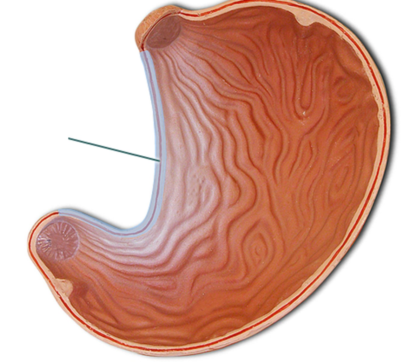
body of the stomach
main portion of the stomach
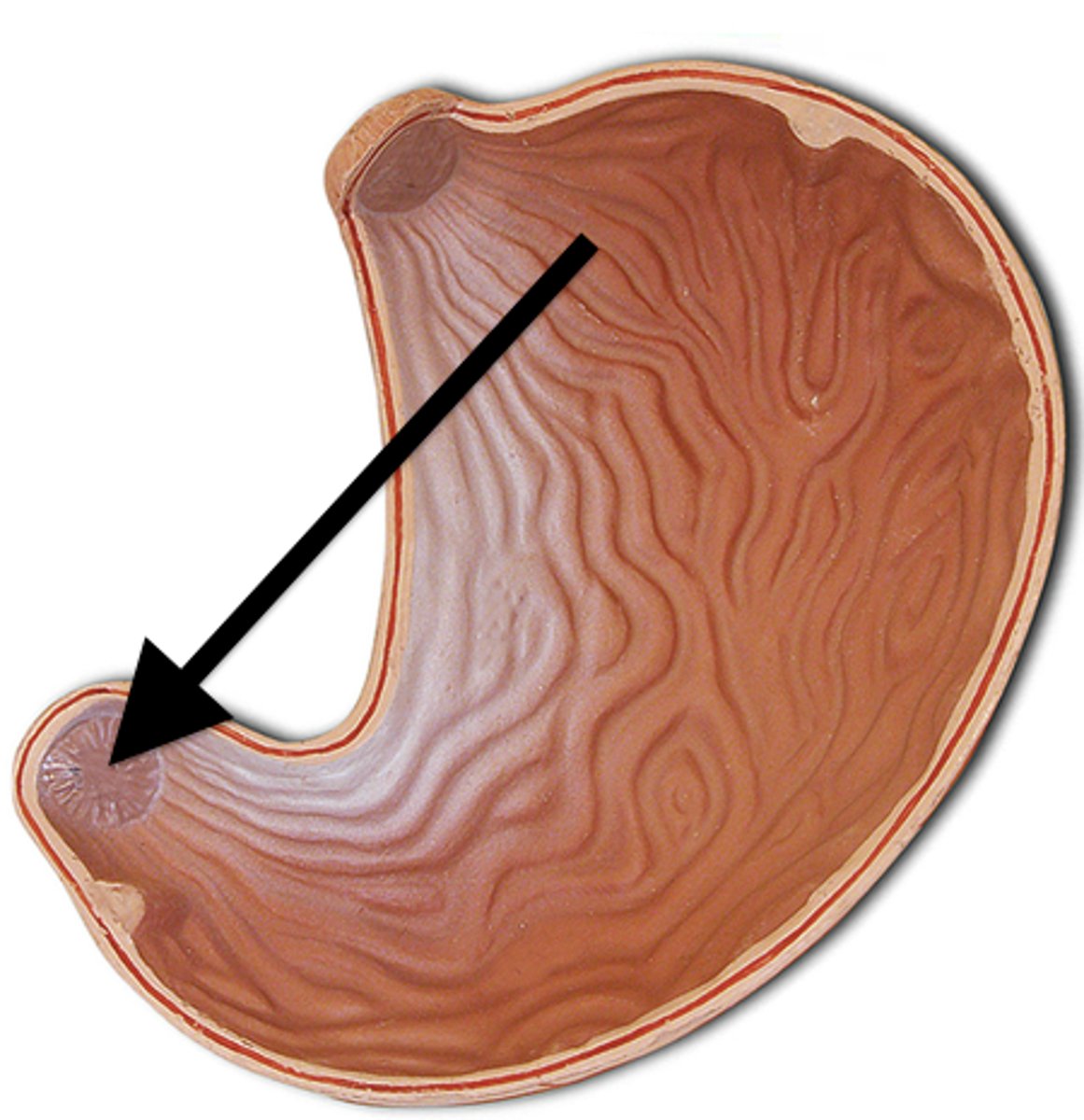
pylorus of the stomach
region
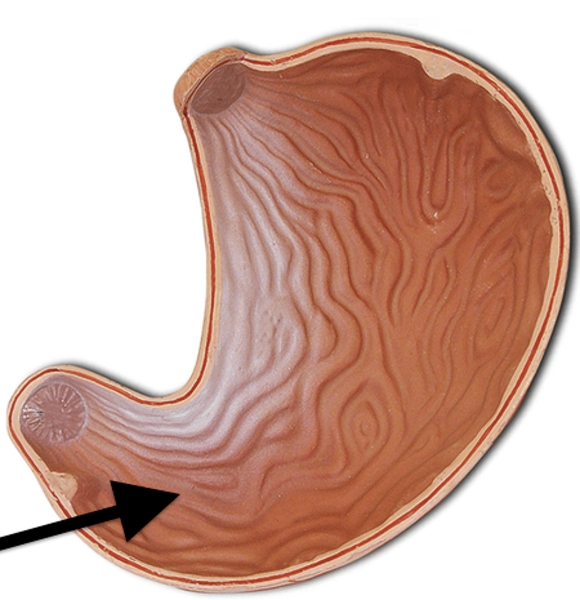
pyloric sphincter
sphincter
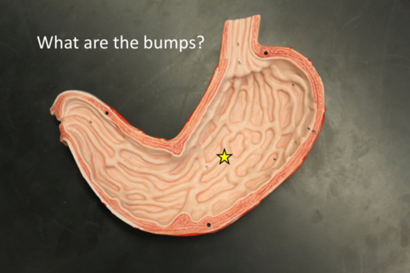
rugae of the stomach
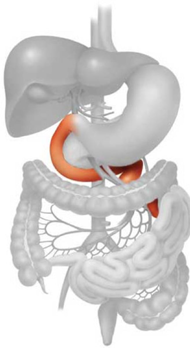
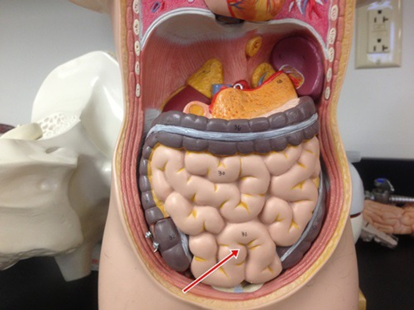
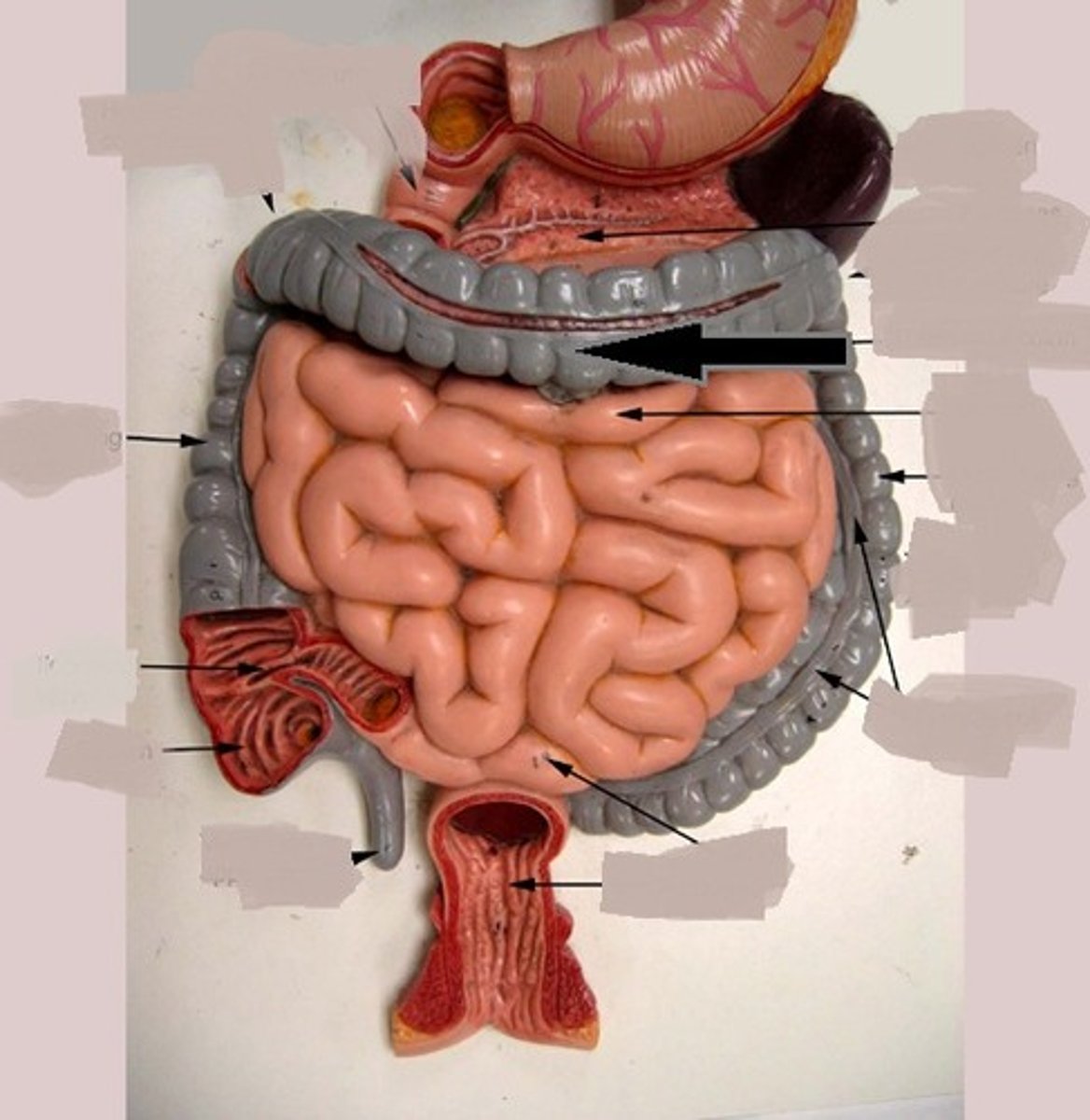
duodenum
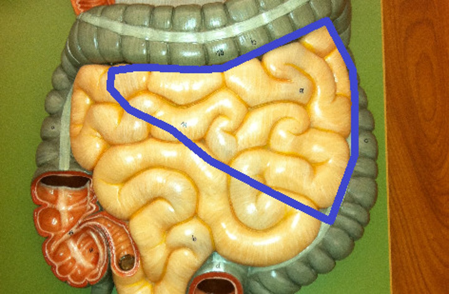
jejunum
superior portion of small intestines
ilieum
inferior portion of small intestines
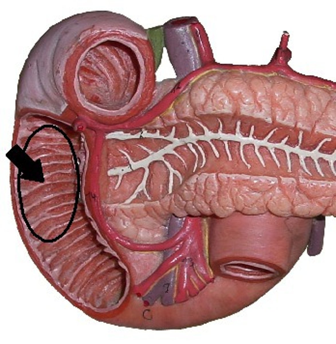
plicae circulares
rings that LINE inside of small intestines
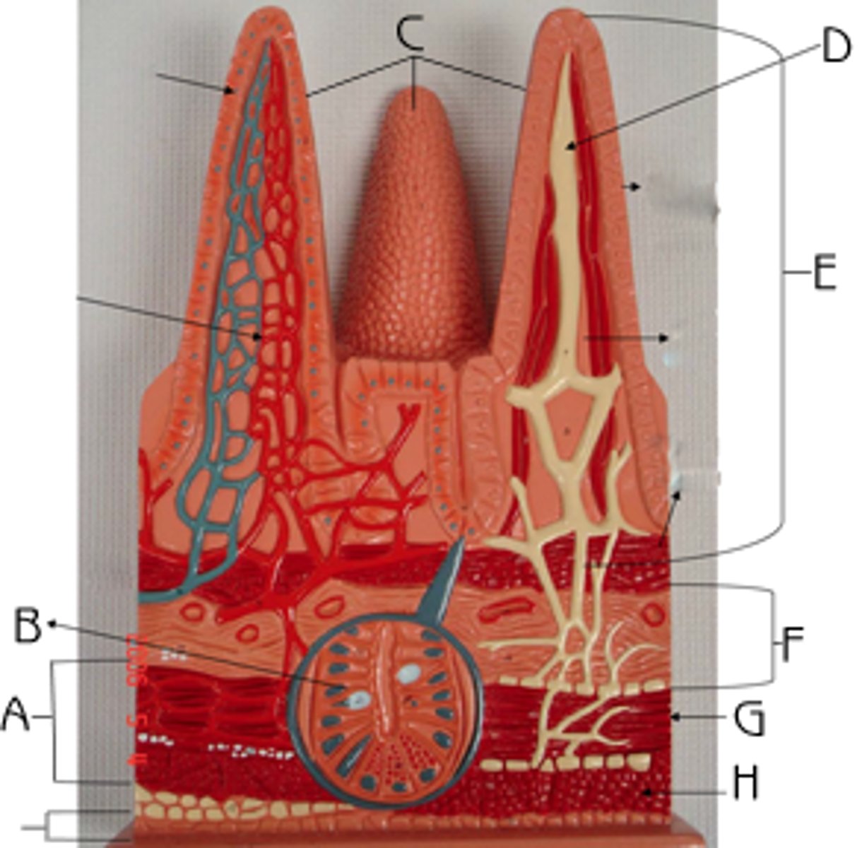
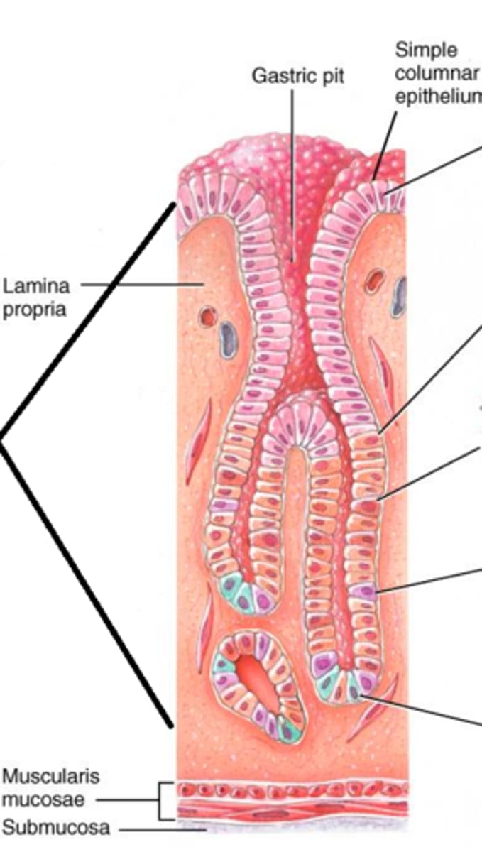
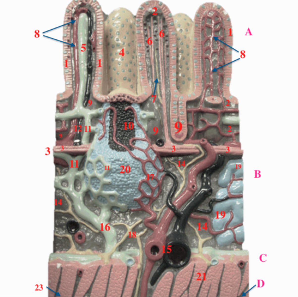
mucosa
E
mucosal epithelium
surface layer of mucosa
mucous (goblet) cells
of villi
indentations
white cells
lamina propria
background orange
muscularis mucosae
in mucosa
lower portion of mucosa
intestinal crypt
indentations next to upped portions

submucosa
F
under mucosa

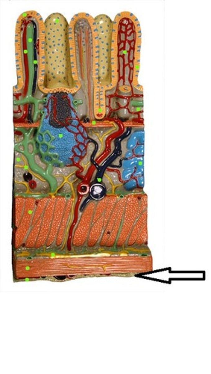
muscularis externa
A

circular muscle layer
G

longitudinal muscle layer
layer between serosa and circular muscle layer
serosa
outer layer of large model of stomach/small intestinal lining
gastric pit
stomach-specific
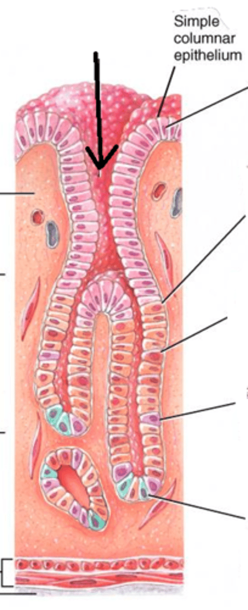
gastric gland
additional indentations after 1st
stomach-specific
oblique muscle layer
darker colored-layer
stomach-specific
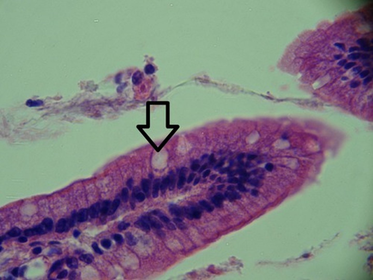
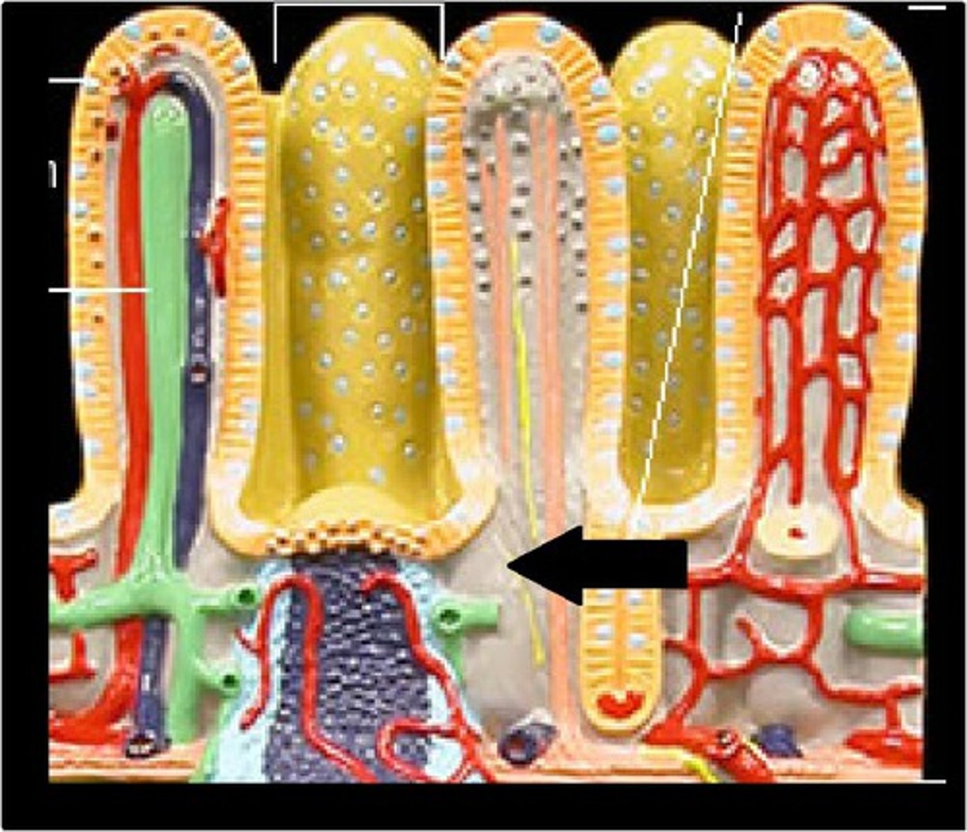
intestinal villi
villi of these
specific to small intestine

lacteal
specific to small intestine
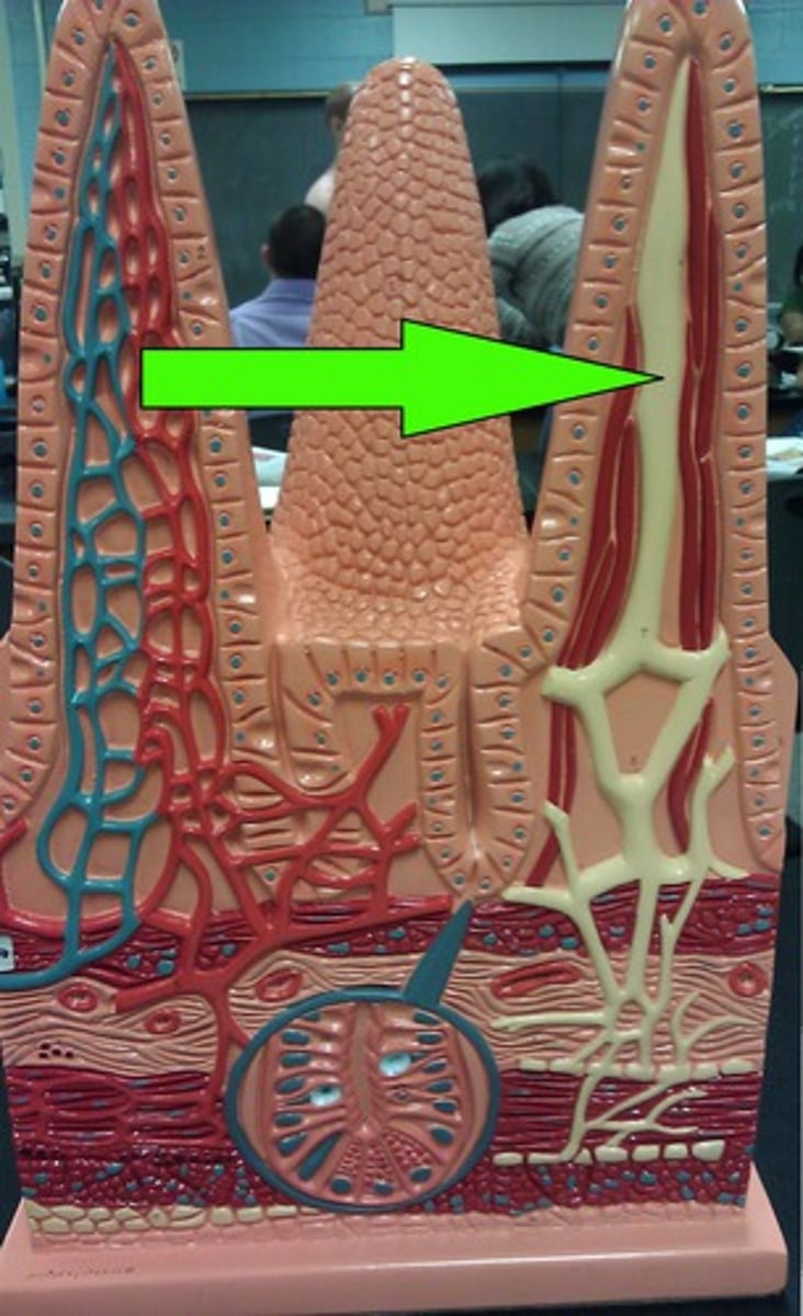
capillary network
#8 on right
specific to small intestines
ileocecal valve
valve
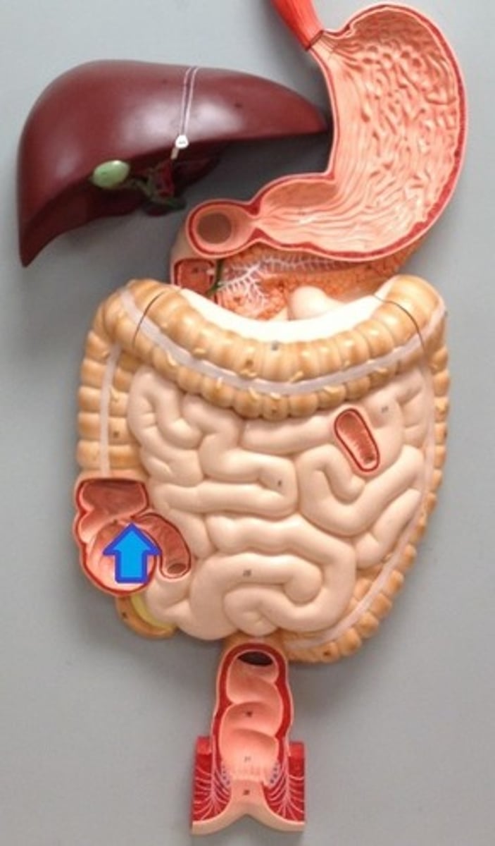
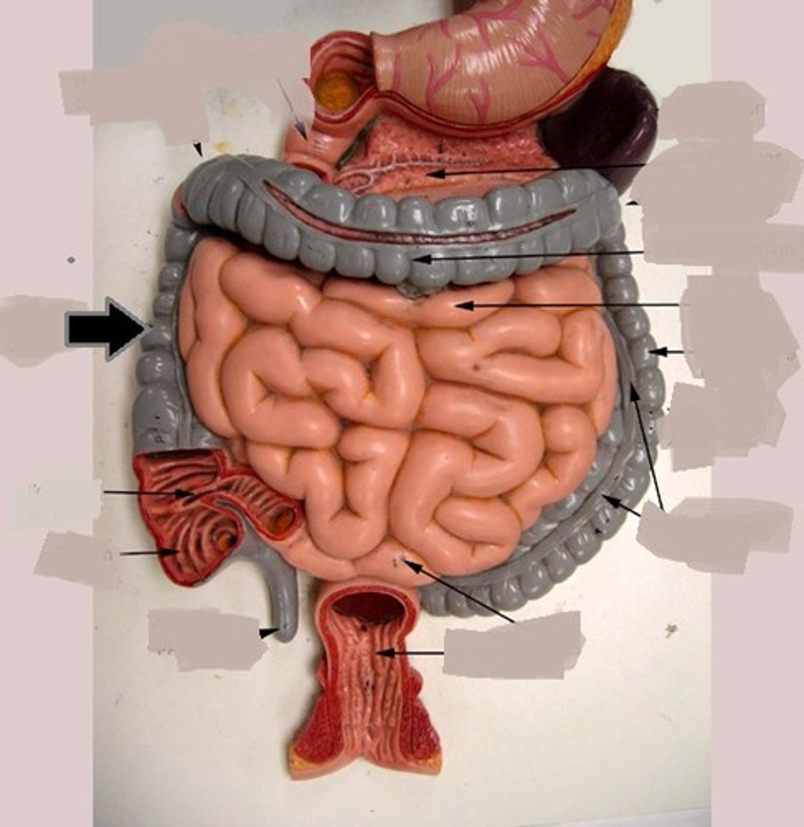
cecum
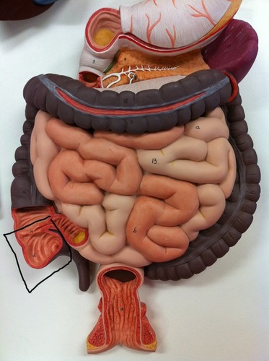
appendix
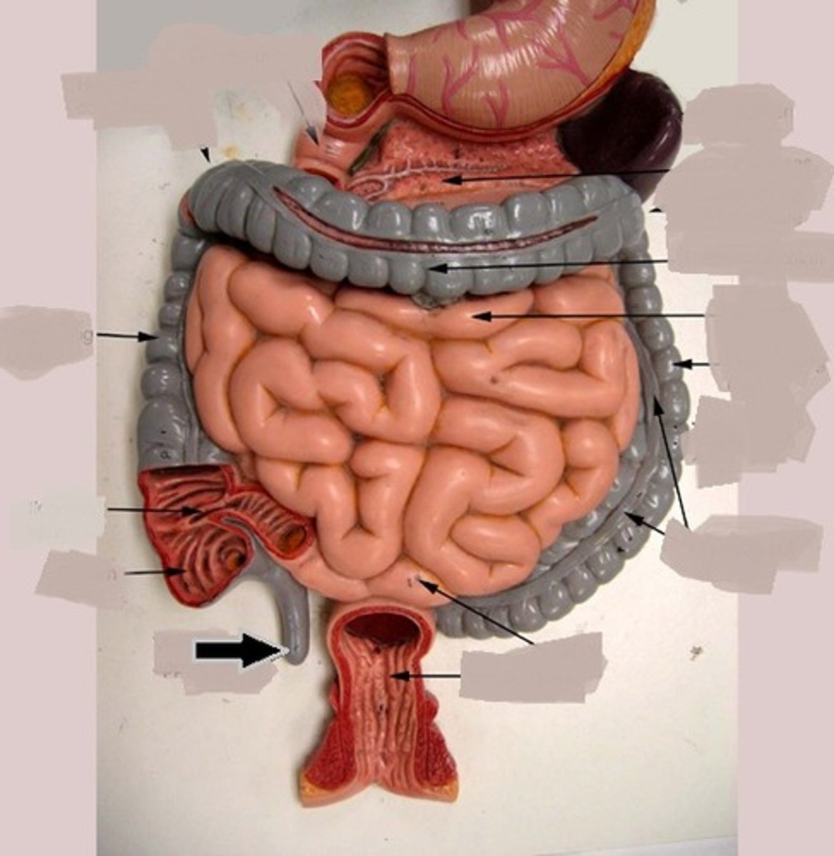
ascending colon
right colic flexure
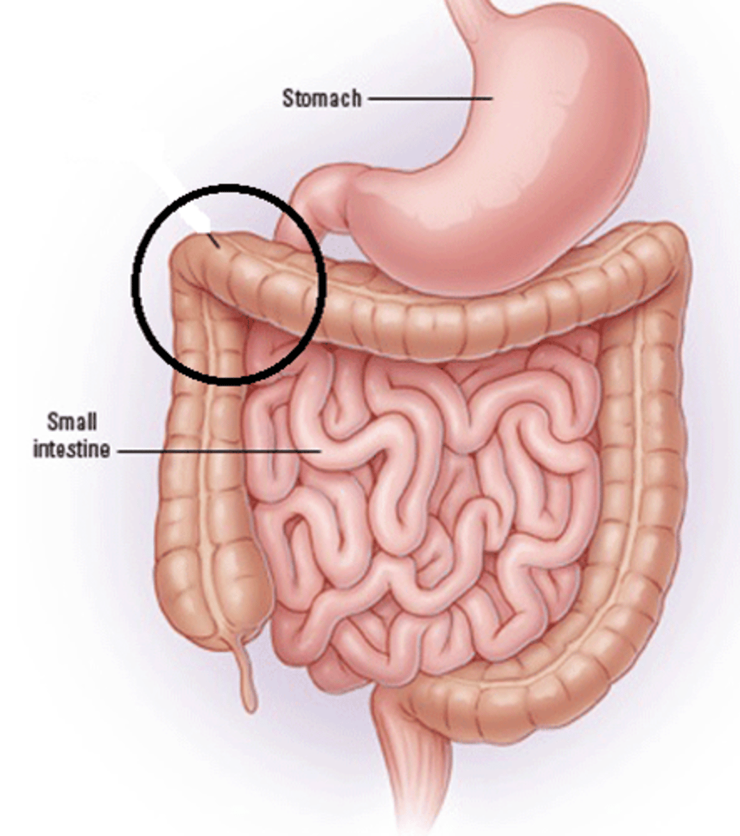
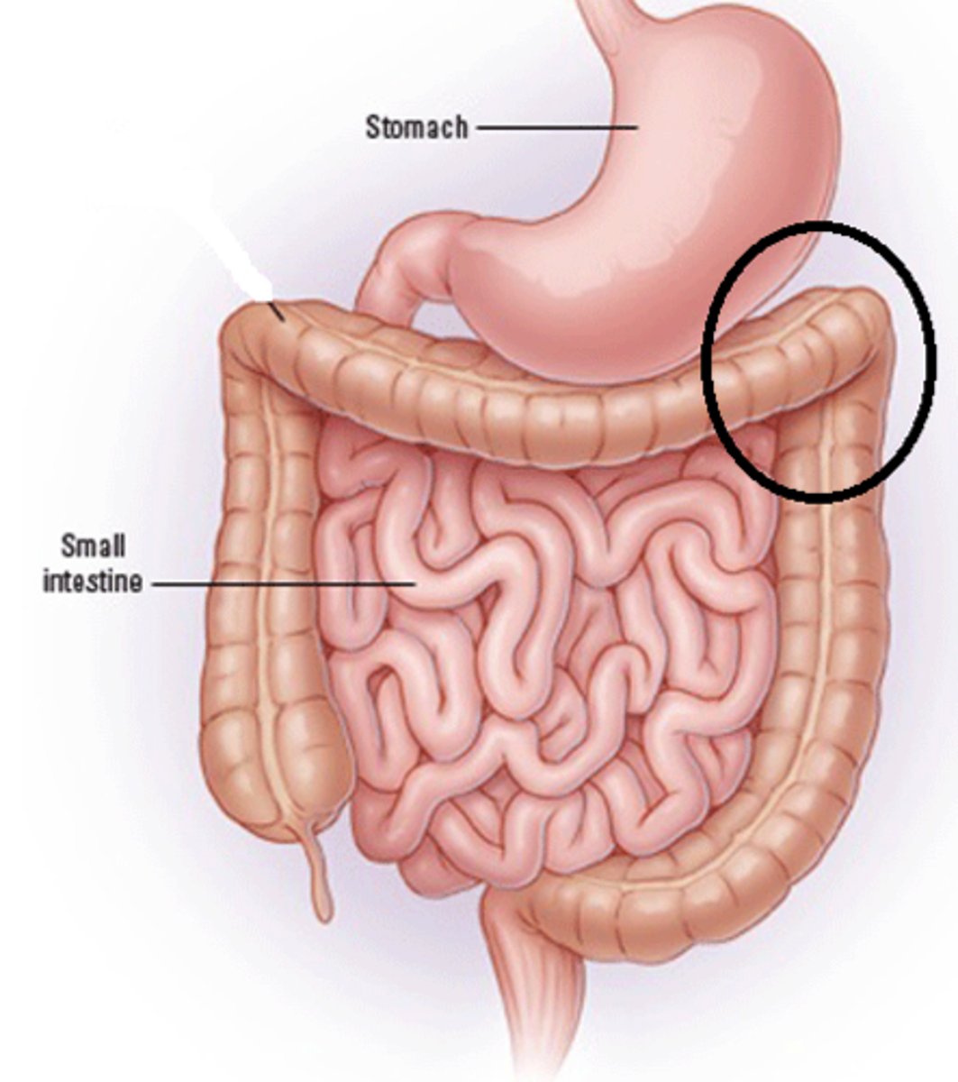
transverse colon
left colic flexure
descending colon
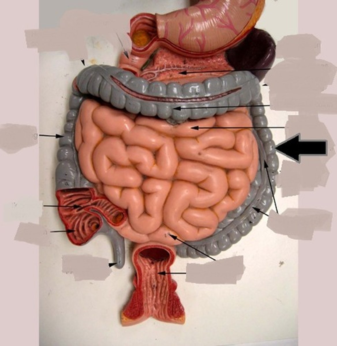
sigmoid colon
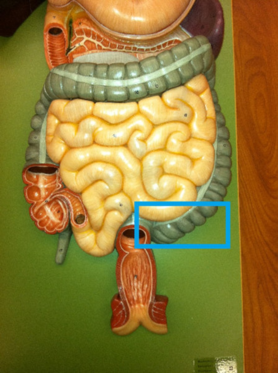
haustra
circled portions
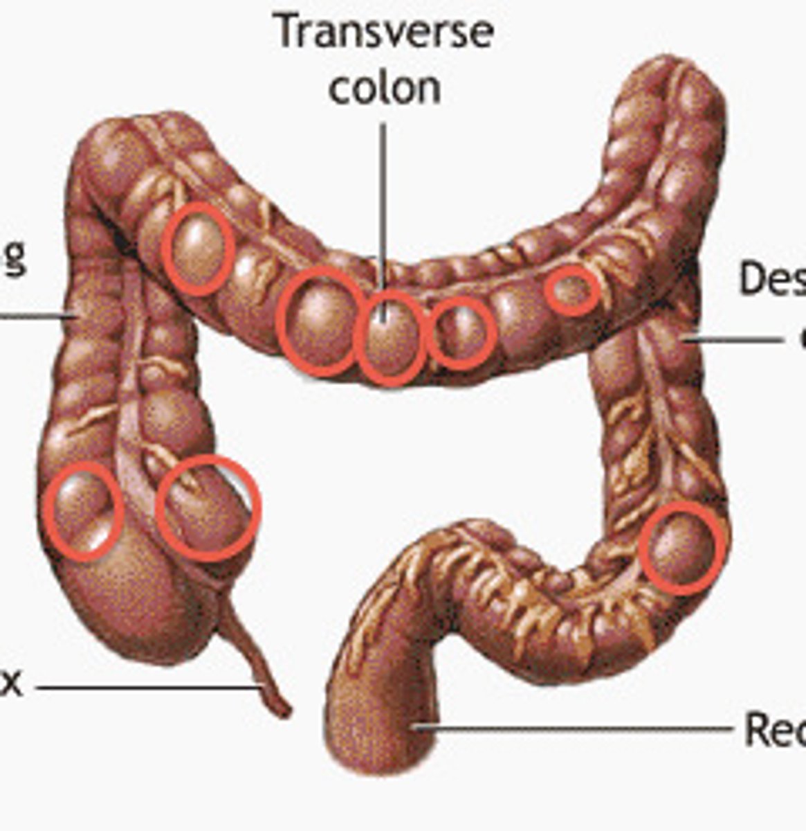
taeniae coli
muscle along
rectum
end muscle after sigmoid colon
anus
opening at end of rectum
internal anal sphincter
internal muscles controlling opening of anus (involuntary)
external anal sphincter
external muscles controlling opening of anus (voluntary)
falciform ligament
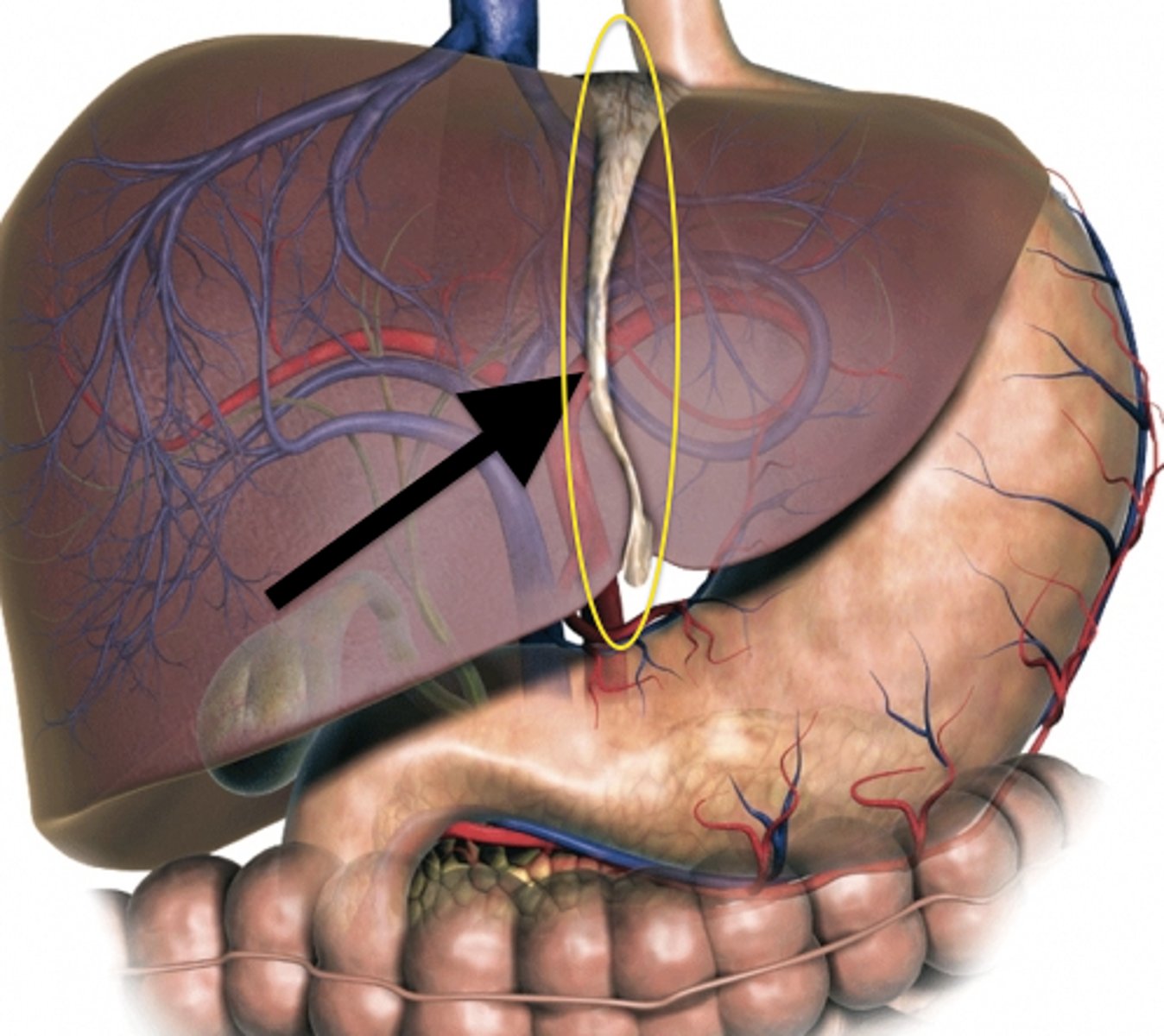
round ligament
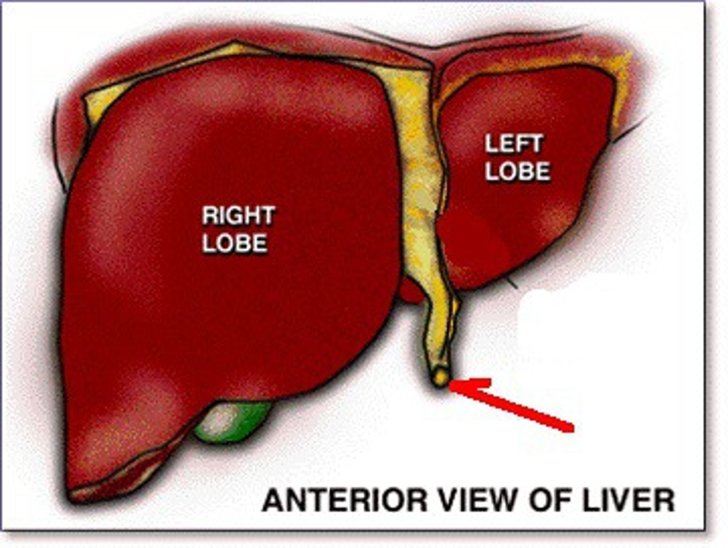
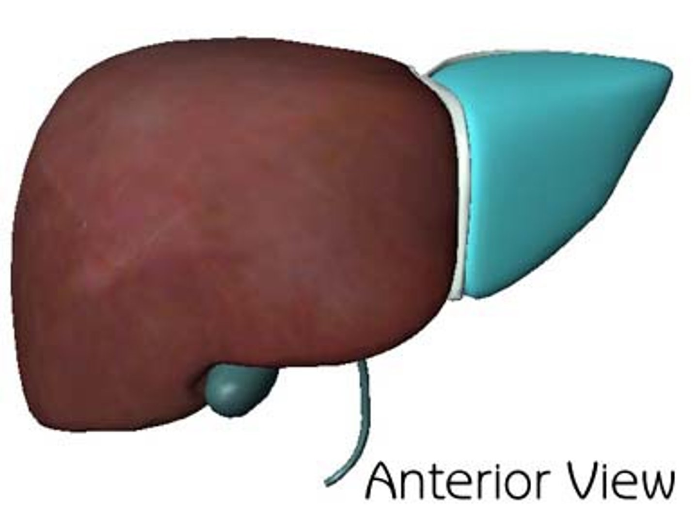
right lobe of the liver
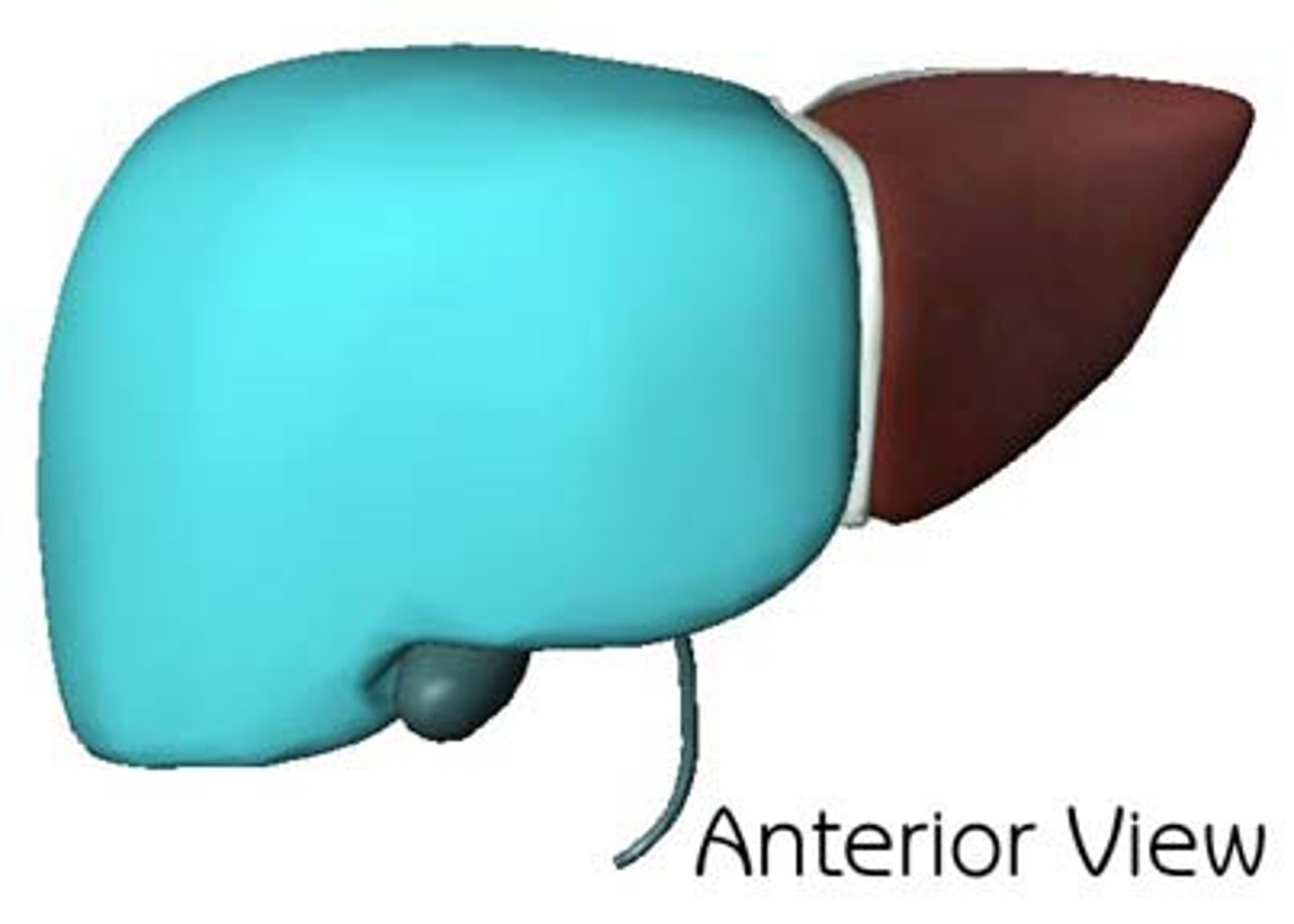
quadrate lobe
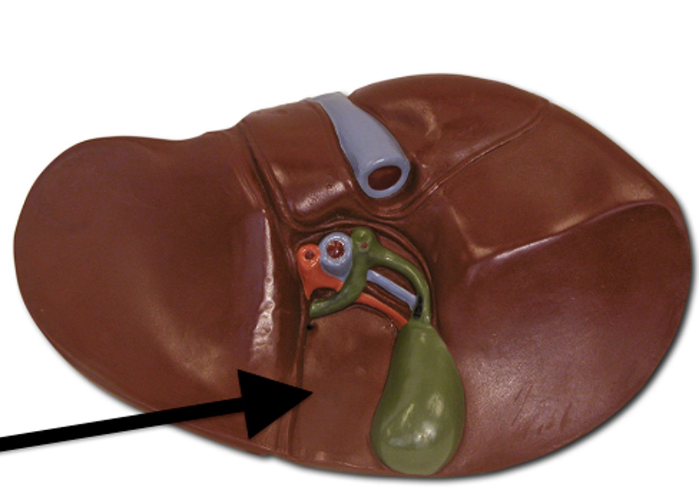
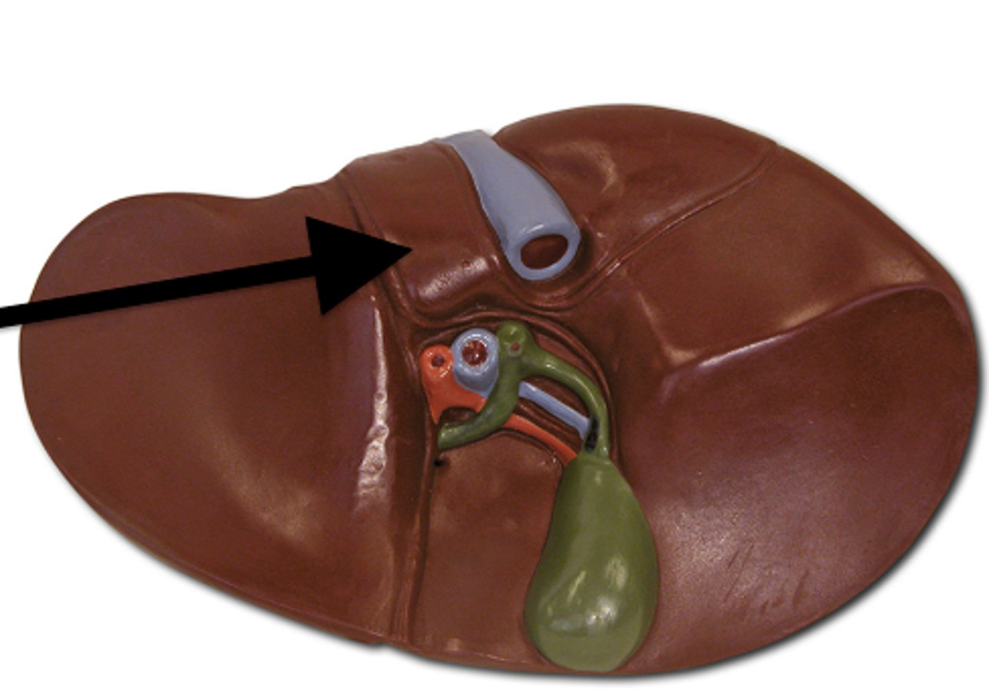
caudate lobe
left lobe of the liver
common hepatic duct
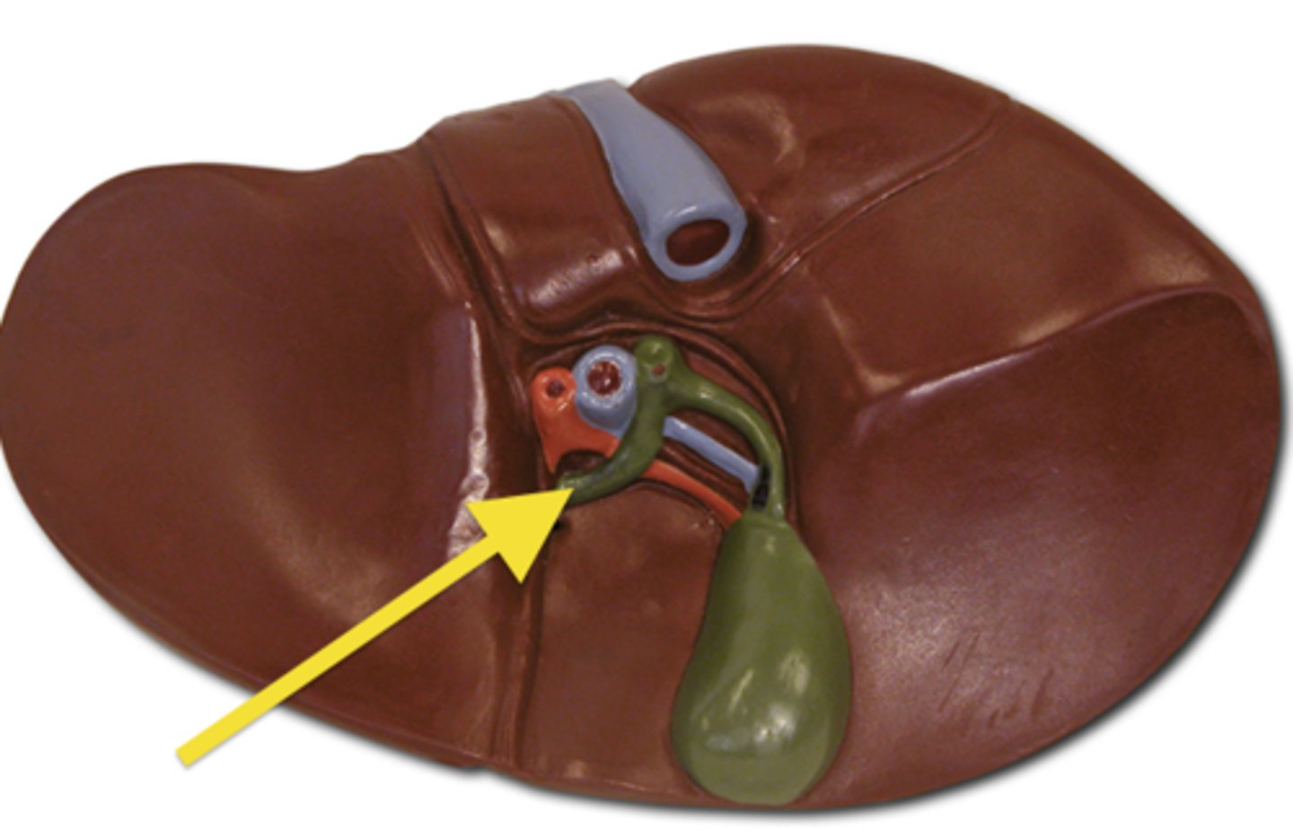
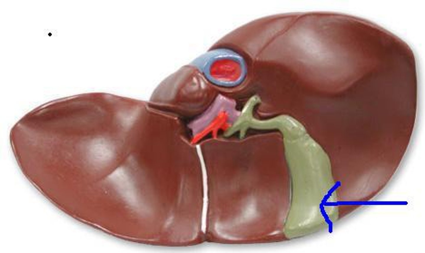
gallbladder
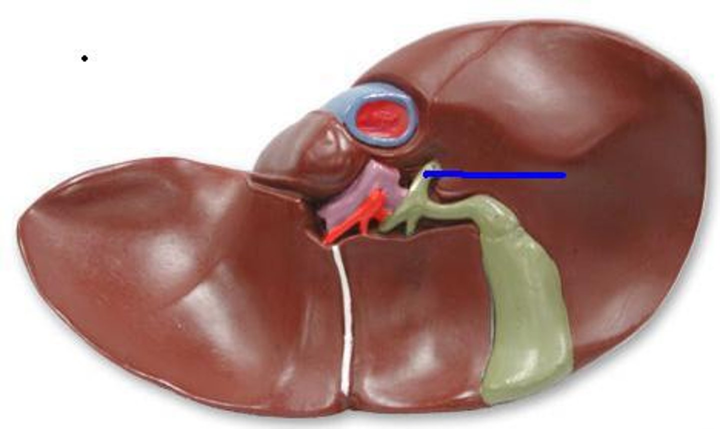
cystic duct
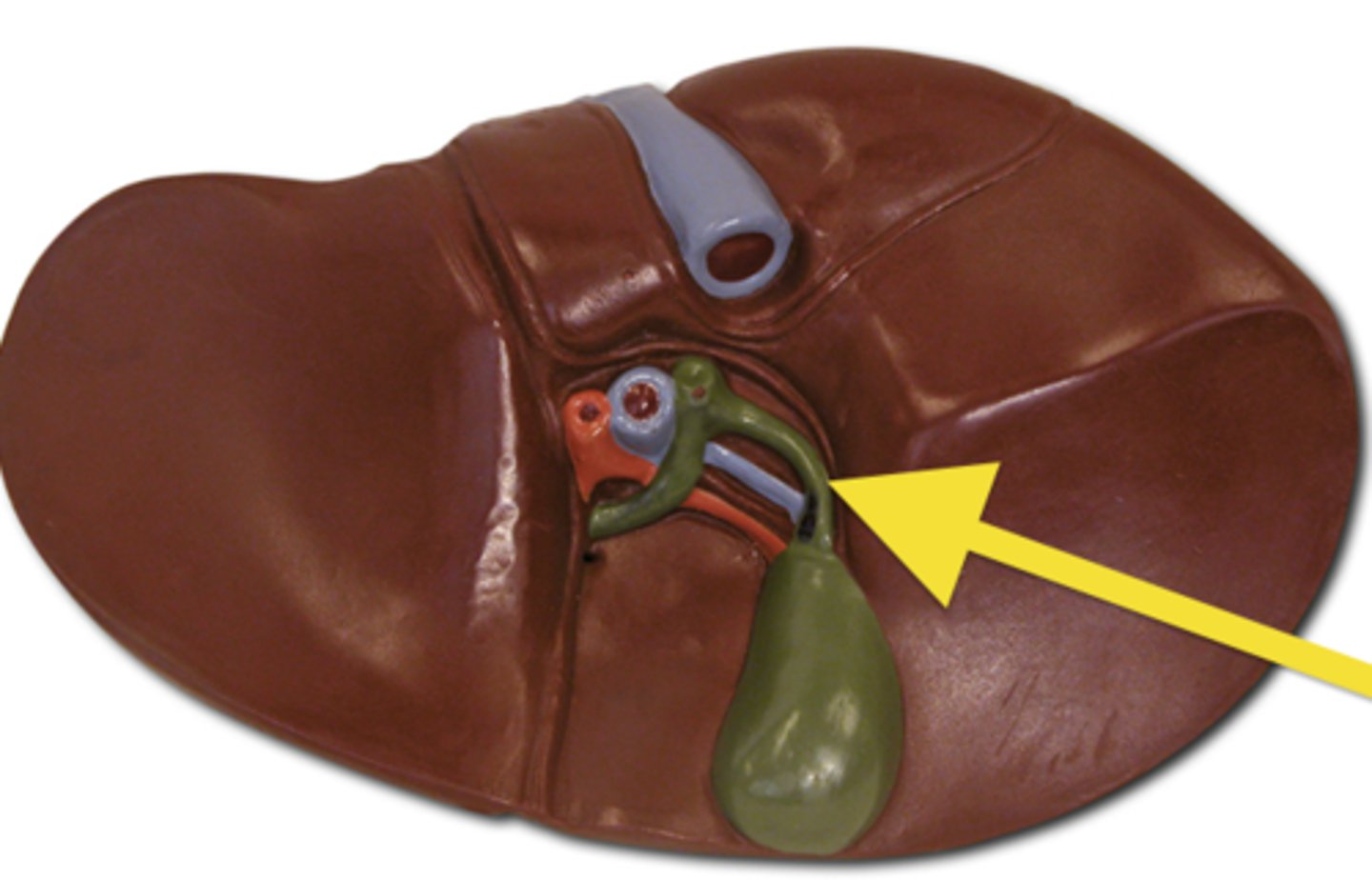
common bile duct
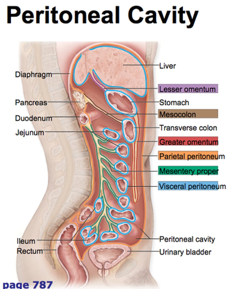
parietal peritoneum
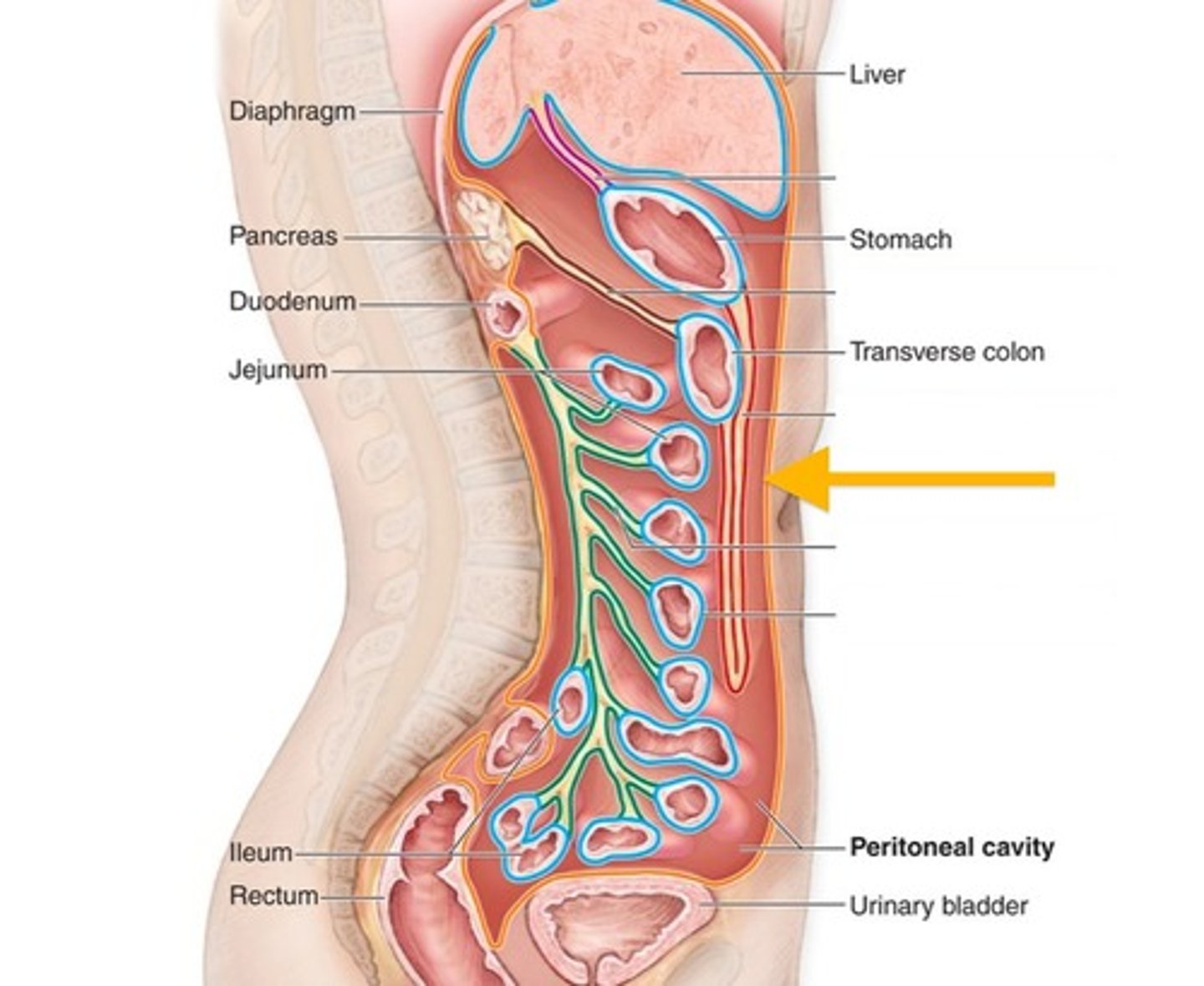
visceral peritoneum
lining of internal organs
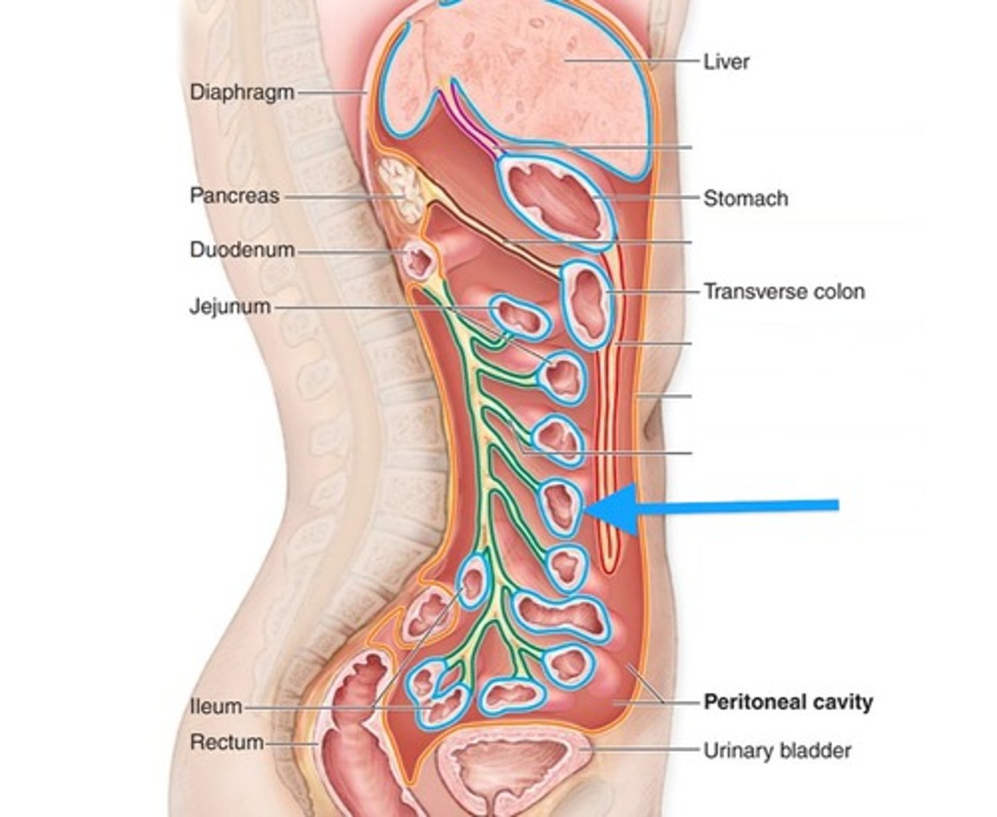
mesentery proper
proper
falciform ligament
above liver in midsagittal section of torso connecting liver to above
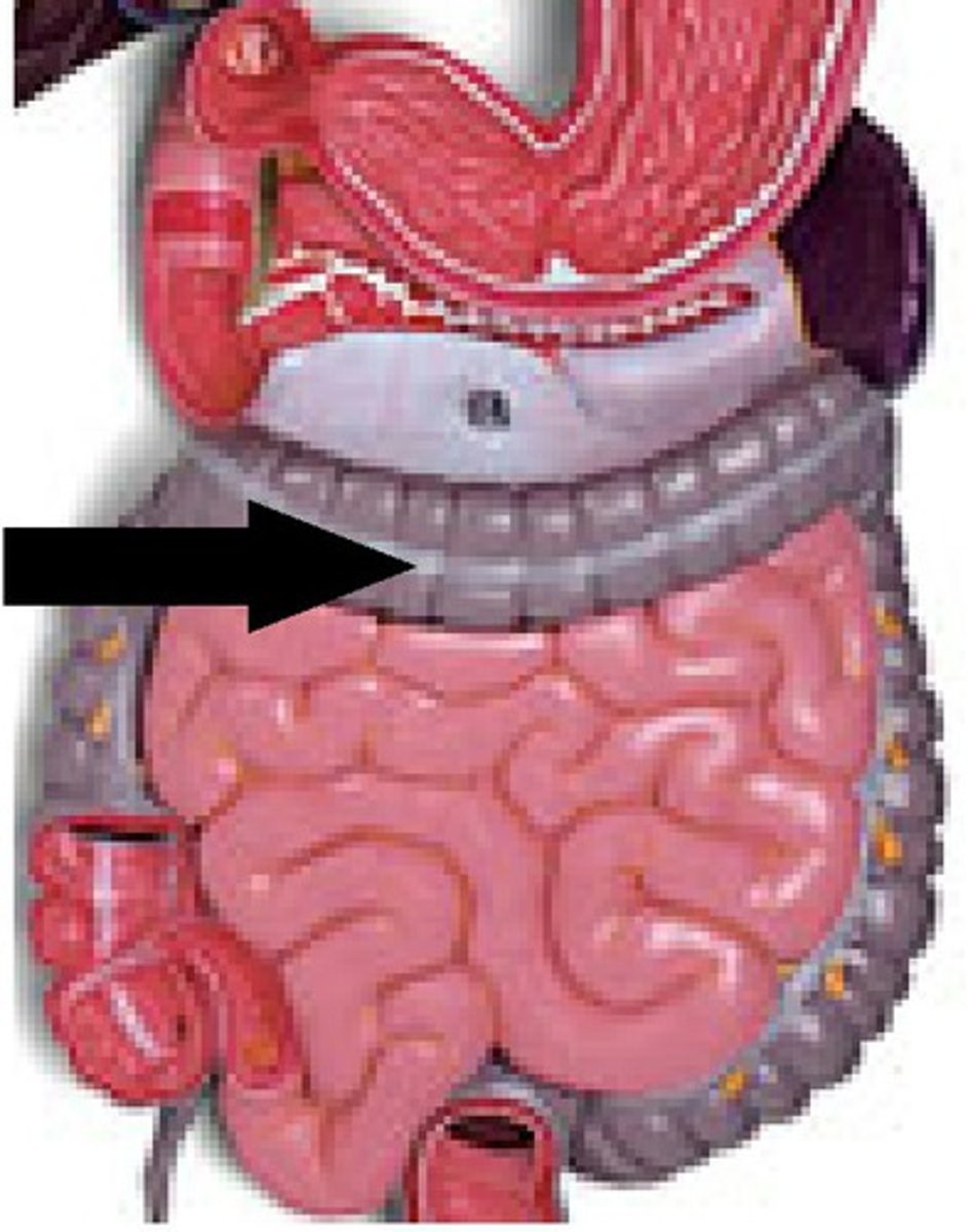
lesser omentum
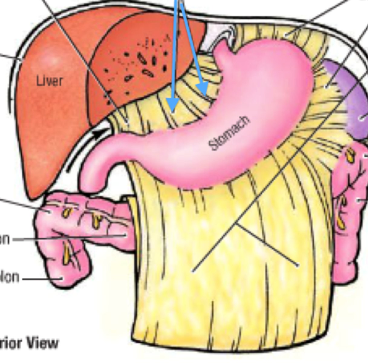
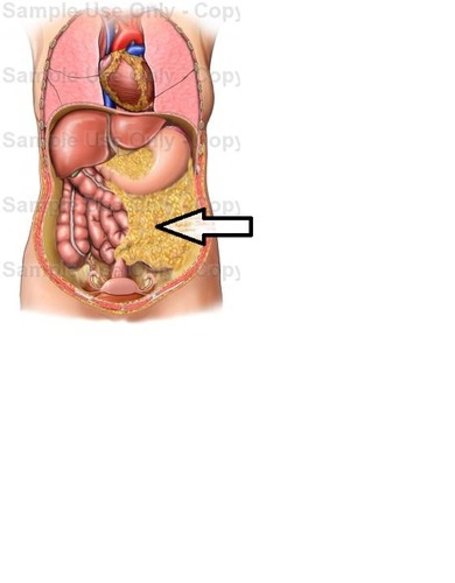
greater omentum
pancreatic duct
E
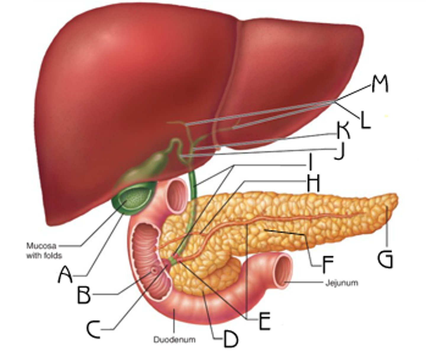
accessory pancreatic duct