Exam 3- Patho
1/121
There's no tags or description
Looks like no tags are added yet.
Name | Mastery | Learn | Test | Matching | Spaced |
|---|
No study sessions yet.
122 Terms
Hematopoiesis: process of blood cell production
All blood cells arise from a small number of underdeveloped, precursor cells called pluripotent stem cells in the bone marrow
Erythropoiesis
Development of RBCs
Erythrocytes are derived from erythroblasts (normoblasts)
Maturation is stimulated by erythropoietin
Each step HgB increases + nucleus decreases in size
120-day life cycle
Substances needed for synthesis of healthy RBCs- protein, iron, Vit B12, folic acid
Iron is the main nutritional element needed for HgB synthesis
Hypoxia stimulates the kidney to release erythropoietin (EPO)
EPO stimulates bone marrow to synthesize RBCs
Volume of circulating RBCs in healthy individuals remain constant
The spleen
A highly vascular organ, the “graveyard of RBCs” + an organ of immunity
⭐️In spleen, RBCs are broken down into component parts which are recycled to make NEW RBCs
Spleen is a secondary lymphoid organ containing lymphocytes + resident macrophages
It isolates abnormally shaped + hemolyzed RBCs and destroys them
Splenomegaly occurs when there’s a large amnt of RBC breakdown occurring
If spleen is unable/absnet, Kupffer cells remove older RBCs in liver
Anemia
Reduction in total number of erythrocytes in circulating blood or in the quality/quantity of HgB
Insufficient delivery of oxygen → tissues
Impaired erythrocyte production (Bone marrow dysfunction, lack of nutrients- iron, folate, B12)
Acute or chronic blood loss
Increased erythrocyte destruction (hemolysis)
Combination of above
Women more likely to have anemia bc of menstrual cycle
Classifications of Anemia
Etiology
Morphology- Size ; Identified by terms then end in -cytic
Macrocytic, microcytic, normocytic
HgB content; Identified by terms that end in -chromic
Normocytic + hypochromic
Microcytic hypochromic Anemia
Disorders of HgB synthesis (iron deficiency)
Macrocytic Anemia
arise from abnormalities that hinder maturation of erythroid precursors in bone marrow
Normocytic–Normochromic Anemia
shape of RBCs help determine cause
Ex: sickle cell anemia
Complete Blood Count
MCV (mean corpuscular volume)- size of RBC
MCH (mean corpuscular HgB)- color of RBC
MCHC (mean corpuscular HgB concentration)- concentration of RBC
Compensatory mechanisms of Anemia (3)
1) Heart rate increased
2) Cardiovascular
3) Capillary dilation
Macrocytic-Normocytic (Megaloblastic) Anemias: Pernicious
Etiology: Congenital/acquired deficiency of intrinsic factor (IF)- in stomach lining
Mechanism: Insufficient influence of Vit B12 on developing cells bc of deficient IF; Abnormal DNA + RNA synthesis; Premature cell death
Vitamin B12 + Neuro Effects
Vit B12 deficiency myelin defects + abnormal neural conduction occur mainly in dorsal horns + corticospinal tract of spinal cord called subacute combined degeneration, manifested as numbness + weakness in extremities (paresthesia) and gait disturbance
Synthesis of serotonin, norepinephrine, and dopamine are affected which is relevant to cognitive changes, such as confusion + memory loss
Iron deficiency Anemia (IDA) Highest risks/problems / who is it found in
Highest risk: toddlers, adolescent girls, women of childbearing age, those living in poverty, infants consuming cow’s milk, older adults on restrictive diets, teenagers on junk food diets
Causes: dietary lack + eating disorders, impaired absorption, increased requirement, chronic blood loss, medications (cause GI bleeding, some surgeries)
Particularly in males and postmenopausal women, due to chronic blood loss from GI bleeding
Fecal occult blood testing (FOBT) recommended w/ Anemia patients for what
To rule out GI Bleeding + potential colon cancer
What is an essential element of Hgb synthesis
Iron- body maintains balance between iron in use for Hgb + iron stored available for future Hgb synthesis
IDA specific signs:
Hair loss, cheilitis, glossitis, nail changes (koilonychia), cold/numb fingertips
⭐️Pica- craving for nonfood substances such as ice, clay, starch, chalk, dirt/other material
Normo-Normochromic Anemias:
Sickle Cell Anemia: autosomal recessive disease
Causes structural fragility of the sickle cell anemias RBCs, upon exposure to hypoxia/stress, the RBC contorts into a sickle shape
Vaso-Occlusive Crises in Sickle Cell Anemia
Sickled RBCs cannot pass thru capillaries
Become trapped, blocking blood flow + creating ischemia + consequent tissue hypoxia, leads to organ damage + possible infarction
Episodes of ischemia are painful vaso-occlusive crises
Chest, abdomen, long bones, and joints affected. Multiple sites often involved simultaneously
Splenic dysfunction occurs bc of excess RBC death
Myeloproliferative RBC Disorders
Polycythemia- overproduction of red blood cells
Types of Polycythemia (2)
Relative Polycythemia- results of dehydration
Fluid loss results in relative increases of RBC counts + Hgb + Hct values
Minor consequences, resolved w/ fluid admin. + treatment
Absolute Polycythemia-
Primary (polycythemia vera): hyperproliferation of all blood cells; abnormality of stem cells in bone marrow
Secondary (erythrocytosis): increase in erythropoietin as normal response to chronic hypoxia/ inappropriate response to erythropoietin-secreting tumors
Composition of blood: Leukocytes
(WBCs)- defend body against infection + remove debris
Composition of blood: Granulocytes
Membrane-bound granules in their cytoplasm; Granules contain enzymes capable of destroying microorganisms; Inflammatory + immune func.
Neutrophils- phagocytes in early inflammation
Eosinophils- ingest antigen-antibody complexes; induced by IgE hypersensitivity; increase in parasitic infections
Basophils- structurally + functionally similar to mast cells
Composition of blood: Agranulocytes
Monocytes: immature macrophages
Macrophages: in tissues
Lymphocytes: mature to T cells, B cells, plasma cells
Natural Killer Cells
Composition of blood: Platelets
Essential for blood coagulation + control of bleeding
Disk-shaped cytoplasmic fragments
Infectious Mononucleosis / Caused by?
Mono/kissing disease
Acute, self-limiting infection of B lymphocytes transmitted by saliva thru personal contact
Caused by Epstein-Barr virus (EBV)- 85%
B cells have EBV receptor site
Other: CMV, hepatitis, influenza, HIV
Symptoms: fever, sore throat, swollen cervical lymph nodes, increased lymphocyte count, atypical lymphocytes
Hematologic Neoplasms + 2 main characteristics
Types of cancer that affect blood, bone marrow, and lymph nodes
Either located in blood (leukemia) or lymph nodes (lymphoma)
1) ⭐️Cancerous WBCs proliferate uncontrollably, leading to overcrowding
Leukemia primarily occurs in bone marrow
Lymphoma affects lymphoid tissues such as lymph nodes
2) ⭐️Genetic susceptibility to mutagens often factor in both
Leukemia- excess cancerous WBCs suppress normal blood cell development in bone marrow
Lymphoma- cancerous WBCs primarily affect lymphatic system
Clinical manifestations of Hematologic Cancer (symptoms/what physical exam reveals)
Symptoms include:
⭐️Anemia (fatigue, weakness, pallor)
⭐️Leukopenia (increased susceptibility to infection)
⭐️Thrombocytopenia (increased susceptibility to bleeding + bruising)
⭐️Bone pain (bc of proliferating cancerous blood cells putting pressure in bone marrow)
Physical exam may reveal enlarged lymph nodes, splenomegaly, or both
Enlarged lymph node from proliferative neoplastic cells
Splenomegaly result of excessive infiltration of neoplastic blood cells /excessive hemolysis by overactive spleen
Leukemia
Uncontrolled proliferation of malignant leukocytes causing overcrowding of bone marrow and decreased production + function of normal hematopoietic cells
Classification based on
Predominant cell of origin- myeloid or lymphoid
Degree of differentiation that took place before cell became malignant
Acute with rapid growth of immature (blast) cells
Chronic with slow growth of more differentiated cell
Acute Lymphocytic Leukemia (ALL)
Rapid, >30% lymphoblasts + B cells
Chronic Lymphocytic Leukemia (CLL)
Slow; Monoclonal B
Acute Myeloid Leukemia (AML)
Rapid; precursor myeloid cells
Chronic Myelogenous Leukemia (CML)
Slow; neutrophilic/eosonphilic/clonal; arise from hematopoietic stem cells
Lymphadenopathy
Enlarged lymph nodes that become palpable + tender
During infection, macrophages + lymphocytes are proliferating
Local lymphadenopathy
drainage of inflammatory lesion located near the enlarged node
General lymphadenopathy
occurs in presence of malignant/nonmalignant disease
Lymphoma- Hodgkin
Nodal involvement |
Extranodal movement |
Spread |
Fever, night sweats, weight loss |
Reed Sternberg cells |
Extent of disease |
Localized to single axial group of nodes |
Rare |
Orderly spread |
Common |
Present |
Often localized |
Lymphoma- Non-Hodgkin
Nodal involvement |
Extranodal movement |
Spread |
Fever, night sweats, weight loss |
Reed Sternberg cells |
Extent of disease |
Multiple peripheral nodes |
Common |
Non Contagious |
Uncommon |
Not present |
Rarely localized |
Burkitt Lymphoma
Highly aggressive B-cell non-Hodgkin lymphoma
Very fast growing tumor of the jaw + facial bones (Africa); rare in US
EBV more than 90% of cases
Abdominal swelling for people affected in US
Biopsy/bone marrow findings
Multiple Myeloma: Arise where/ what does it produce
Arises in B lymphocytes, causing proliferation of abnormal plasma cells in bone marrow
Malignant plasma cells produce abnormally large amounts of one class of immunoglobulin (M protein: abnormal antibody molecule)
Multiple Myeloma: Causes
Cause bone destruction, bone marrow failure, renal failure, neurological complications, amyloidosis
Bone pain, especially in back common (result of lytic destruction + formation of plasmacytomas)
Lytic lesions are rounded, punched-out areas of bone found in vertebra, skull, ribs, humerus, and femur
Platelets: Thrombocytopenia what does it cause?
Low number of platelets
<100,000/uL (can cause bleeding)
Prolongation of normal clotting may result
If <20,000, spontaneous bleeding may occur
Platelets: Thrombocytosis what does it cause?
>750,000/uL (can cause excessive clotting)
Risk for spontaneous blood clots (thrombosis), stroke, <3 attack increases
Platelet formation stimulated by…, sythesized by the …
Thrombopoietin; liver
Thrombocythemia (Cause/Types/Causes)
Too much platelet count
Cause: accelerated platelet production in bone marrow
Types: primary/secondary (reactive)
Causes: intravascular clot formation (thrombosis), hemorrhage, other
Essential (primary) thrombocytopenia (ET)
⭐️Chronic myeloproliferative disorder
Characterized by excessive platelet production, resulting from a defect in megakaryocyte progenitor cells
⭐️Clinical: microvascular thrombosis, erythromelalgia, possible bleeding
Distribution of Body fluids
Total body water (TBW)
Intracellular fluid (ICF)- ⅔ of TBW
Extracellular fluid (ECF) ⅓ of TBW
Interstitial fluid (ISF), Intravascular fluid (blood plasma), Lymph, synovial, intestinal, CSF, sweat, urine, pleural, peritoneal, pericardial, and intraocular fluids
Distribution of Body fluids: Peds + 2 examples of decreased TBW
Peds: 70-80% of body weight; susceptible to significant changes in body fluids
️Aging: decreased % of TBW
1) Increased fat mass + Decreased muscle mass
2) Renal decline; diminished thirst perception
Obesity: less TBW because fat is water repelling!
Water Movement Between Fluid Compartments (Osmosis )
How water moves btwn ICF and ECF compartments
Water Movement Between Fluid Compartments (Osmolality)
Concentration of solutes per kg of fluid
Water crosses cell membranes freely so osmolality of TBW normally equilibrium
Water Movement Between Fluid Compartments (Law of Osmosis)
Water moves from area of lower solute concentration to areas of higher solute concentration to balance solute levels across a semipermeable membrane
Water Movement Between Fluid Compartments (Osmotic forces/2 Types)
Osmotic forces: pressure exerted by solutes in solution
Sodium for ECF
Potassium for ICF
Fluid Homeostasis Maintained by:
Osmoreceptors in the hypothalamus- respond changes in blood osmolarity + blood fluid volume
Sensation of thirst at hypothalamus
Antidiuretic hormone (ADH, arginine vasopressin)
Renin-angiotensin-aldosterone system (RAAS)
Natriuretic hormones-excretion of both Na+ and H2O by kidneys in response to excess ECF volume
Edema + Causes
Accumulation of fluid within interstitial spaces
Causes:
Increase in hydrostatic pressure
Decrease in oncotic pressure
Oncotic pressure- force exerted by albumin in bloodstream
Increase in capillary permeability
Lymph obstruction (lymphedema)
Localized vs generalized
Pitting edema
Acid-Base Balance
Carefully regulated to maintain normal pH via multiple mechanisms
pH: negative logarithm of H+ concentration
Acids are formed as end products of protein, carb, and fat metabolism
To maintain body’s normal pH (7.35-7.45) H+ must be neutralized/excreted
Bones, lungs, kidneys= major organs involved in regulation of acid-base balance
Buffering Systems - Whats buffer? Most important plasma buffering systems?
Buffer is a chemical that can bind excessive H+/OH- w/o significant change in pH
Most important plasma buffering systems= Carbonic Acid, Bicarb, Hgb
Carbonic Acid-Bicarb Buffering
Buffering Systems - Where does it operate?
Operates in lung and kidney *
Greater the PaCO2, the more carbonic acid is formed
At pH of 7.4, ratio of bicarb-carbonic acid is 20:1
Carbonic Acid-Bicarbonate Buffering
Lungs can decrease carbonic acid
Kidneys reabsorb/regenerate bicarb but do not act as fast as lungs
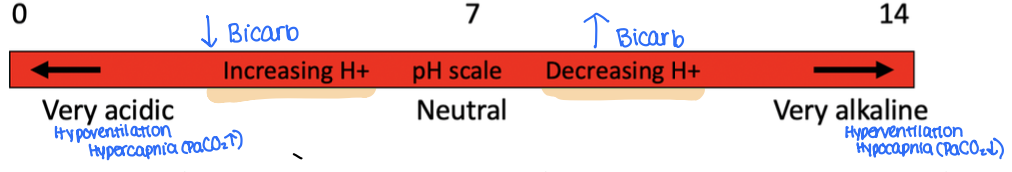
Carbonic Acid-Bicarbonate Buffering: If bicarb decreases…. + explain compensation
…Then pH decreases, causes acidosis
pH can be returned to normal if carbonic acid also decreases (aka compensation)
Carbonic Acid-Bicarbonate Buffering: Respiratory compensates by…
… increasing ventilation to expire CO2 (acidosis) or by decreasing ventilation to retain carbon dioxide (alkalosis)
Carbonic Acid-Bicarbonate Buffering: Renal system compensates by…
Producing acidic/alkaline urine (kidneys)
Protein buffering (+2 types)
Proteins carry negative charges, allow them to bind w/ H+ and act as buffers
Intracellular buffering: Hgb serves as primary buffer, helping to maintain pH by binding with excess H+
Plasma buffering: while Bicarb is the major plasma buffer, proteins (Hgb + albumin) contribute to buffering in bloodstream
Cellular ion exchange
Exchanges of K+ for H+ in acidosis + alkalosis
Acute acidosis accompanied by hyperkalemia
Acid-Base Imbalances (normal pH + Pressure + Acidosis/Alkalosis)
Normal arterial blood pH: 7.35-7.45
Obtained by arterial blood gas (ABG) sampling (pressure of gases in bloodstream)
PaO2: pressure of oxygen in arterial blood (80-100 mm Hg)
PaCO2: pressure of carbon dioxide in arterial blood (35-45 mm Hg)
HCO3-: amnt of bicarb in the blood (22-26 mEq/liter)
SaO2: saturation of Hgb with oxygen (95-100%)
Acidosis: systemic increase in H+ concentration / decrease in bicarbonate (base)
Alkalosis: systemic decrease in H+ concentration / increase in bicarbonate
Respiratory Acidosis: Etiology
Failure of respiratory system to remove/exhale CO2 from body fluids as fast as it is produced by cells- excess of CO2 in blood (hypercapnia)
Respiratory Acidosis: Caused by
Caused by any interference with breathing (COPD, respiratory muscle weakness/paralysis, brainstem trauma, over-sedation)
Respiratory Acidosis: Compensation
Kidneys attempt to REABSORB BICARB and EXCRETE H+
Respiratory Alkalosis: Etiology
Loss of CO2 from lungs faster than it is produced by cells; can be breathing too fast; hypocapnia
Respiratory Alkalosis: Caused by
High altitudes, hypermetabolic states, early salicylate intoxication, anxiety or panic disorder, improper use of mechanical ventilators
Respiratory Alkalosis: Compensation
Kidneys attempt to REABSORB max H+ and EXCRETE BICARB
Metabolic acidosis: Etiology
Abnormal accumulation of non carbonic acids/abnormal loss of bases
Metabolic acidosis: Occur in.. / Clinical…
Occur in: lactic acidosis, diabetic ketoacidosis, renal failure causing acid buildup, diarrhea or vomiting with loss of bicarb
Clinical: Headache, lethargy, Kussmaul’s breathing (deep rapid respirations)
Metabolic acidosis: Compensation
Hyperventilation and renal excretion of excess acid
Metabolic Alkalosis: Etiology
Bicarb concentration increased, from loss of metabolic acids (Cl-)
Metabolic Alkalosis: Caused by
Vomiting, gastric suctioning, excessive bicarb intake, hyperaldosteronism w/ hypokalemia, diuretic therapy
Metabolic Alkalosis: Compensation
Hypoventilation, the lungs retain CO2
Overview of Electrolytes
Electrolytes are in both ECF + ICF compartments but different concentrations
All electrolytes move across compartments but must be in balance for health
Overview of Electrolytes: Intracellular
Cation
⭐️Potassium (K+)
Magnesium (Mg)
Calcium (Ca+)
Anion
Phosphate (HPO-)
Overview of Electrolytes: Extracellular
Cation
⭐️Sodium (Na+)
Calcium (Ca+)
Anion
Chloride (Cl-)
Bicarbonate (HCO3-)
Isotonic alterations
TBW change with proportional electrolyte change
Isotonic fluid loss (dehydration + hypovolemia)
Isotonic fluid excess (hypervolemia)
Isotonic (isoosmolar imbalance) Mechanism?
Gain/loss of ECF resulting in concentration = 0.9% sodium chloride (salt) solution (normal saline); no shrinking or swelling of cells
Hypertonic (hyperosmolar imbalance) Mechanism?
Imbalance that results in an ECF >0.9% salt solution, water loss/solute gain; cells shrink
Hypotonic (hypoosmolar imbalance Mechanism?
Imbalance that results in an ECF <0.9% salt solution; water gain/solute loss; cells swell
Hypertonic alterations (definition + causes)
Hypernatremia- related to sodium (Na+) gain / water loss
⭐️Inadequate free water intake, inappropriate administration of hypertonic saline solution, oversecretion of aldosterone, decreased ADH secretion (diabetes insipidus), Cushing's syndrome
Water movement from ICF to the ECF - intracellular dehydration
Manifestations: seizures, muscle twitching, hyperreflexia
What is potassium essential for
Transmission + conduction of nerve impulses, normal cardiac rhythms, and skeletal + smooth muscle contraction
What does potassium regulate
ICF osmolality + deposits glycogen in liver + skeletal muscle
What regulates potassium balance?
⭐️Kidneys, aldosterone, and insulin secretion, and changes in pH regulate K+balance
Insulin promotes uptake of K+ by stimulation the Na+, K+, ATPase pump, facilitating the movement of K+ into the liver and muscle cells along with glucose, which helps regulate blood glucose levels after eating
Calcium + Phosphate; what are they controlled by?
PTH: increases plasma calcium levels via kidney reabsorption
Vit D: fat-soluble steroid; increases calcium absorption from GI tract
Calcitonin: Decreases plasma calcium levels
Hypocalcemia (causes + manifestations)
Calcium lower than 9.0 mg/dL / ionized levels lower than 5.5
Causes: inadequate intestinal absorption, decreases in PTH + Vit D, blood transfusions
Manifestations
Increased neuromuscular excitability (partial depolarization)
Muscle spasms (partially in hands, feet, facial muscles), Chvostek + Trousseau signs, convulsions, tetany
Theories of pain (4)
Specificity pain: amount of pain related to the amount of a tissue injury
Accounts for many types of injuries, does not explain psychologic contributions to pain/chronic pain
Pattern theory: describes roles of nerve impulses interpreted by CNS
Does not account for all pain experiences
Gate control theory: explains complexities of the pain phenomenon
Pain is modulated by a “gate” in the cells of the substantia in the spinal cord
Neuromatrix theory (expands on gate control theory)
Brain produces patterns of nerve impulses drawn from various inputs including genetic, psychologic, and cognitive experiences
Illustrates plasticity (the adaptable change in structure + function) of the brain
Sensory inputs to the brain produce patterns of pain, but stimuli may independently originate in the brain with no external input
Pain is modulated by a “gate” in the cells of the ______ in the spinal cord
substantia
Describe the fibers that either block/allow pain signals to reach the brain
Large myelinated A-delta fibers + small unmyelinated C fibers respond to a broad range of painful stimuli, such as mechanical, thermal, and chemical. These nociceptive transmissions open the gate.
Stimuli from nonnociceptive transmissions, such as touch and larger A-beta fibers close the gate
Neuroanatomy of pain: Afferent Neurons/ Interpretive centers/ Efferent neurons
Afferent neurons: sensory nerves
Begin in PNS, travel to spinal gate in dorsal horn, then ascend to higher centers in the CNS (primarily via spinothalamic tract)
Carry temp, touch, proprioception, vibration, and pressure sensations into the spinal cord
Interpretive centers
Located in brainstem, midbrain, diencephalon, and cerebral cortex
Efferent neurons: motor nerves
Descend from CNS to the dorsal horn of the spinal cord, with corticospinal tract contributing to pain modulation through descending pathways from motor cortex
Nociceptors
Free nerve endings in the afferent PNS that selectively respond to chemical, mechanical and thermal stimuli
Processing of potentially harmful stimuli
Nociceptors- A-delta fibers/ C fibers
A-delta fibers are large and lightly myelinated.
Conduct impulses rapidly + cause the first, short-lived acute experience of pain
Causes reflex withdrawal of affected body part from stimulus before pain sensation is perceived
C fibers are small and unmyelinated
Conduct impulses slowly and cause longer lasting, persistent dull, aching, or burning sensations
Neuroanatomy of Pain- Pain threshold/ pain tolerance
Pain threshold- lowest intensity of pain a person can recognize
Intense pain at one location may increase threshold in another location
One with many painful sites may only report most painful
After dominant pain is diminished, one may identify other painful areas
Pain tolerance- Greatest intensity of pain one can endure
Individualized; varies among people and in the same person over time
Acute pain description
Protective mechanisms
Immediate sensation after injury; transient
Alerts an individual to a condition/experience that’s immediately harmful to body
Begins suddenly + relieved after pain stimulus is removed; lasts <3 months
Acute Somatic Pain
Arises from joints, muscle, bone, and skin
A-delta fibers: pain is sharp and well localized
C-fibers: pain is dull, aching, throbbing, and poorly localized
Acute visceral Pain
Arises from internal organs + lining of body cavities
Poorly localized as a result of fewer #’s of nociceptors
Referred Pain
Pain in an area is removed/distant from its point of origin
Area of referred pain is supplied by same spinal segment as actual site
Can be acute/chronic
Chronic pain
No protective purpose
Prolonged pains sensation after injury has healed (>3-6 mo)
Caused by dysregulation of nociception + pain modulation processes
Neuroplasticity: brain’s ability to reorganize itself by forming new neural connects in response to persistent pain signals
May be persistent/intermittent; may be sudden/develop insidiously
May cause behavioral + psychologic changes, such as depression + anxiety