Quiz 1 Anatomy and Histology
 Call Kai
 Learn
Learn Practice Test
 Spaced Repetition
 Match
Match Flashcards
 Knowt Play
1/124

Earn XP
Description and Tags
Epithelium, Connective, Nervous, Muscle, Skeletal Tissue
Name | Mastery | Learn | Test | Matching | Spaced | Call with Kai | Chat |
|---|
No analytics yet
Send a link to your students to track their progress
125 Terms
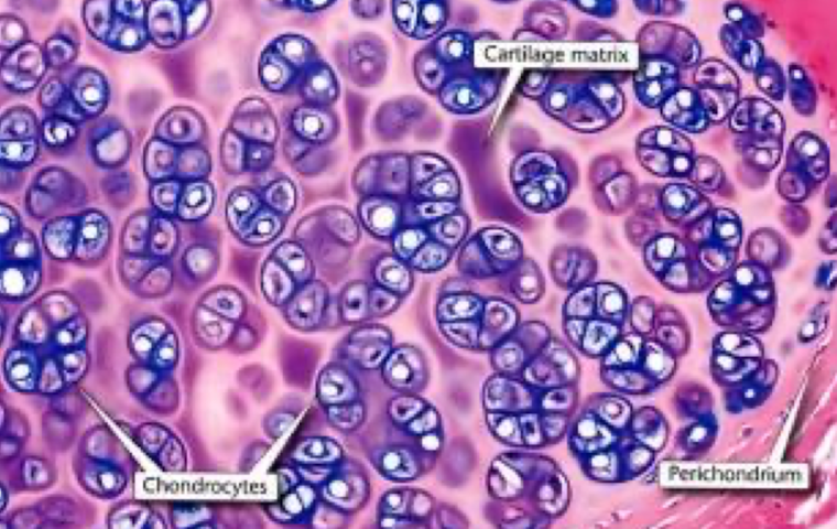
hyaline cartilage
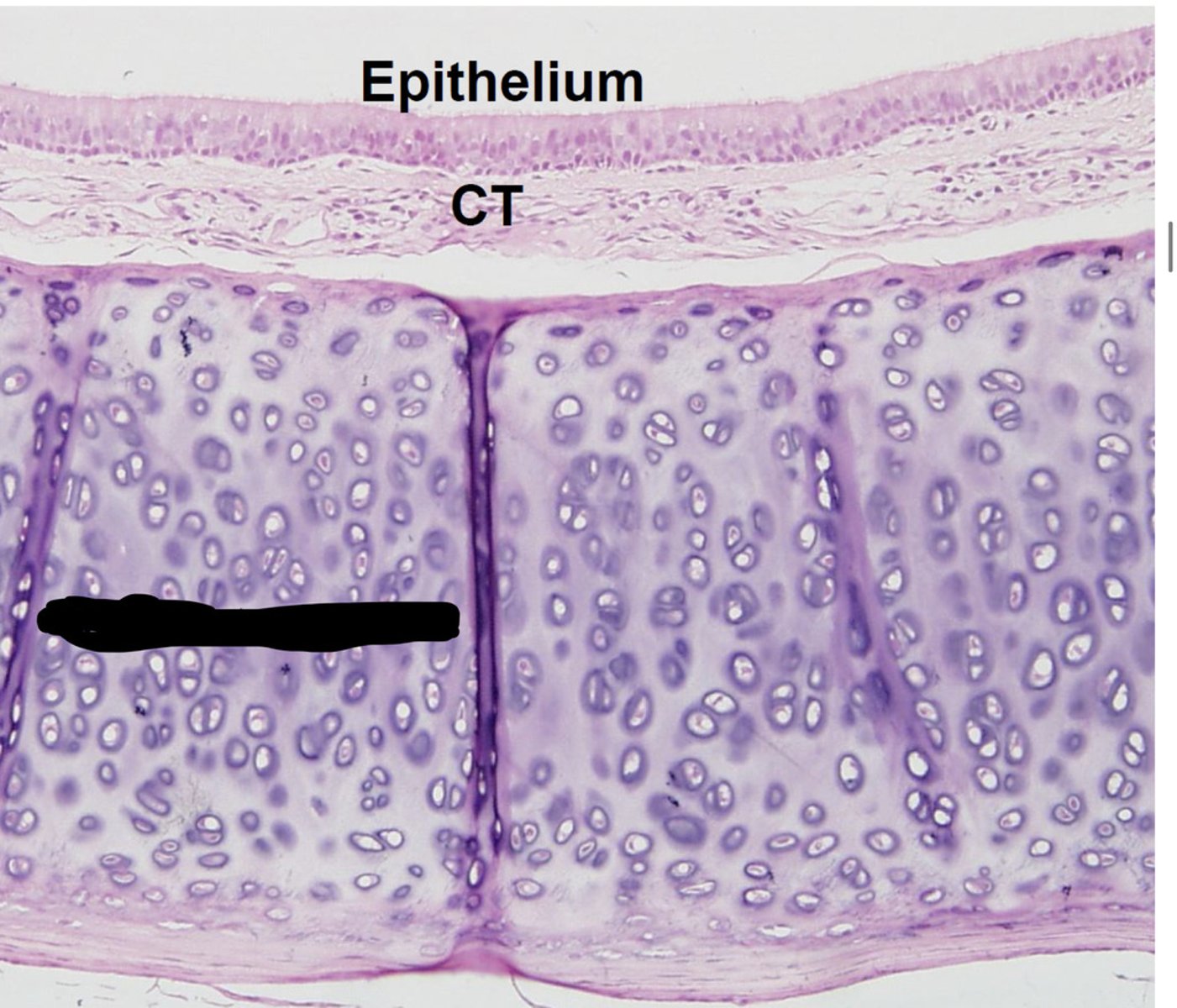
hyaline cartilage
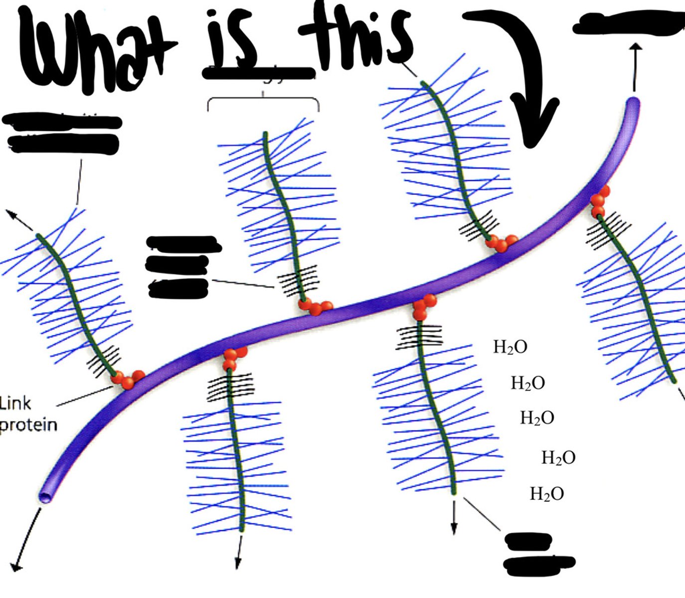
Proteoglycan
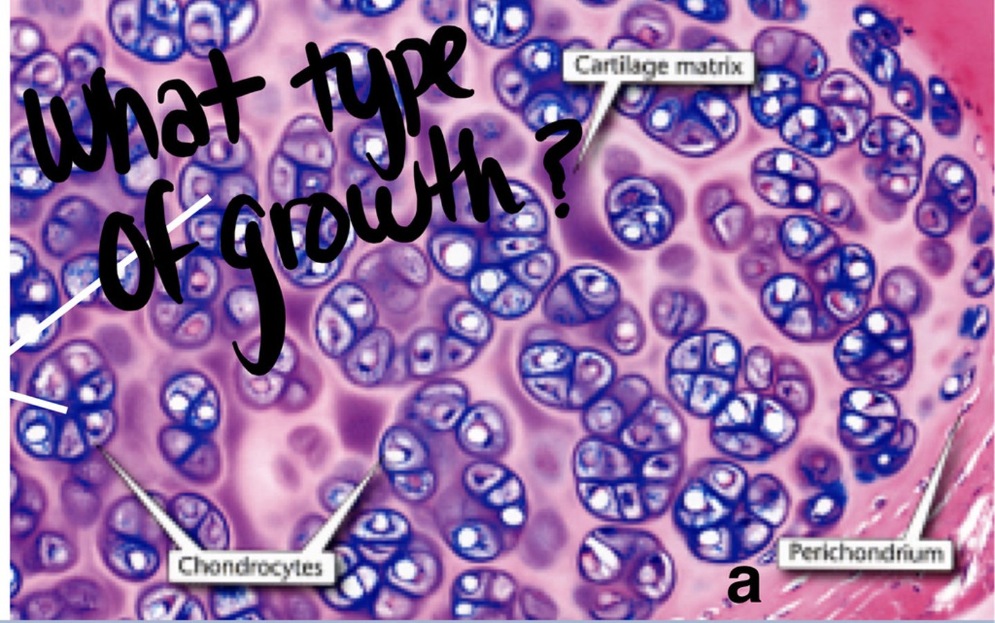
Appositional growth
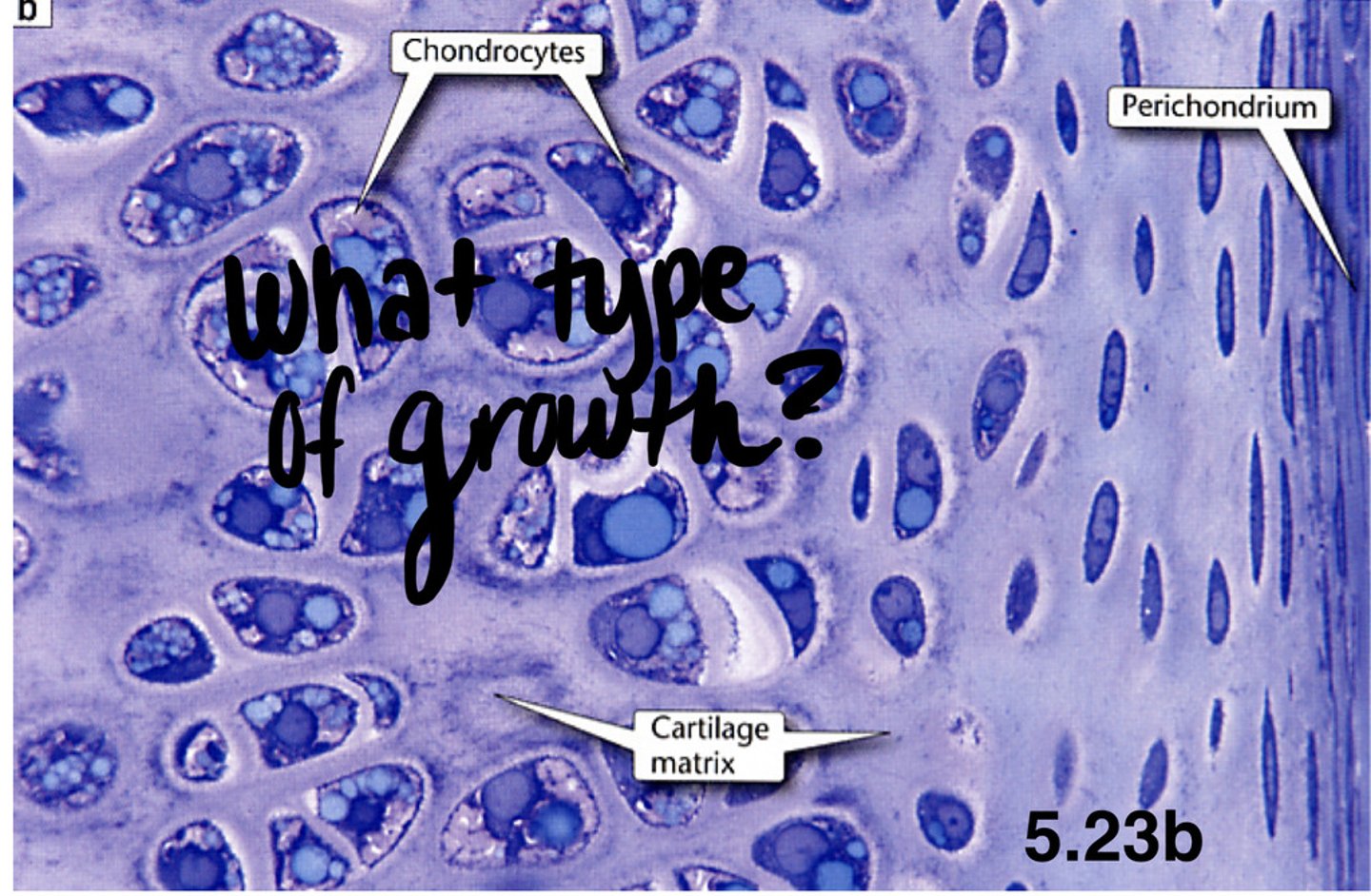
Interstitial growth
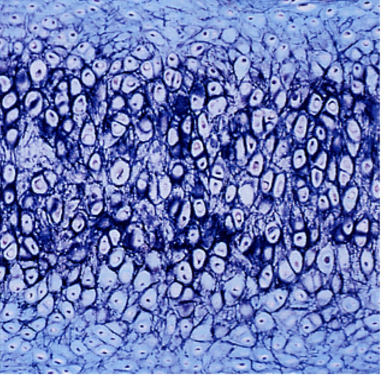
Elastic cartilage
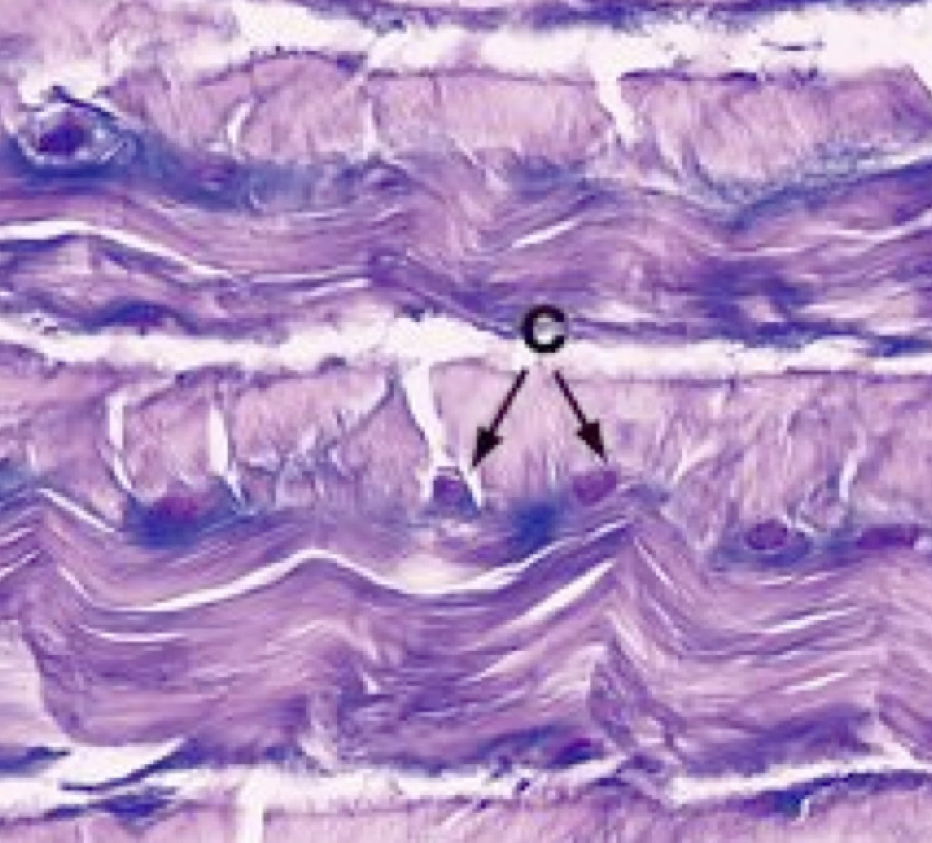
Fibrocartilage
Fibrocartilage
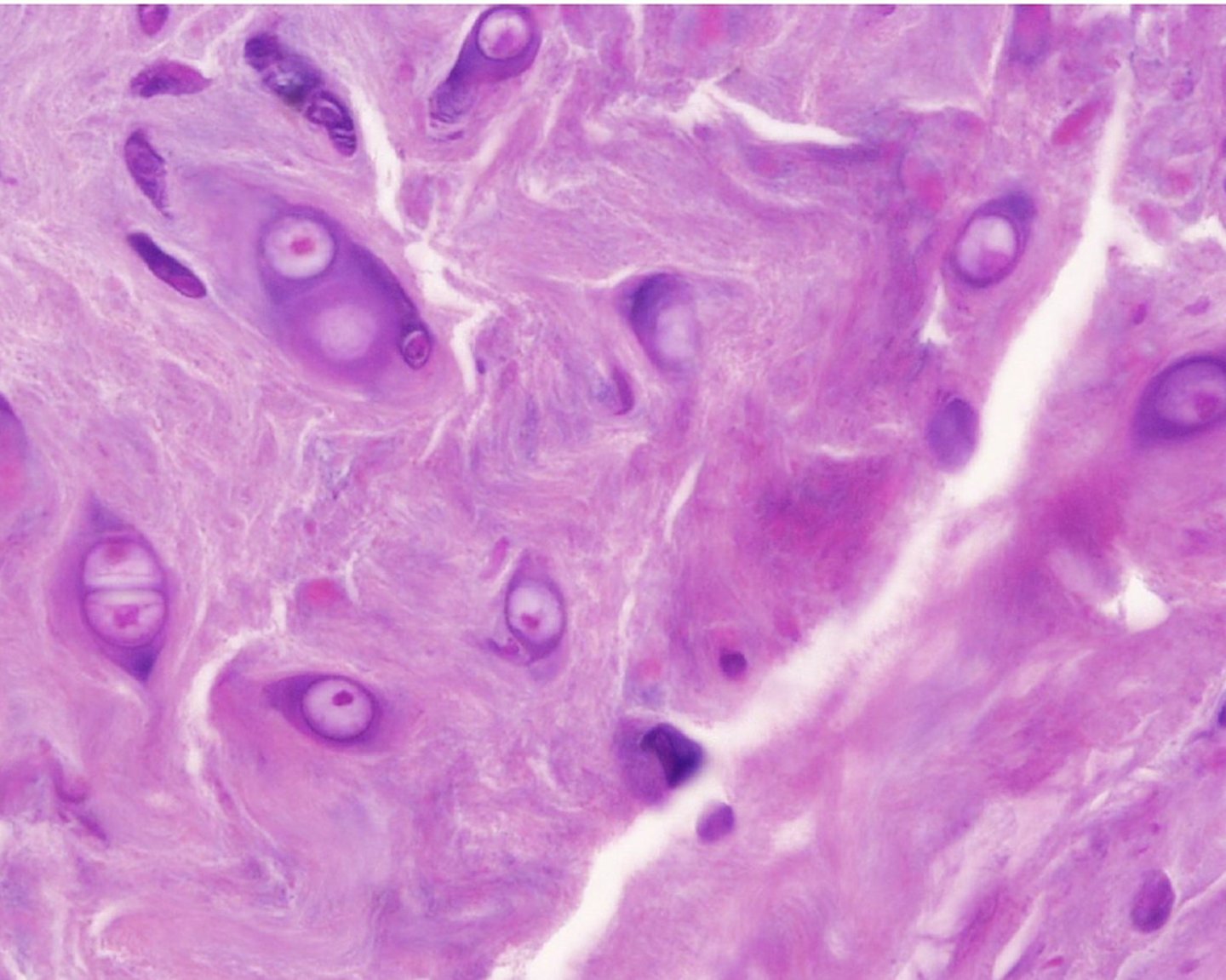
Circumferential lamellae
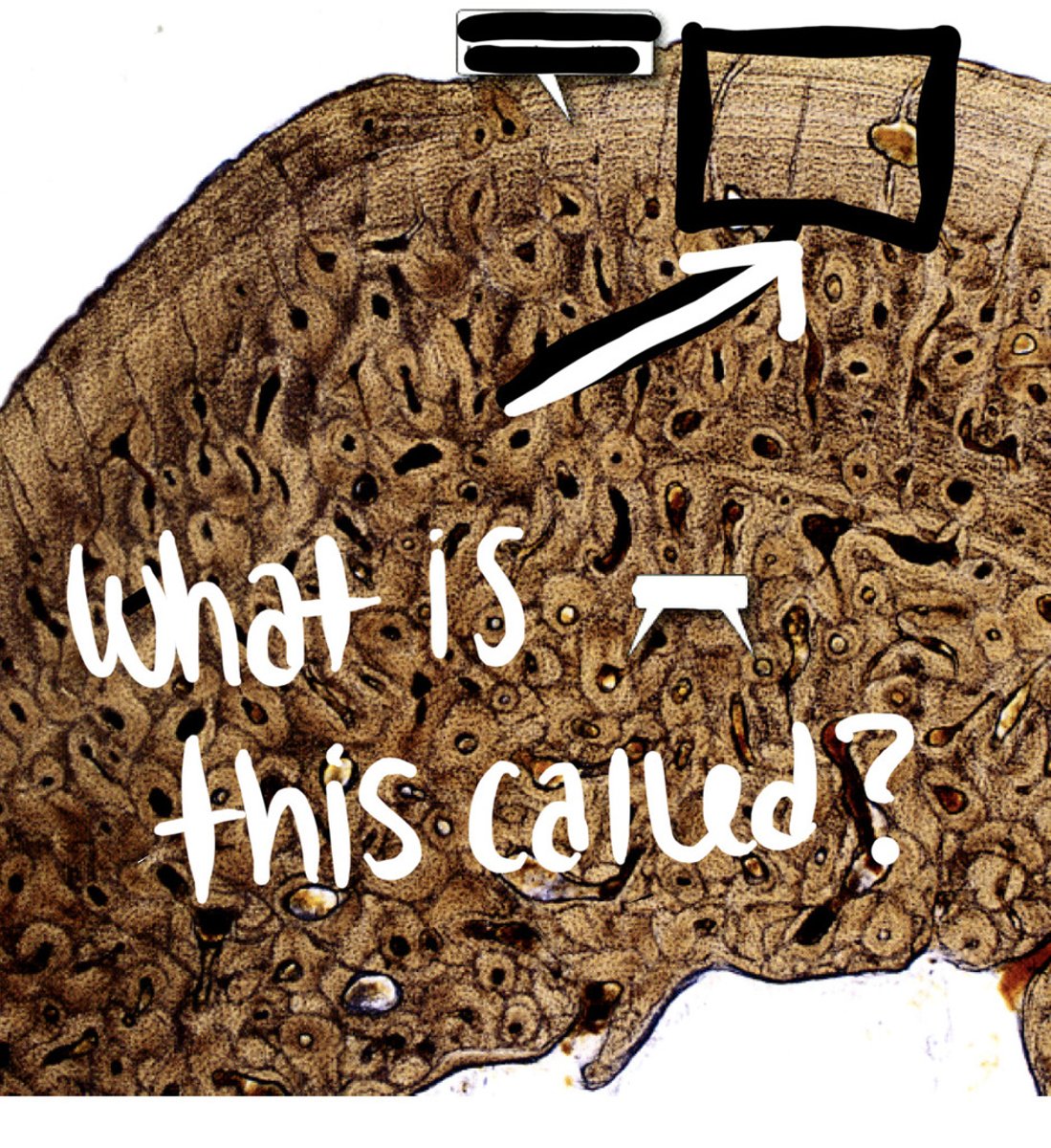
Osteons
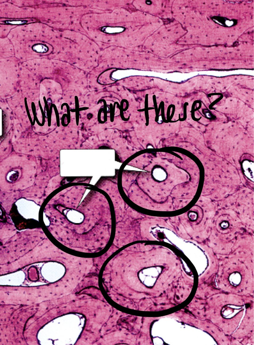
Osteocytes
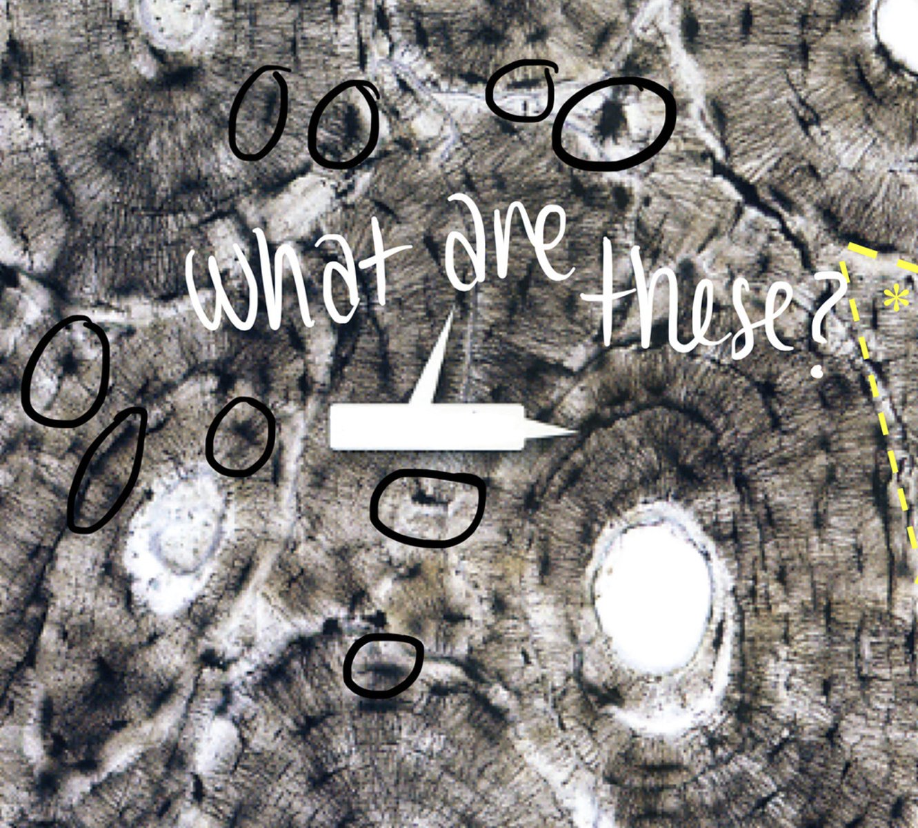
Spongy bone
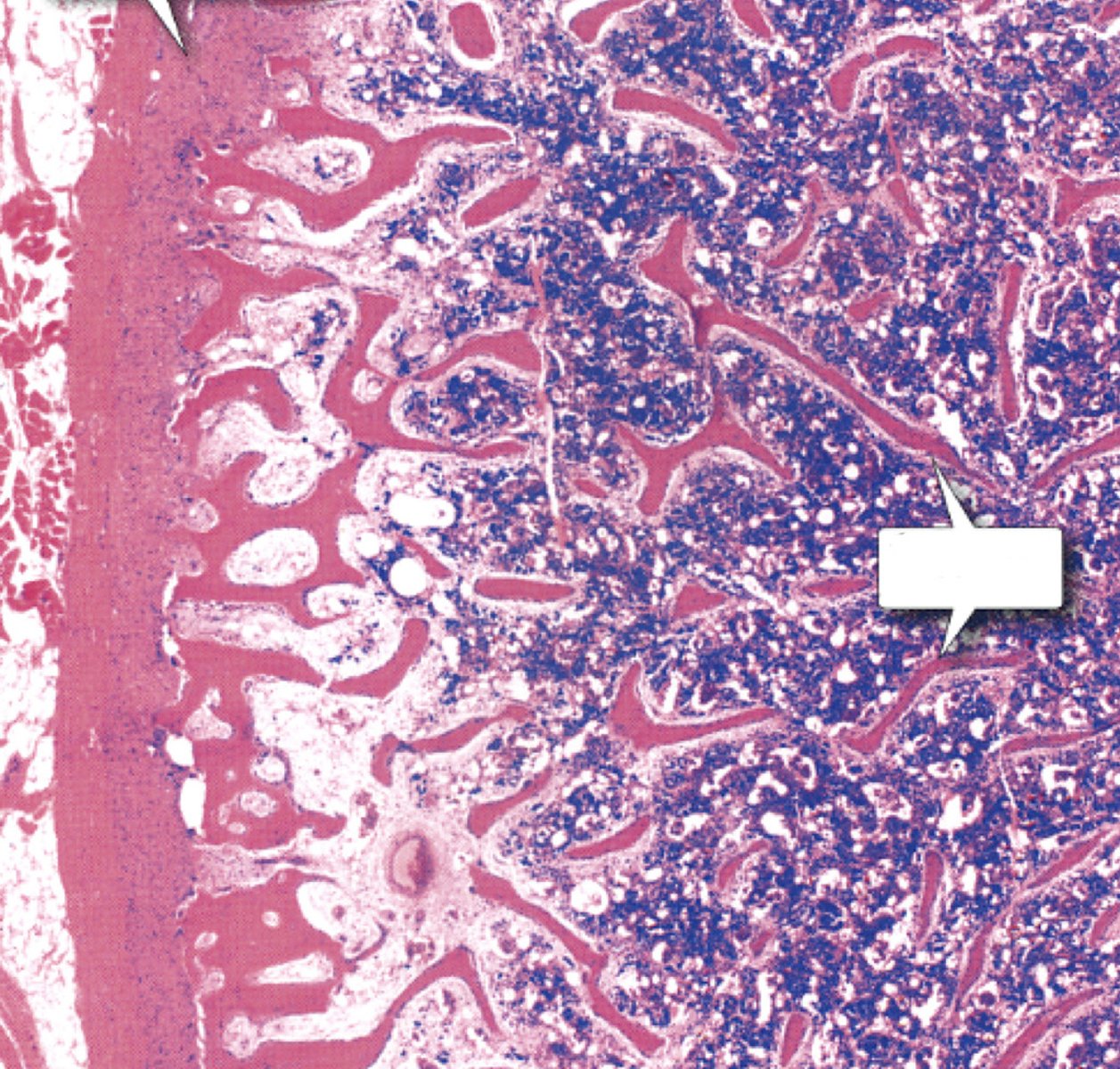
Spongy bone
identify type of bone
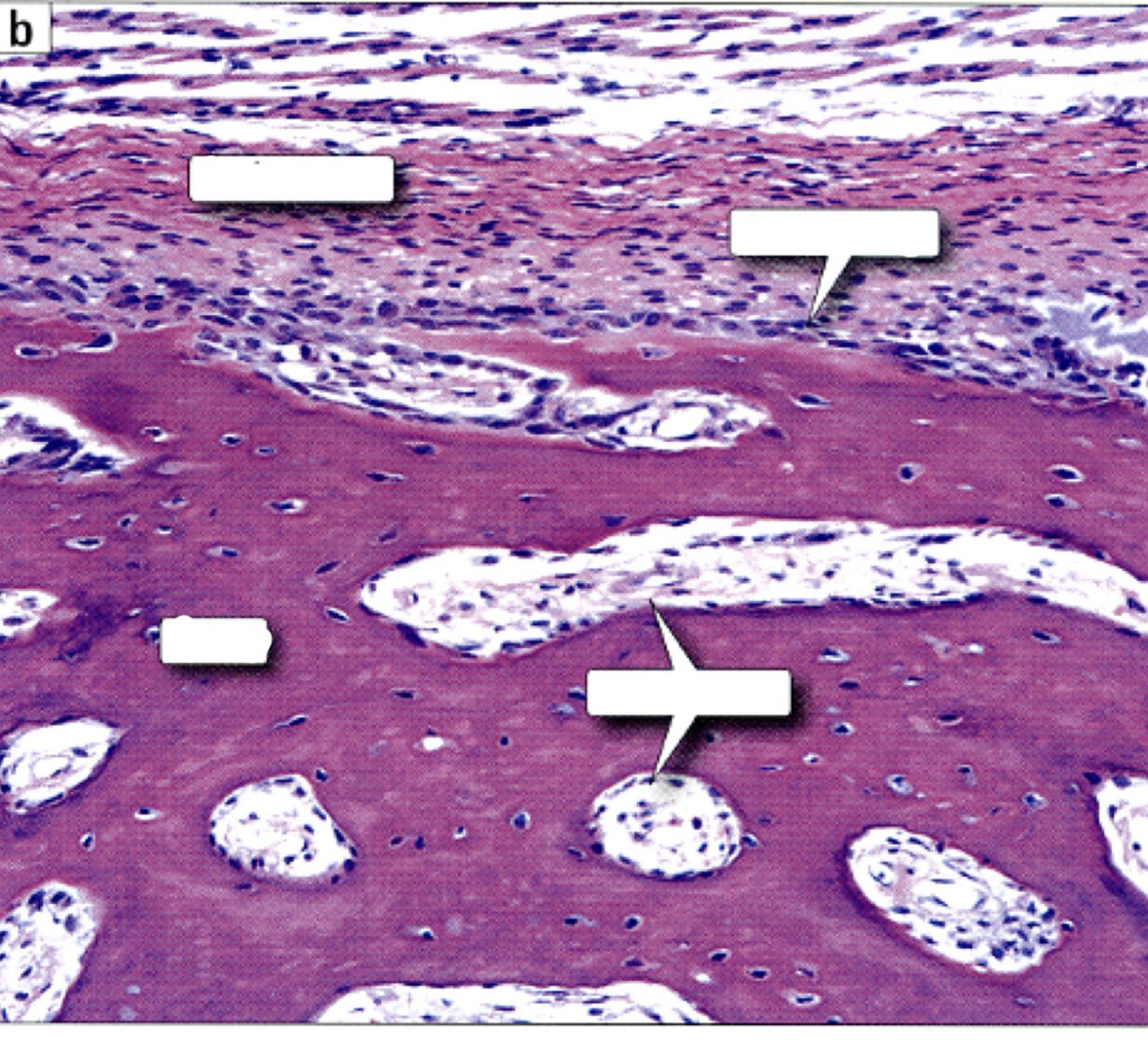
Periosteum
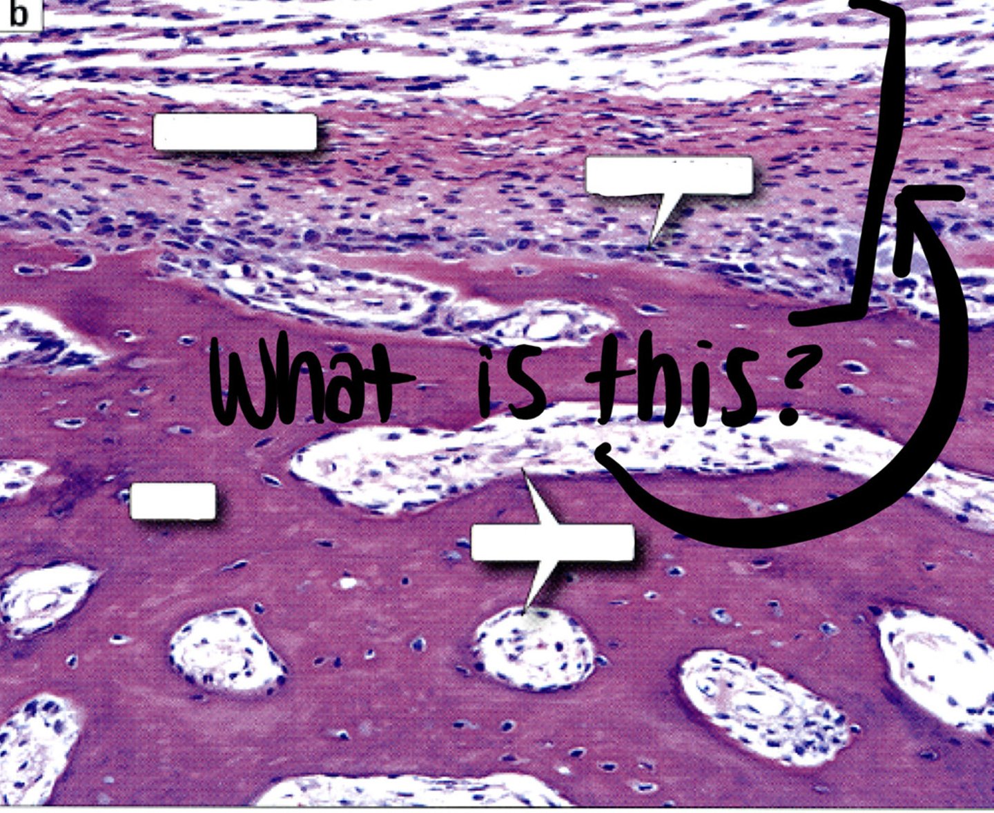
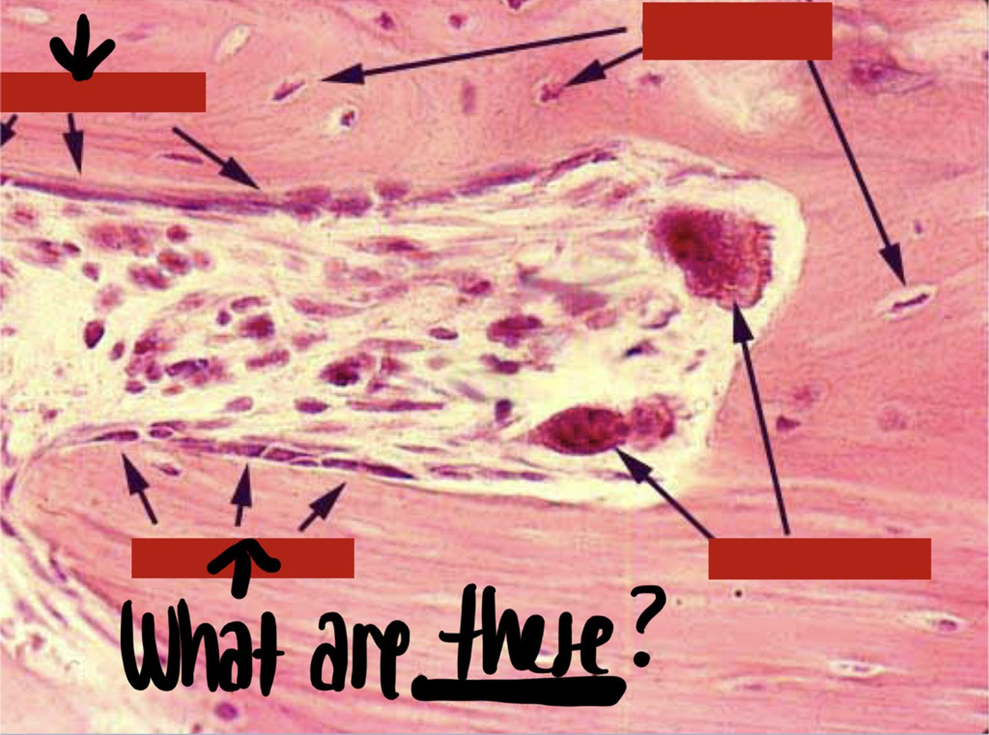
Osteoclasts
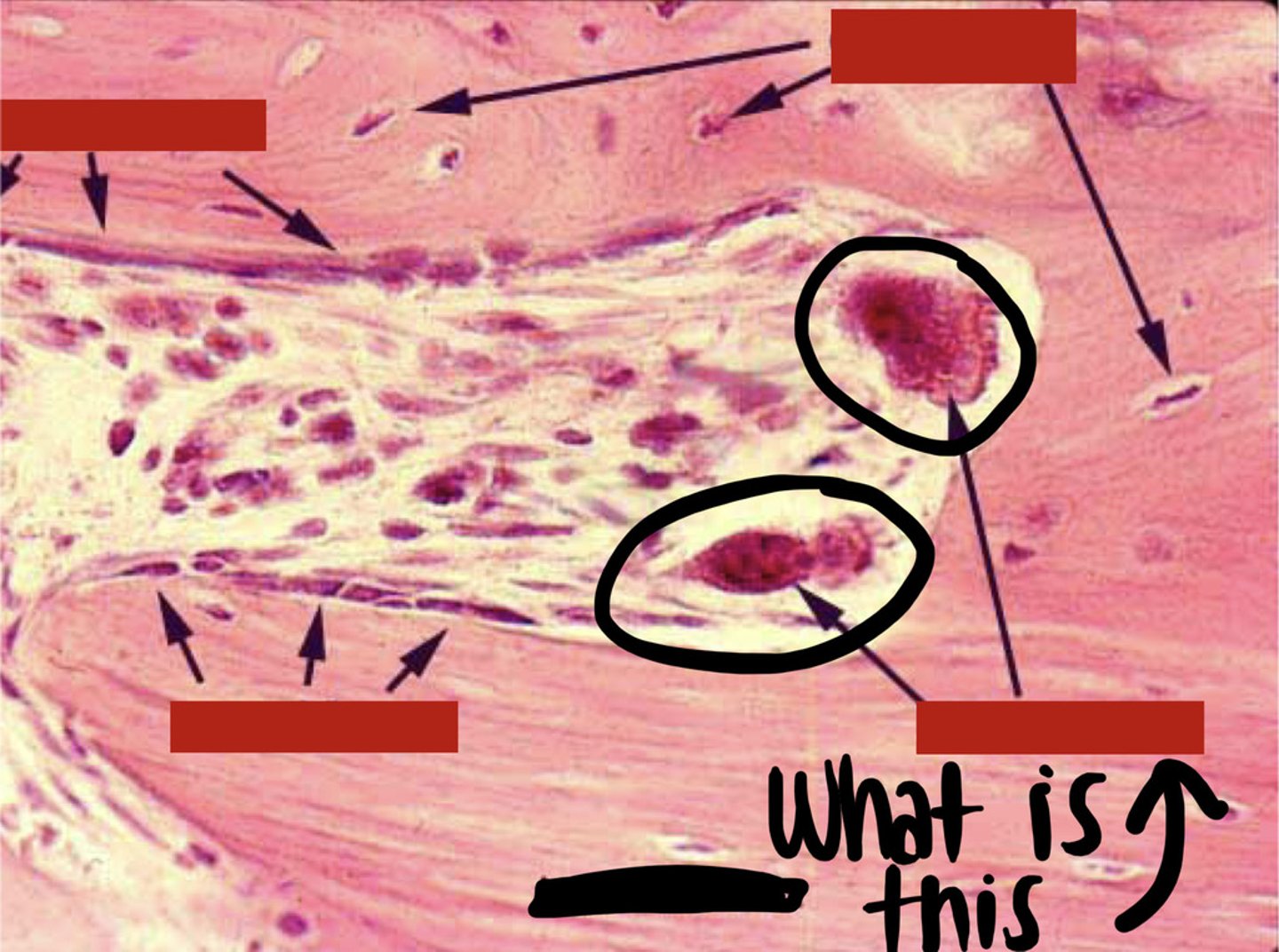
Osteoblasts
Articular cartilage
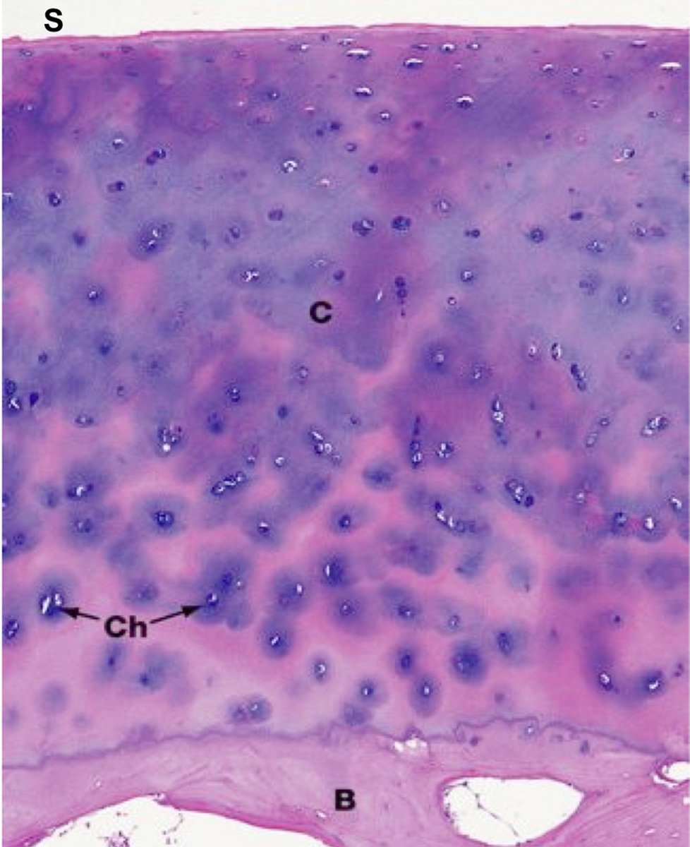
Hyaline cartilage
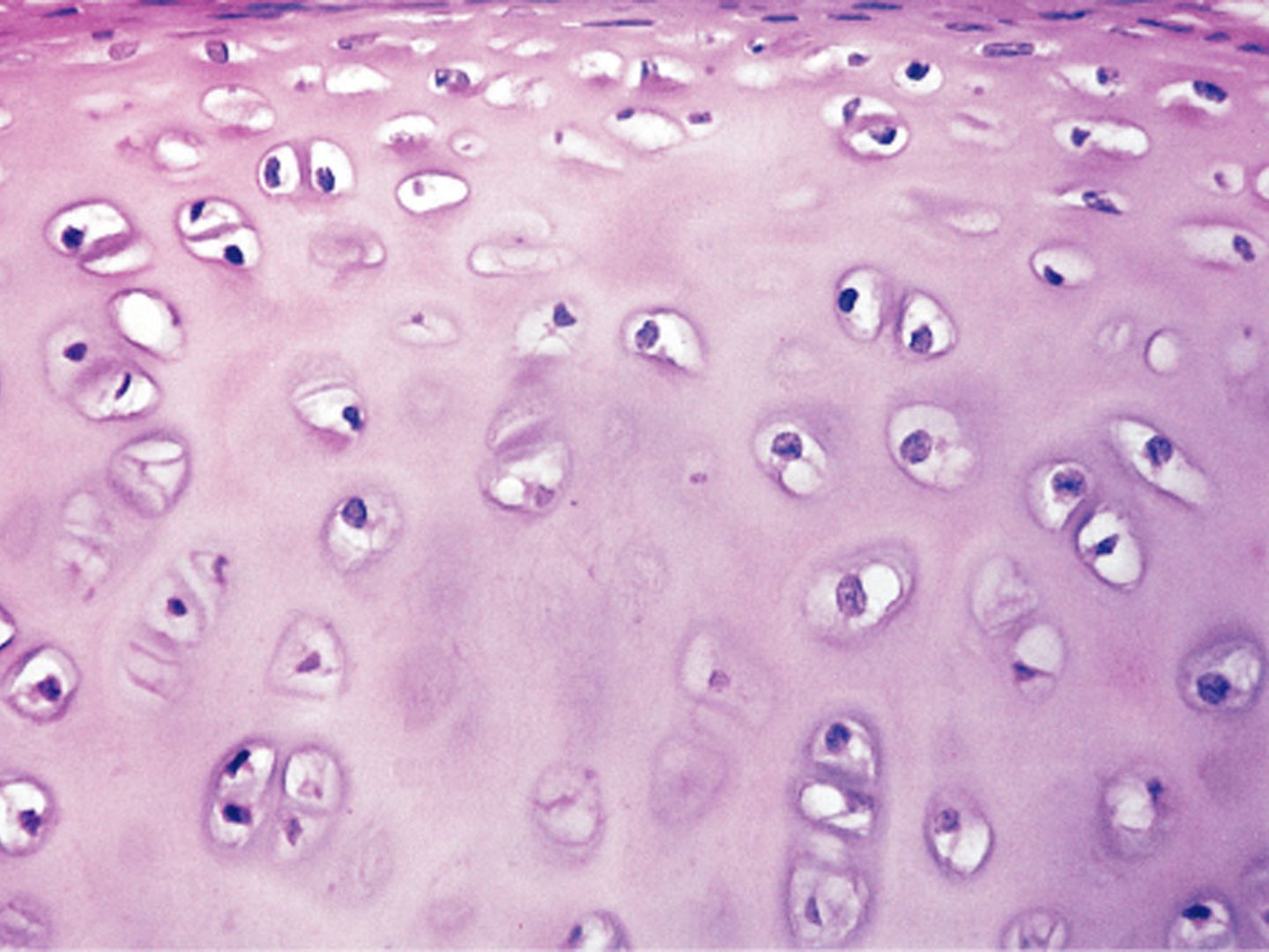
Hyaline cartilage
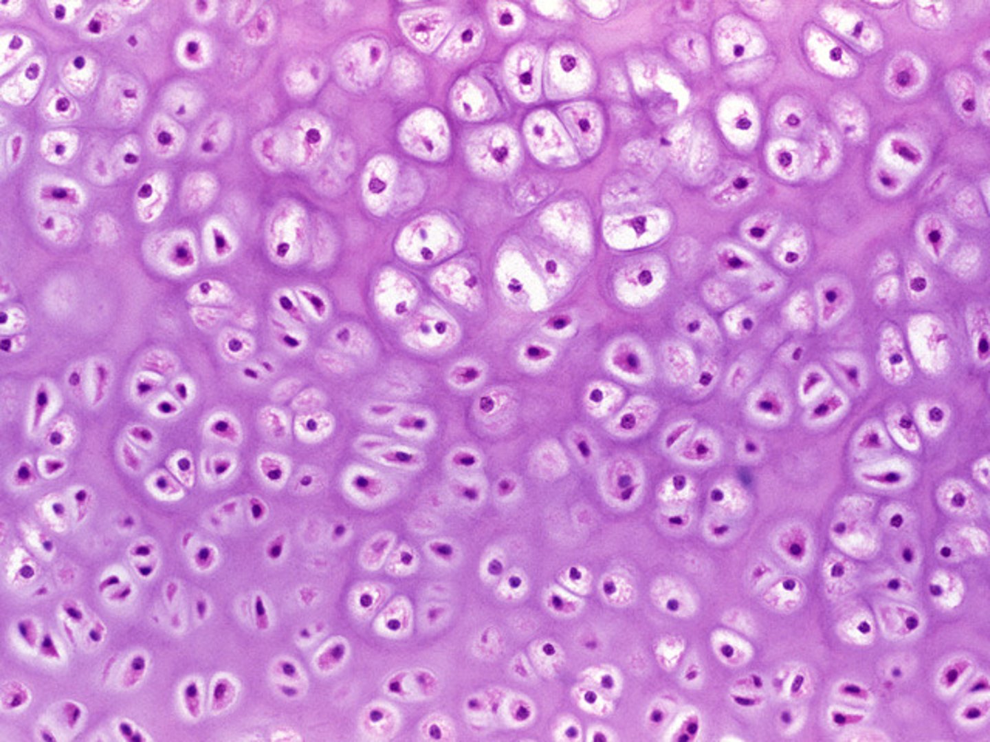
Elastic cartilage
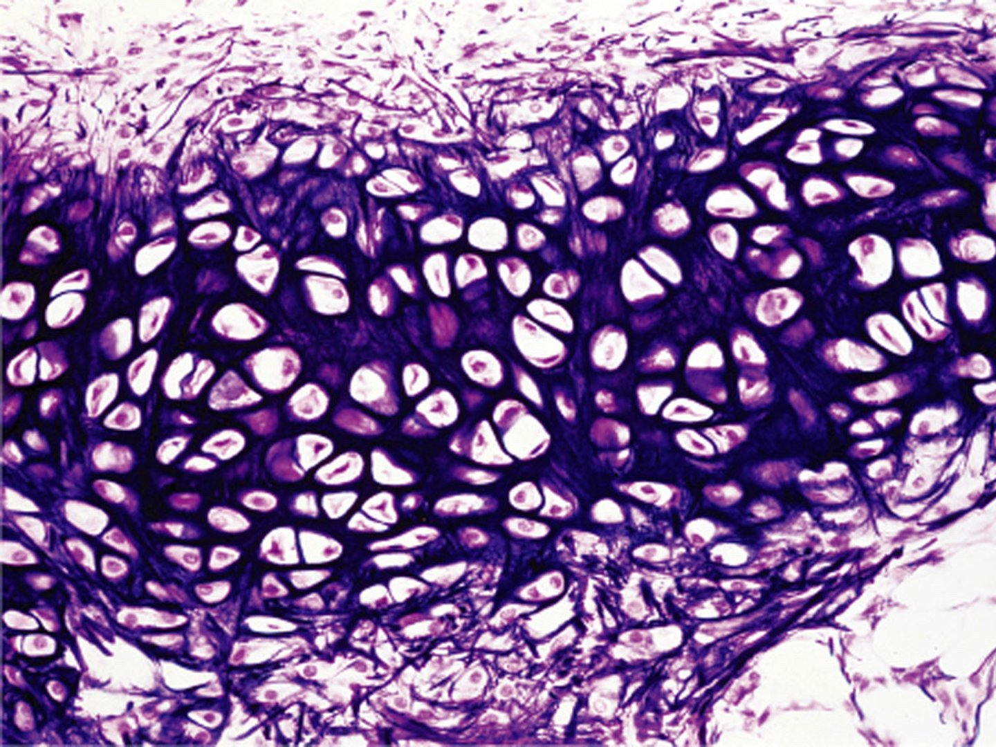
Elastic cartilage
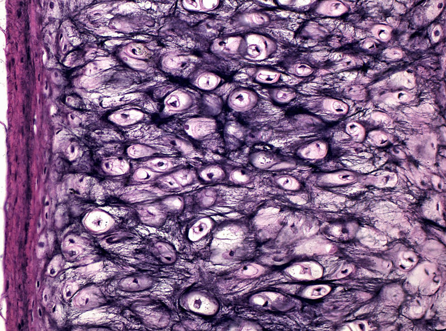
Elastic cartilage
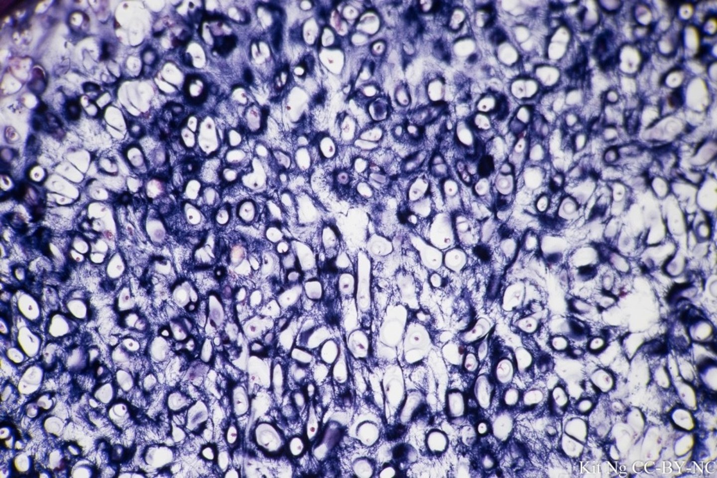
Fibrocartilage
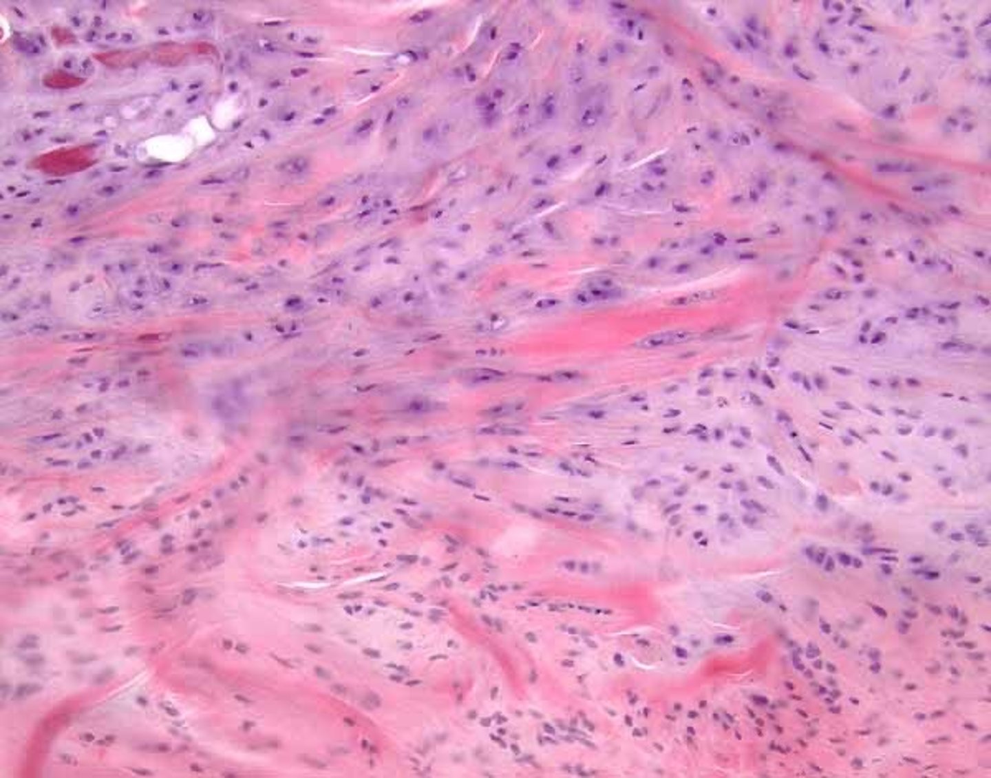
Hyaline cartilage
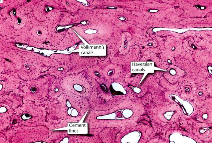
compact bone
identify type of bone
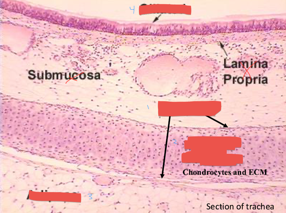
1: perichondrium, 2: hyaline cartilage, 3: adipose, 4: ciliated
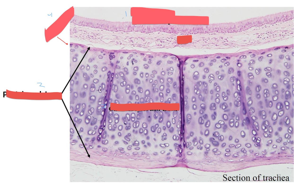
epithelium, 2. perichondrium, 3. chondrocytes and ECM, 4. osteoblast
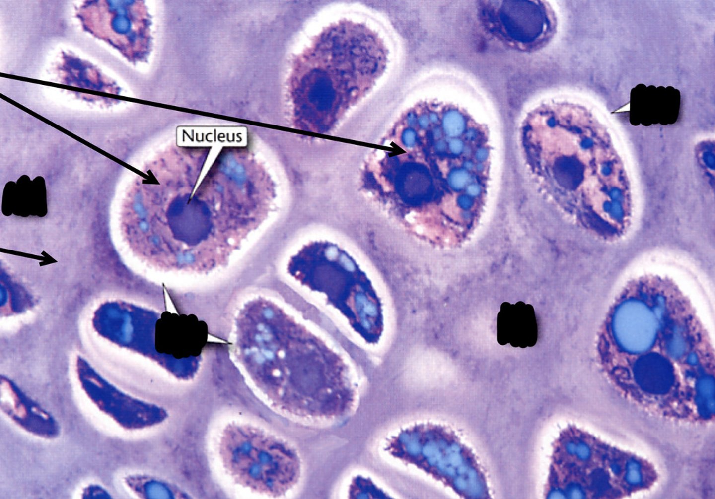
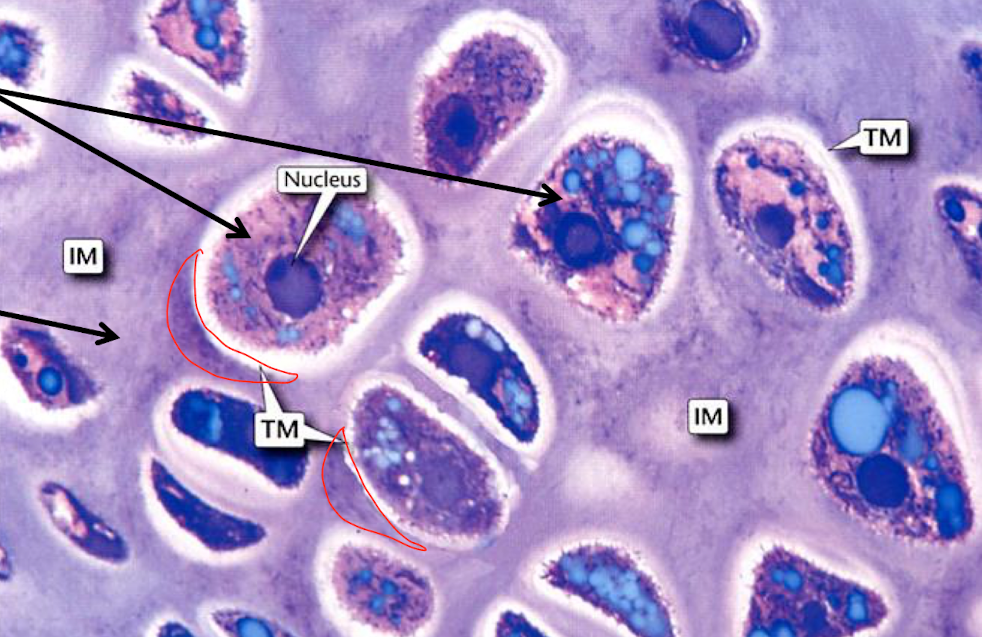
chondrocytes in lacunae, territorial matrix (TM), interterritorial matrix (IM)
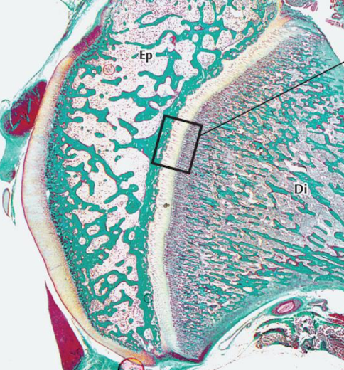
growth plate
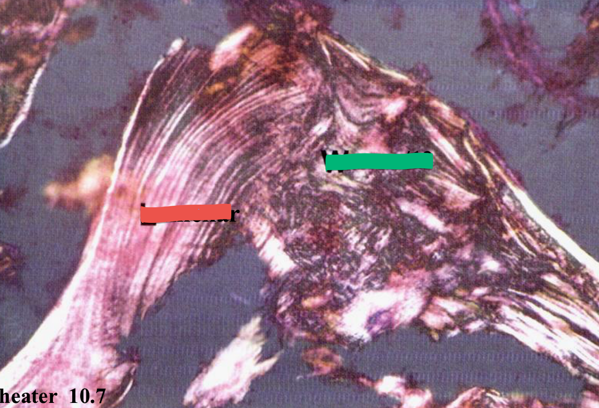
lamellar (red), woven 1 degree (green)
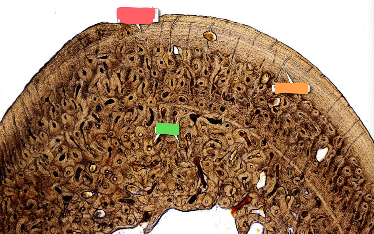
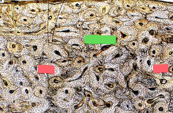
circumferential bone lamellae (red), osteons (green), vasuclar canal (orange)
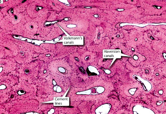
Volkmann’s canals (green), haversian canal (blue)
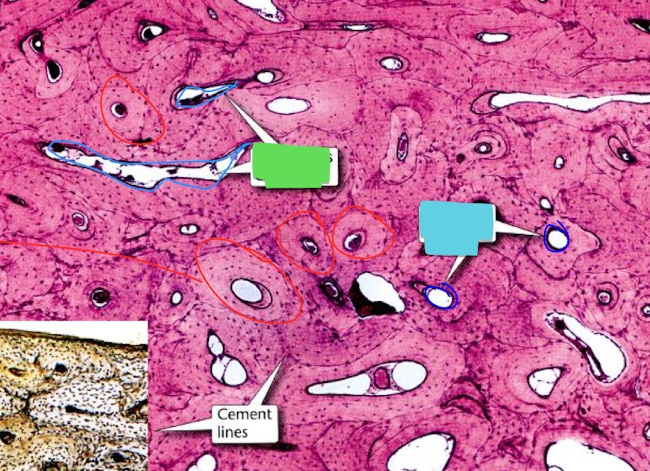
osteon (red), haversian canal (green)
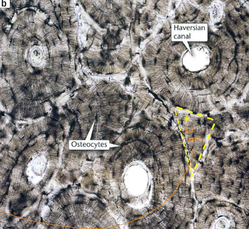
interstitial lamella
yellow triangle
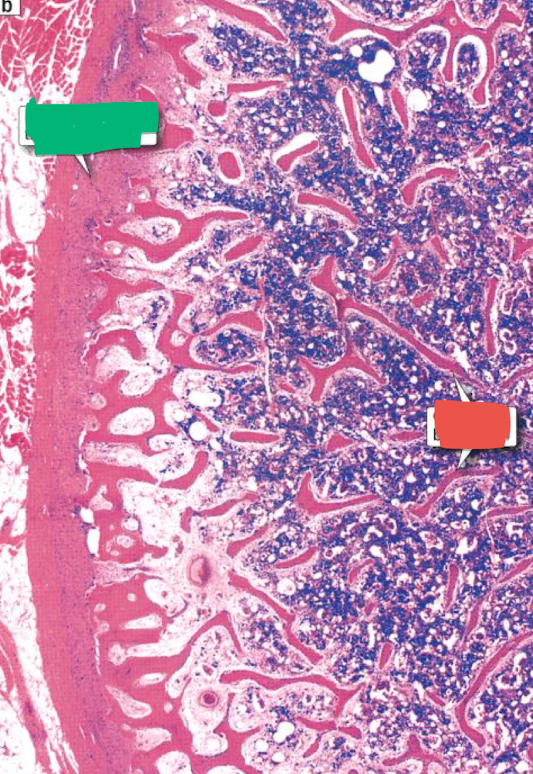
Dense connective tissue (green), Cancellous bone (red)
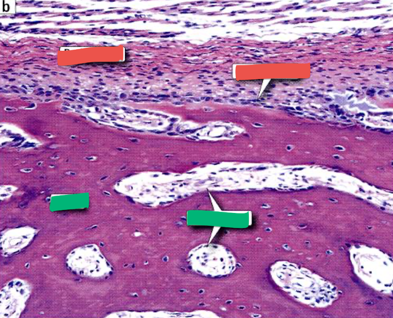
Periosteum (top left red), osteoblast (top right red), bone (left green), endosteum (right green)
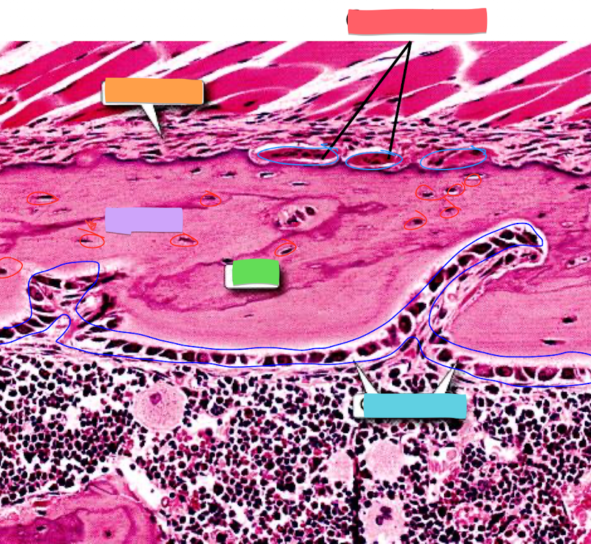
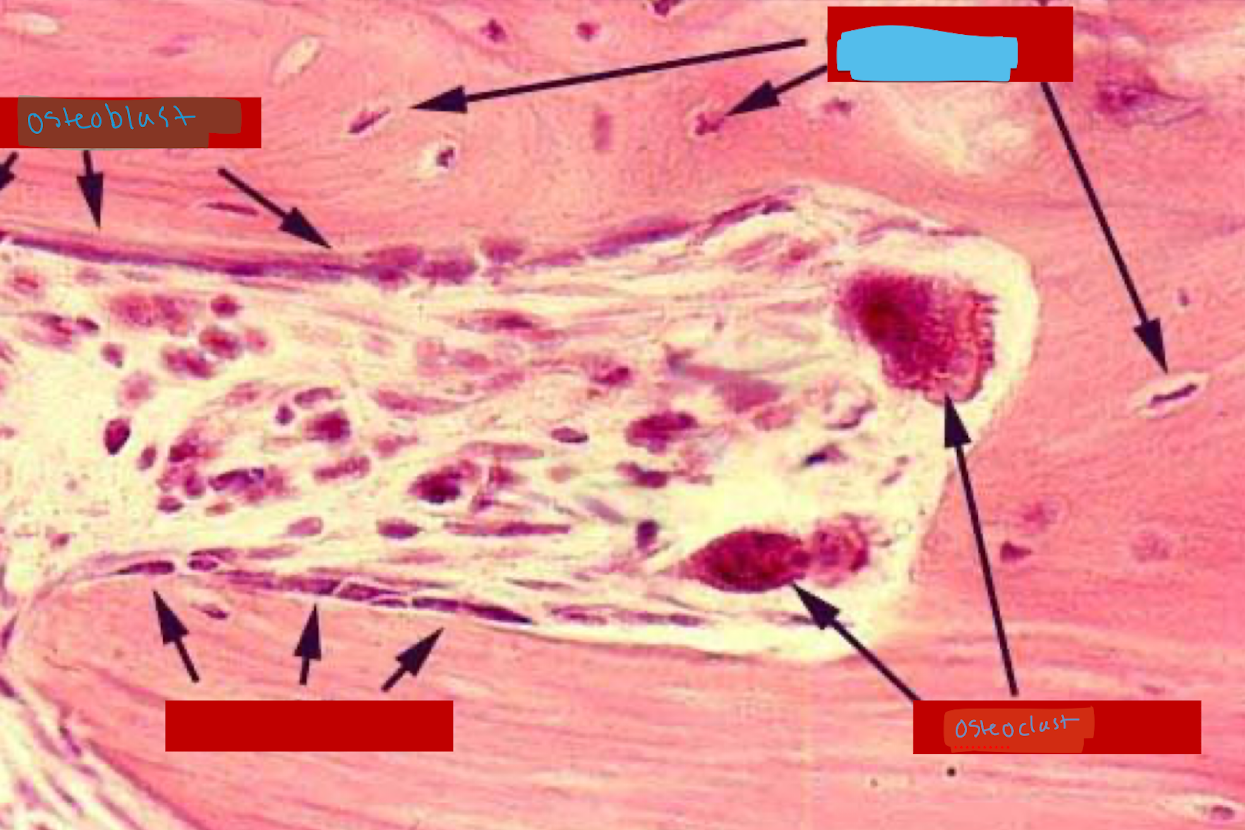
osteoclast (red), periosteum (orange), osteocyte (purple), bone (green), osteoblast (blue)
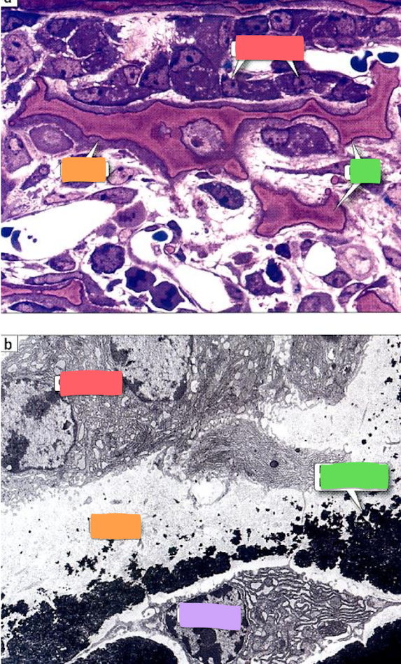
osteoblasts (red), osteoid (orange), bone (green), osteocyte (purple)
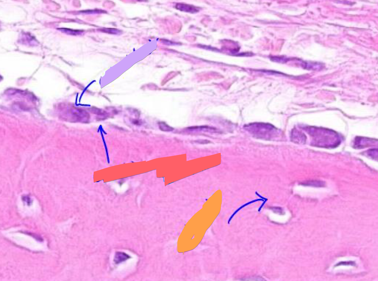
osteoblast (purple), osteoid (red), osteocyte (orange)
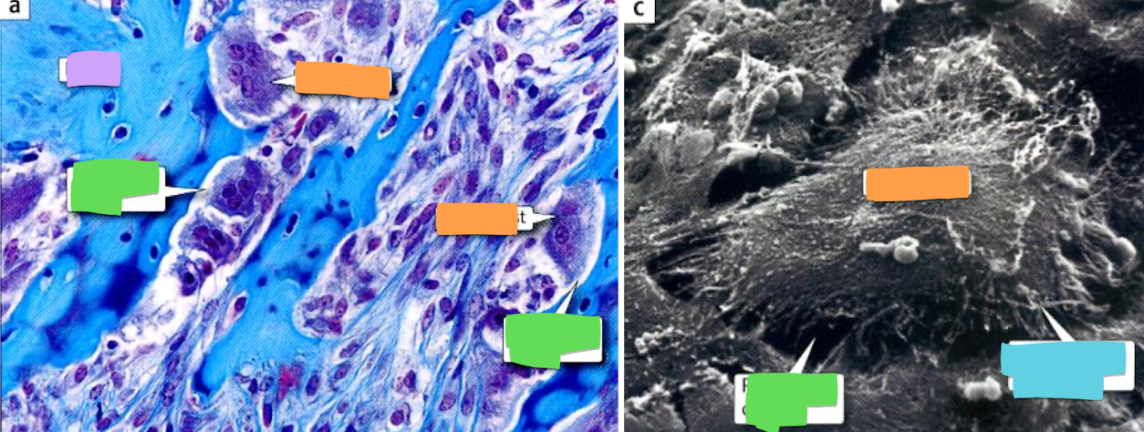
bone (purple), resorption cavity (green), osteoclast (orange), cytoplasmic processes (blue)
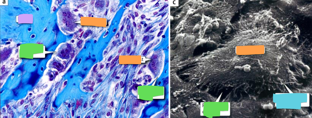
osteocyte
identify blue
compact bone
identify type of bone
Brush Border
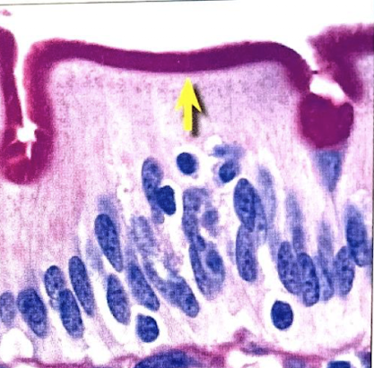
Microvilli
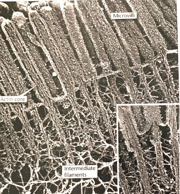
Cilia
cross section
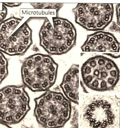
Cilia
top arrow
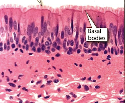
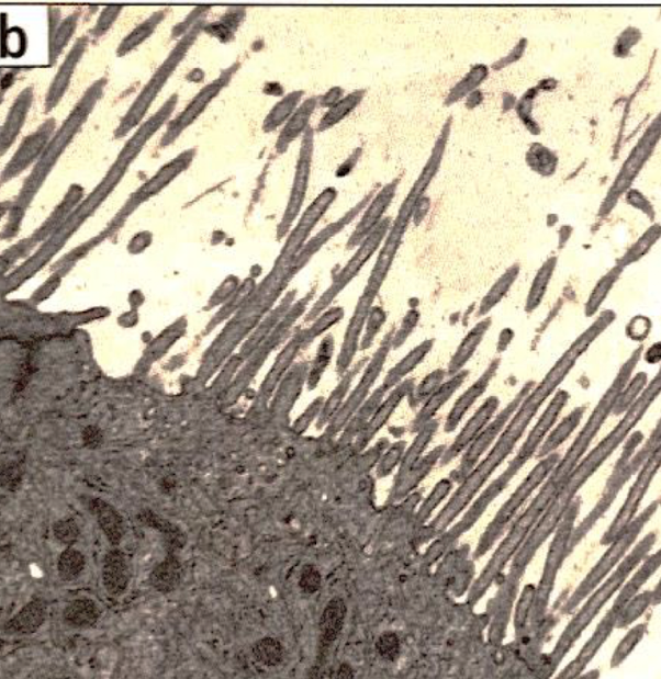
stereocilia (top of epididymis)
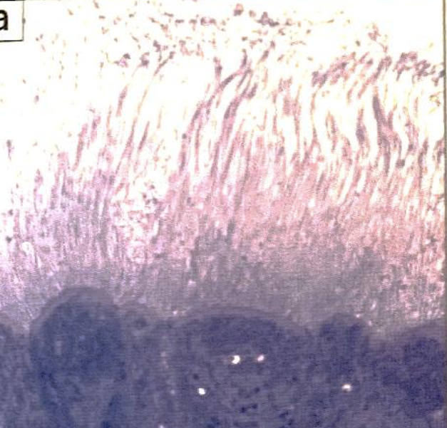
stereocilia (inner ear for balance)
endothelial cell | simple squamous epithelium
function: allows materials to pass by diffusion and filtration in sites where protection is not important
location: epithelium of blood vessels and lymphatic vessls; kidney glomeruli; air sac of lungs
longitudinal
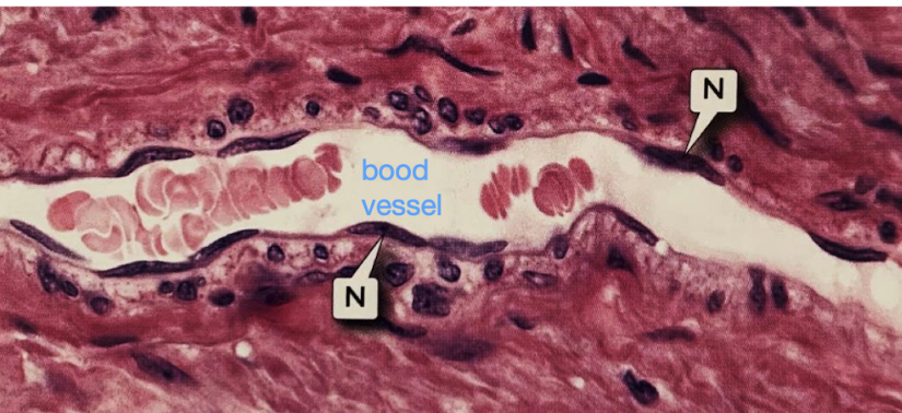
endothelial cell | simple squamous epithelium
function: allows materials to pass by diffusion and filtration in sites where protection is not important
location: epithelium of blood vessels and lymphatic vessls; kidney glomeruli; air sac of lungs
longitudinal
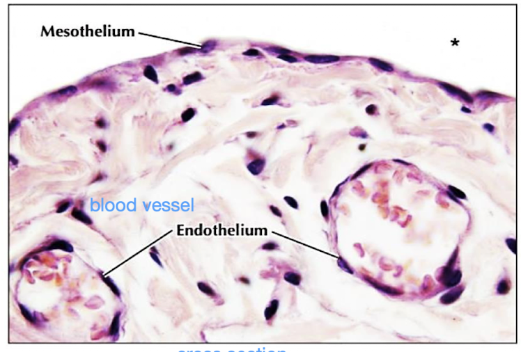
simple cuboidal epithelium
renal tubules
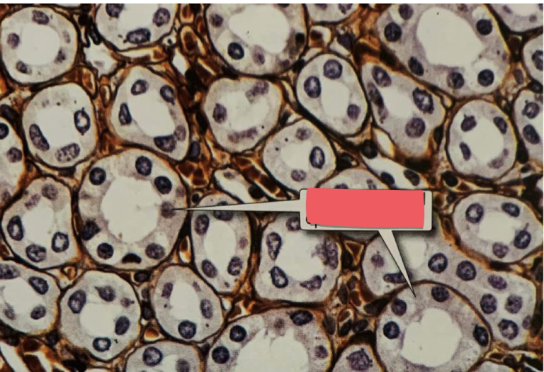
simple cuboidal epithelium
tubules in kidney
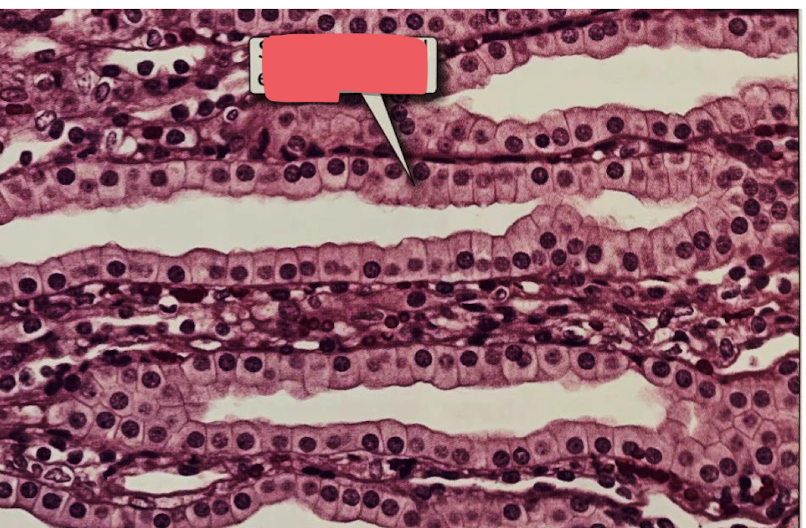
simple columnar epithelium
gallbladder
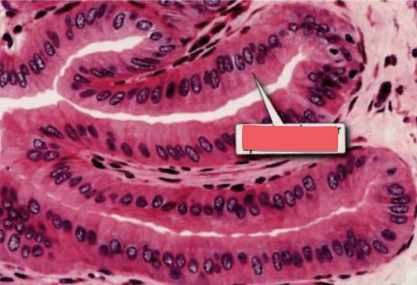
simple columnar epithelium with microvilli
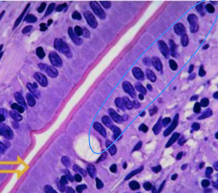
microvilli
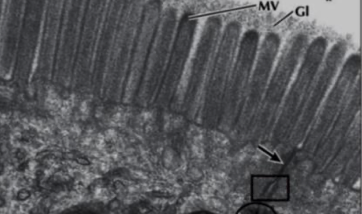
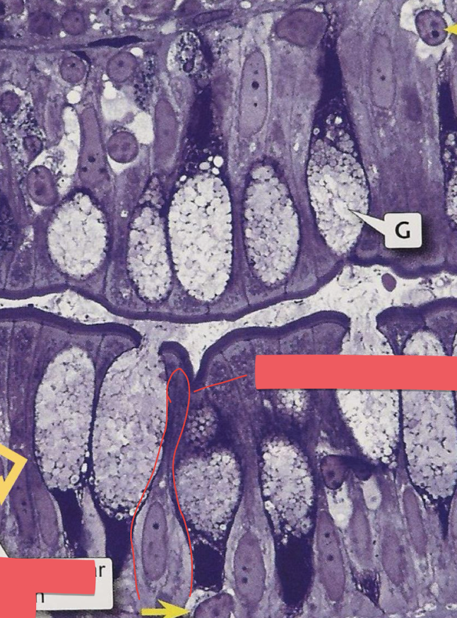
simple columnar cells with goblet cells and microvilli
yellow arrow = goblet cell
simple columnar cell in red border
brush border = thick purple line
pseudostratified ciliated columnar epithelia with goblet cells
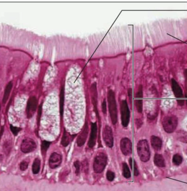
pseudostratified ciliated columnar epithelia with goblet cells
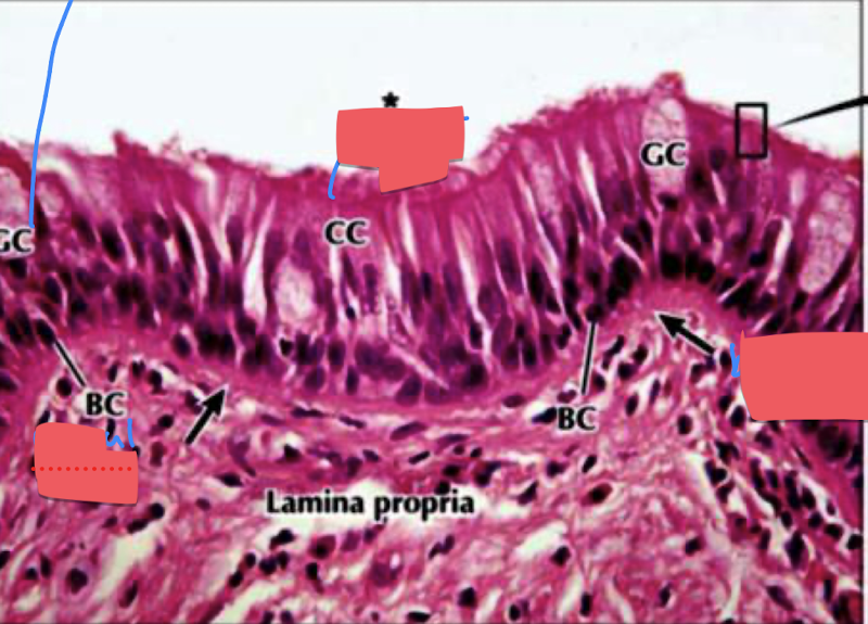
stratified squamous epithelium nonkeratinized
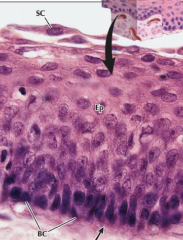
stratified squamous epithelium keratinized
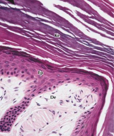
stratified squamous epithelium keratinized
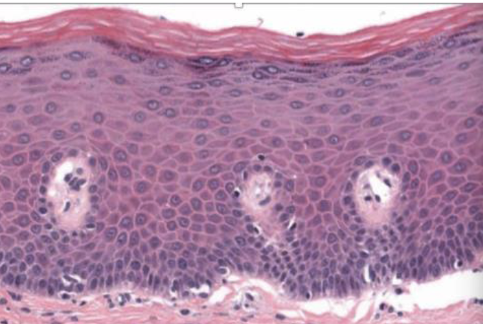
stratified squamous epithelium nonkeratinized
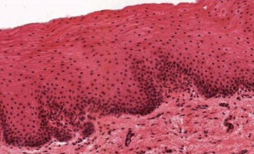
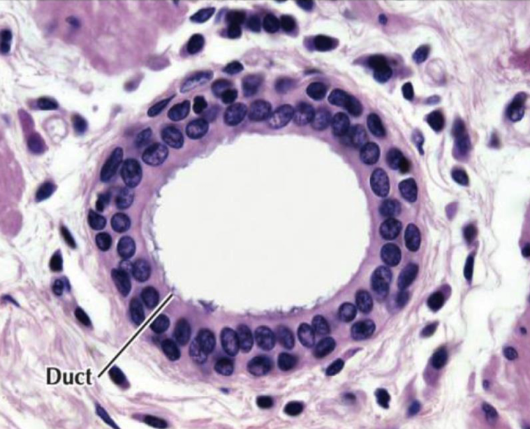
straitifed cuboidal epithelium
duct of salivary glands
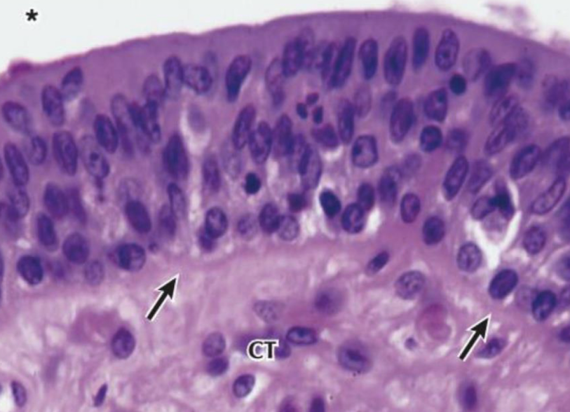
stratified columnar epithelium
male urethra, pharnyx
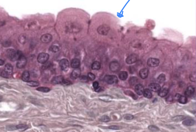
transitional epithelium relaxed
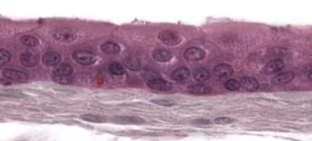
transitional epithelium extended state
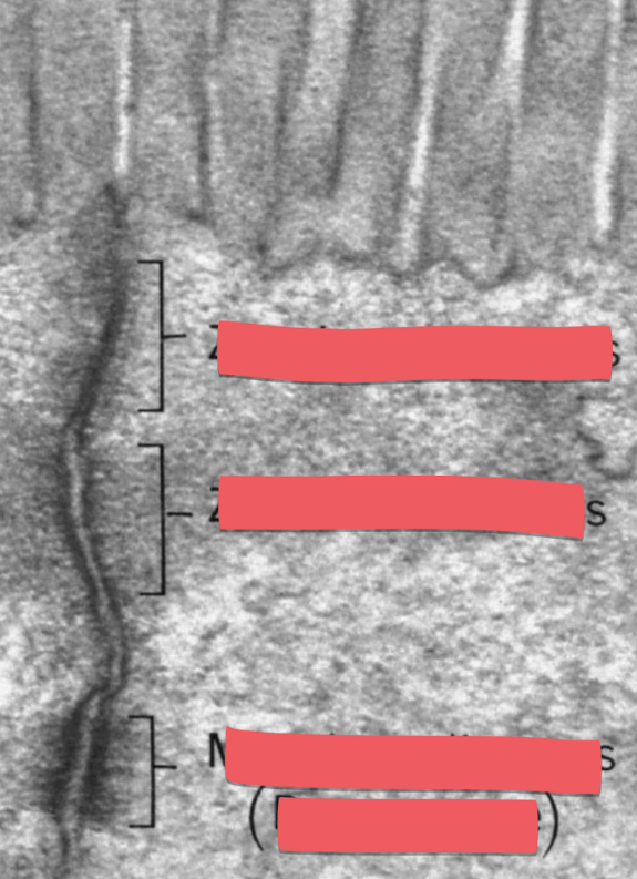
zonula adherens (bottom), zonula occludens (top)
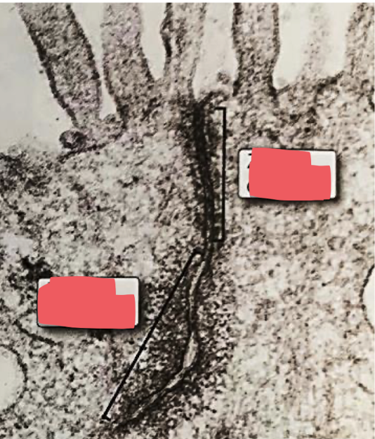
adherens junction
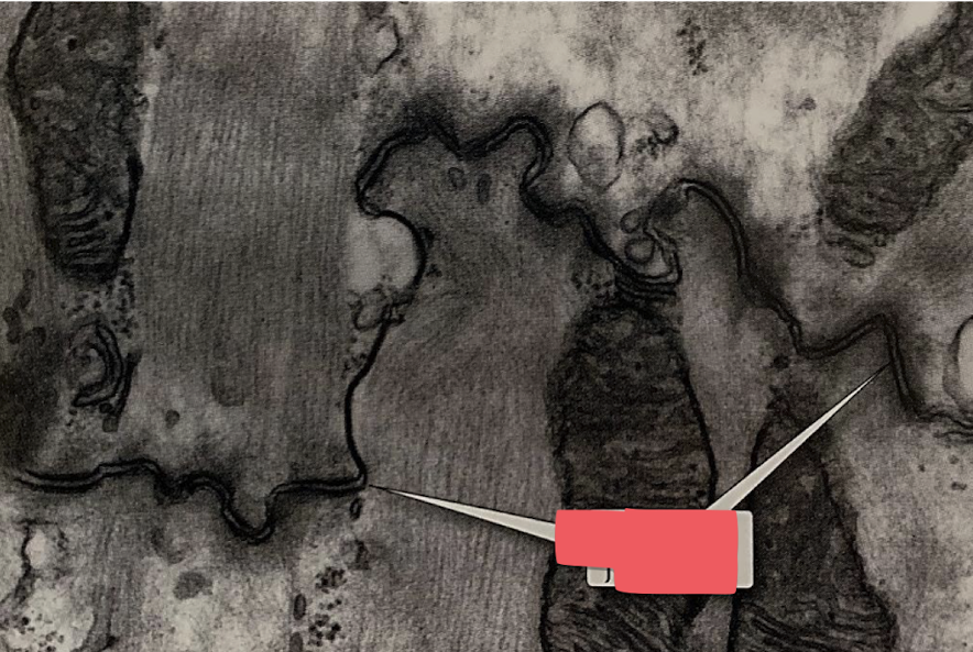
desmosomes
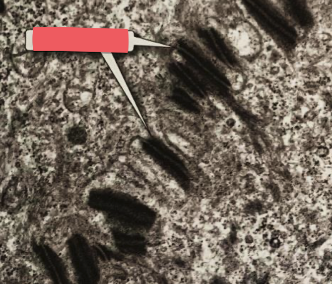
gap junction
lines across very hard to see
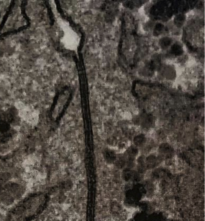
zonula occludens (top), zonula adherens (middle), macula adherens (desmosome bottom)
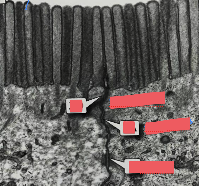
blue arrow (microvilli)
zonula occludens
zona adherens
desmosome
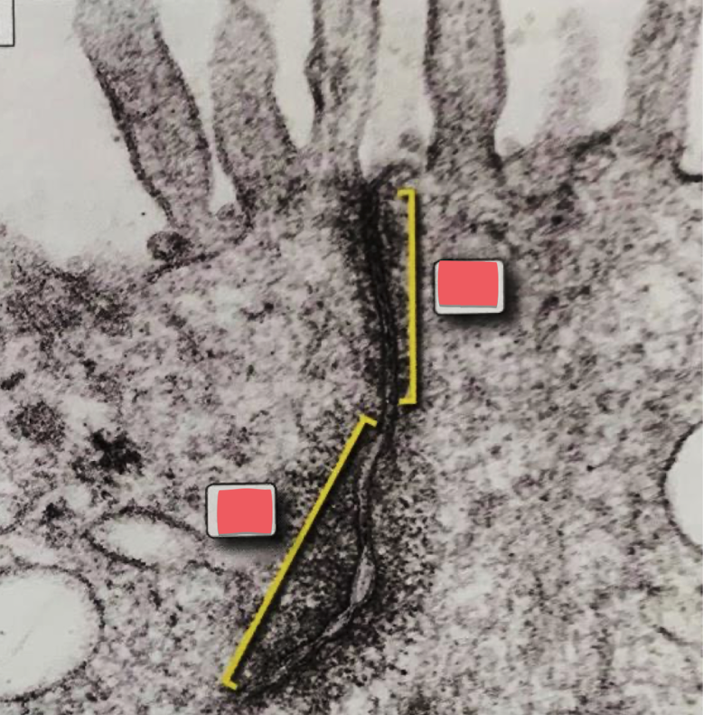
top: zonula occludens (tight junction)
bottom: zonula adherens
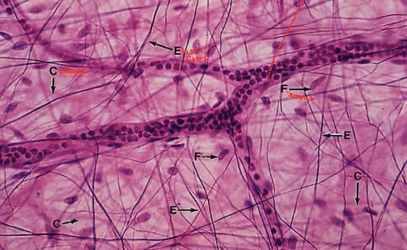
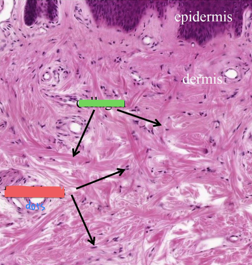
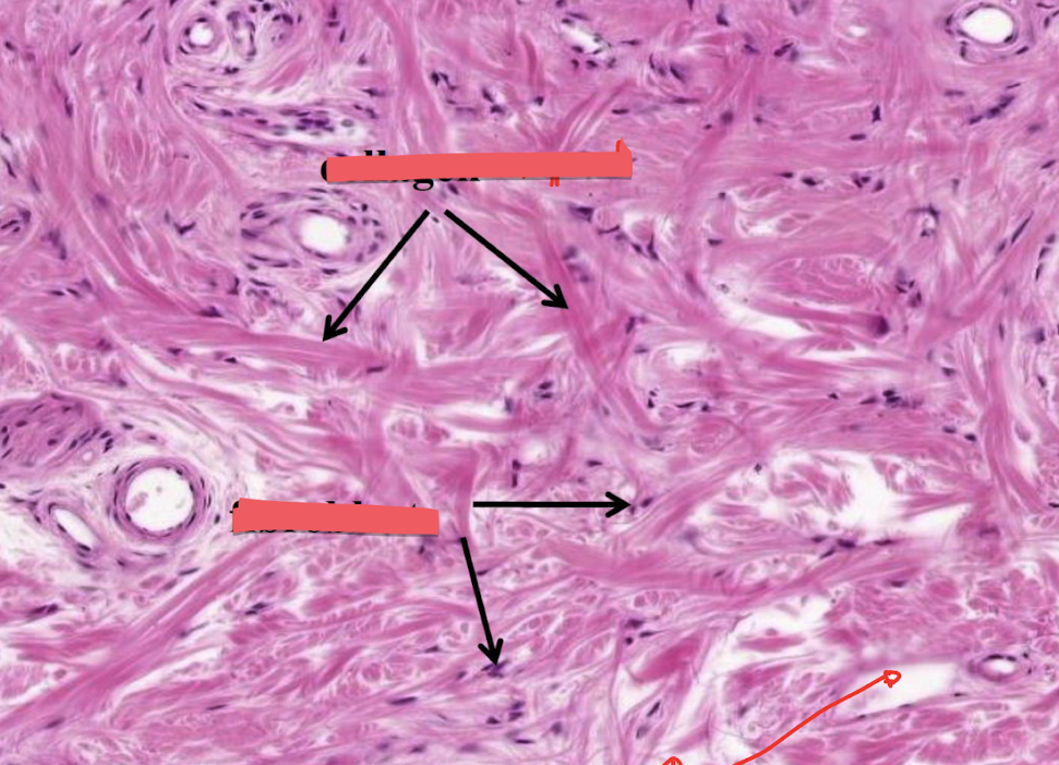
collagen fibers (red box), fibroblast (green box)
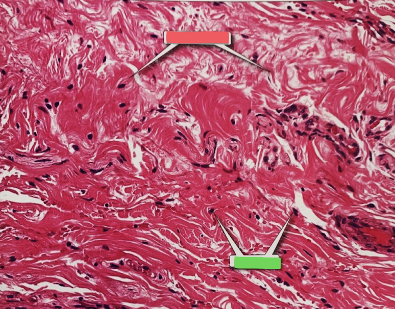
collagen (green box), fibroblasts (red box)
mature fibroblast
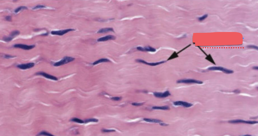
collagen fiber (with mature fibroblast)
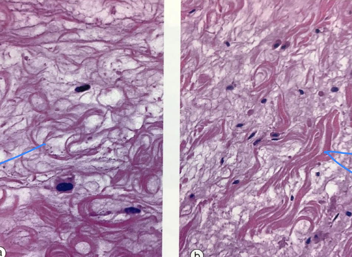
pseudopodium (red box), whole thing is a macrophage
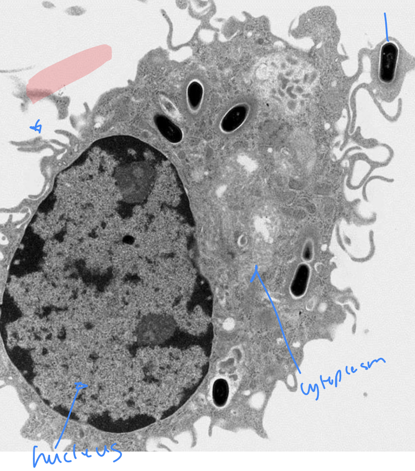
Macrophages
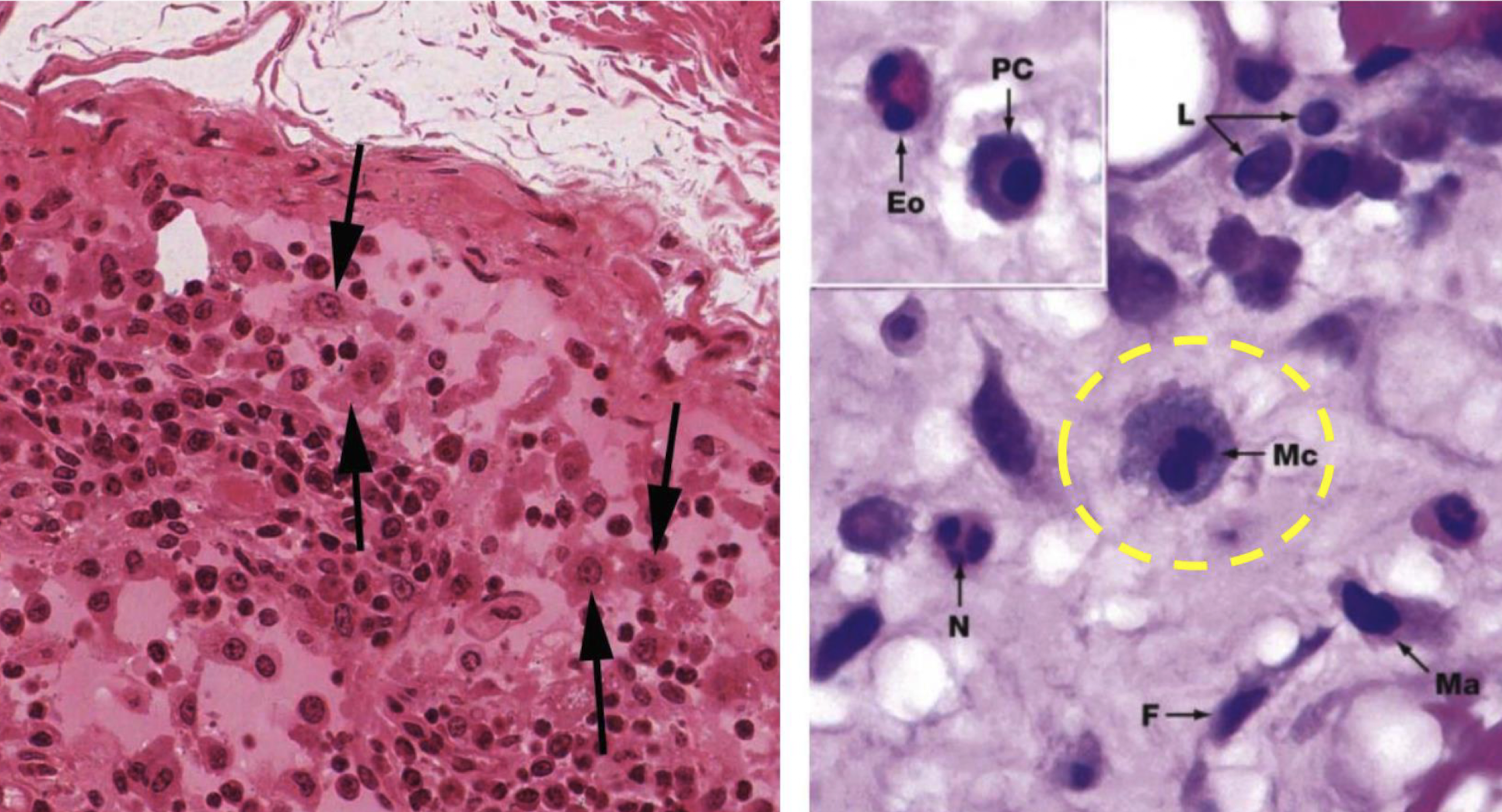
mast cell
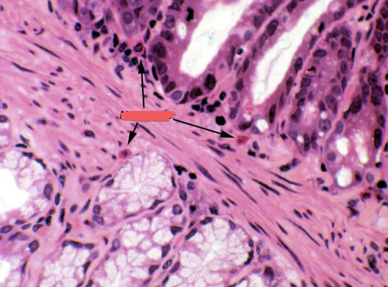
look at the dark pink circles for the right image
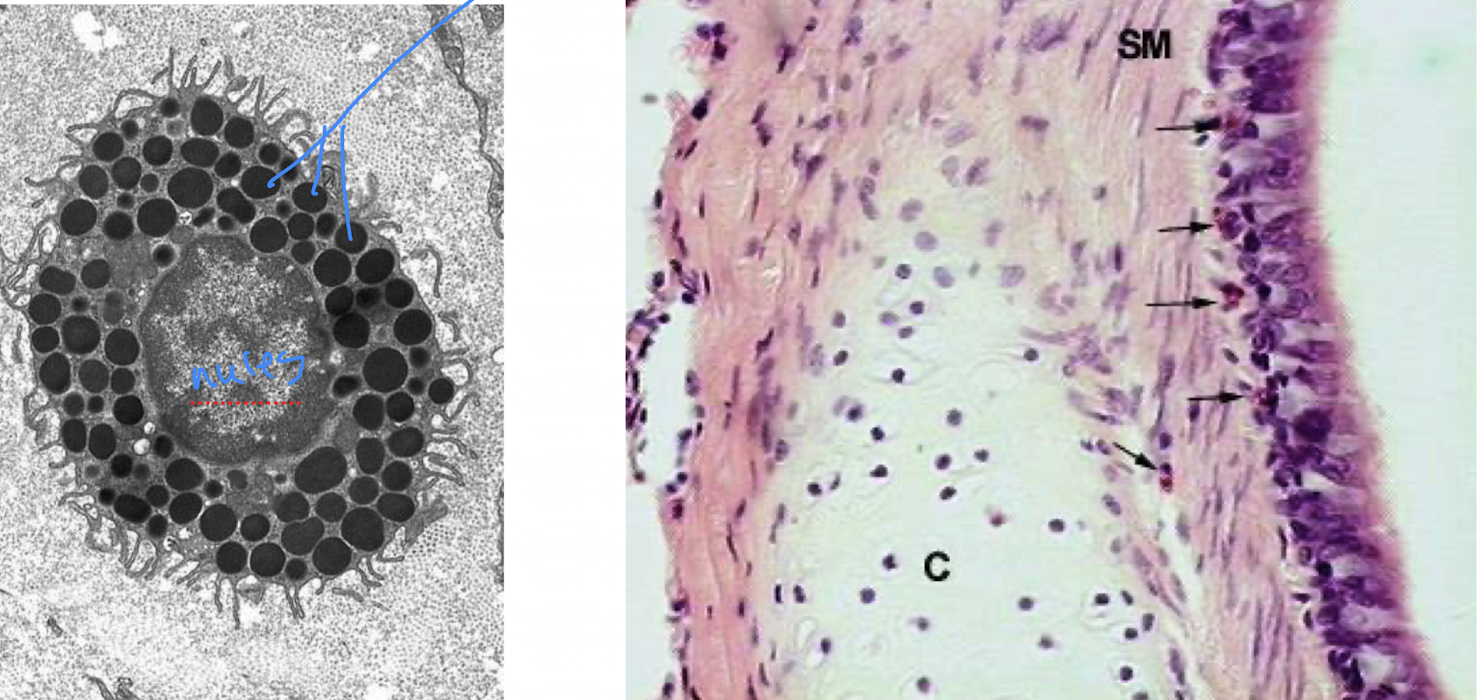
Mast cells
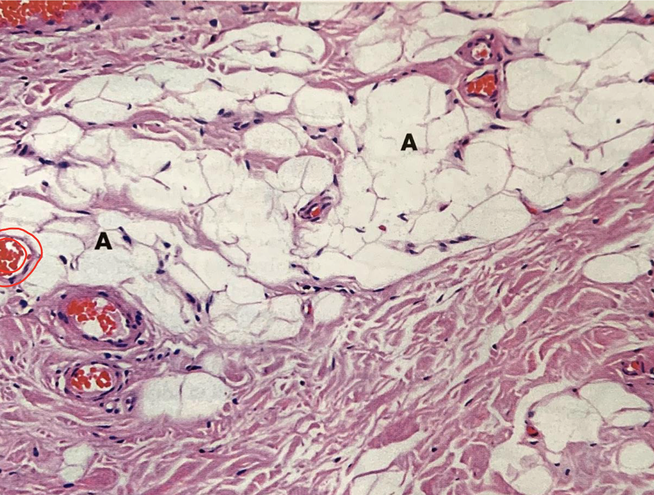
adipocytes
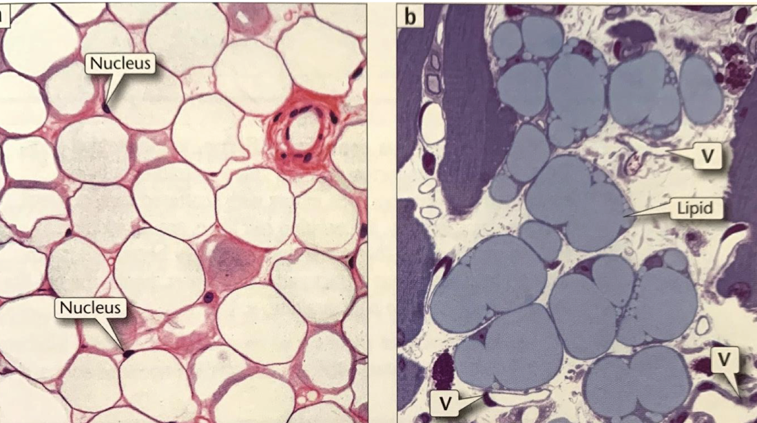
simple squamous epithelium (red border), blood vessels with erythryoctes (blue arrow), aidpocytes (A)
identify red, blue, and A site
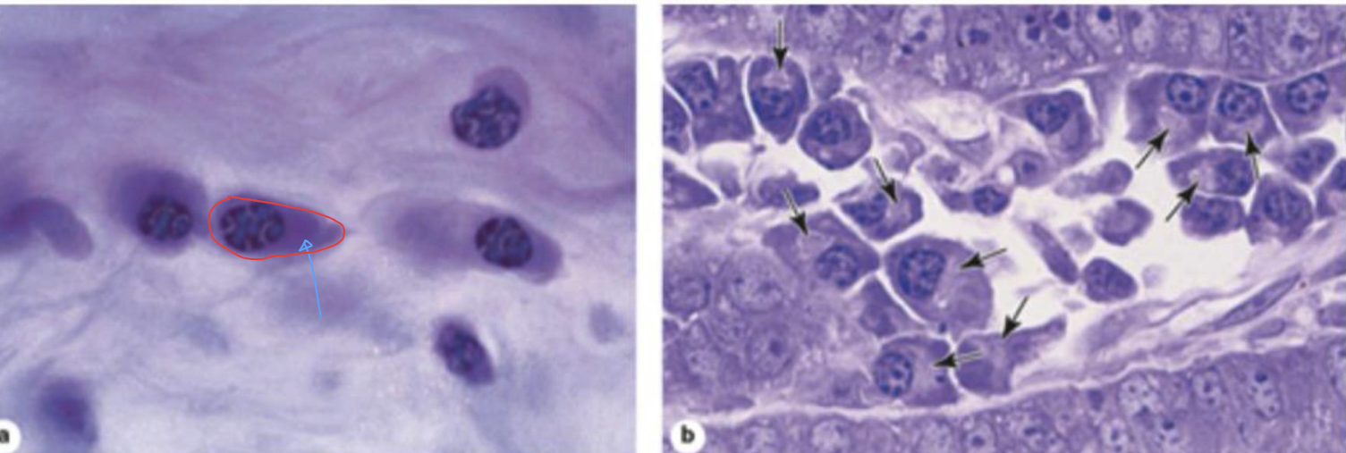
plasma cells, blue arrow (basophilic cytoplasm)
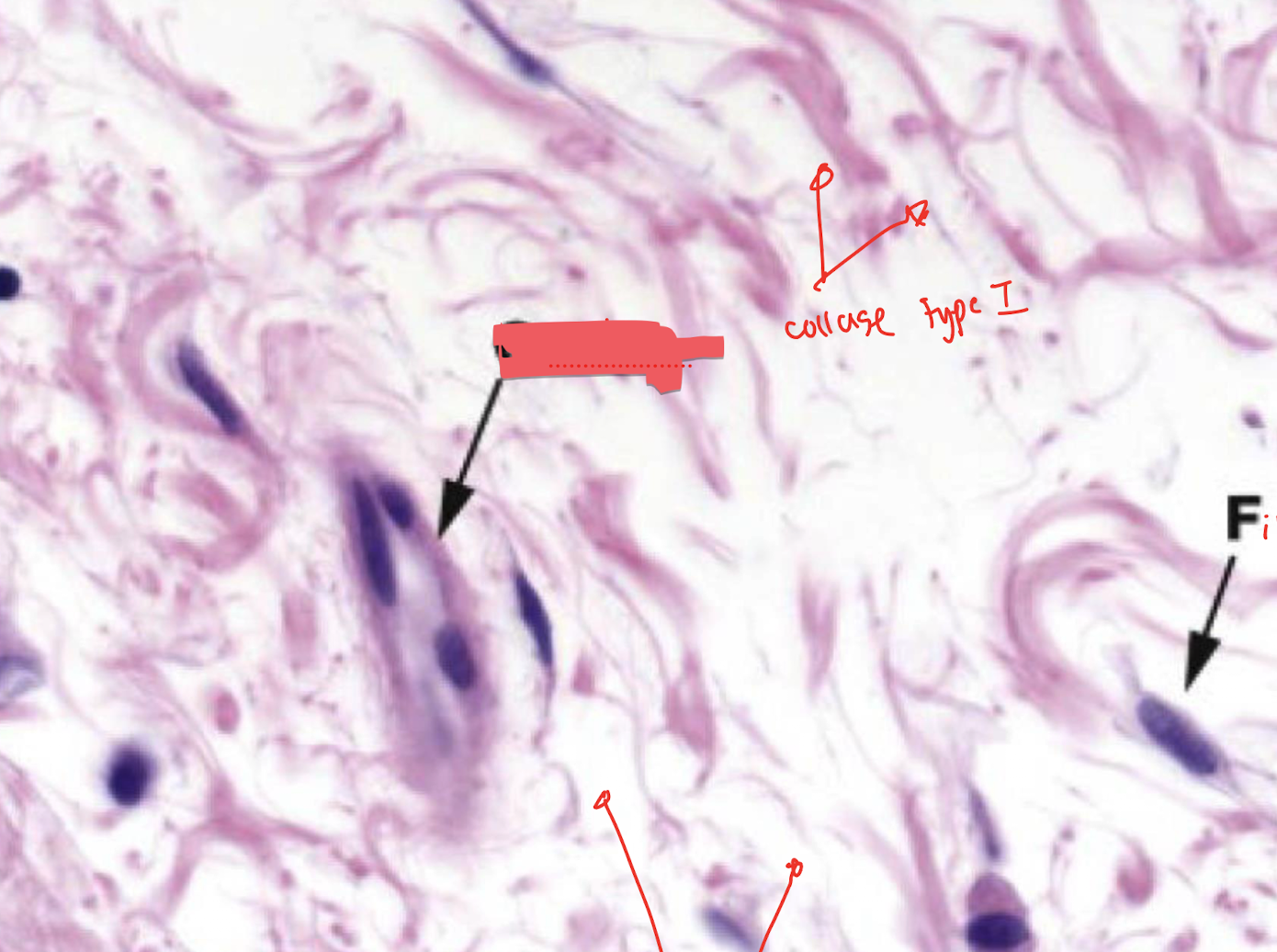
collagen type 1(top), fibroblast, ground substance (bottom)
capillary
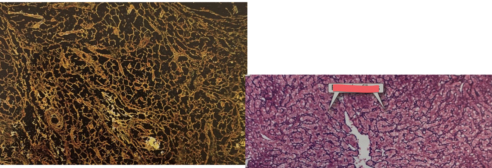
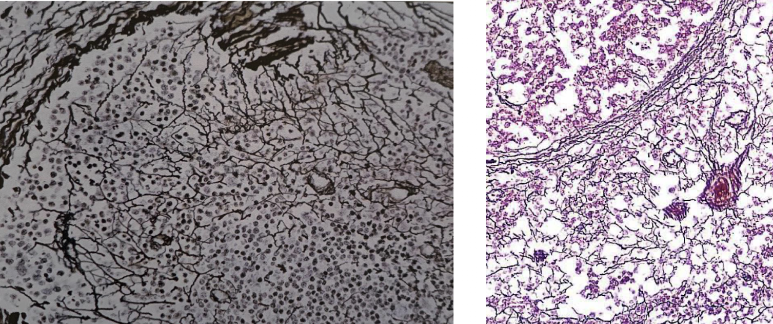
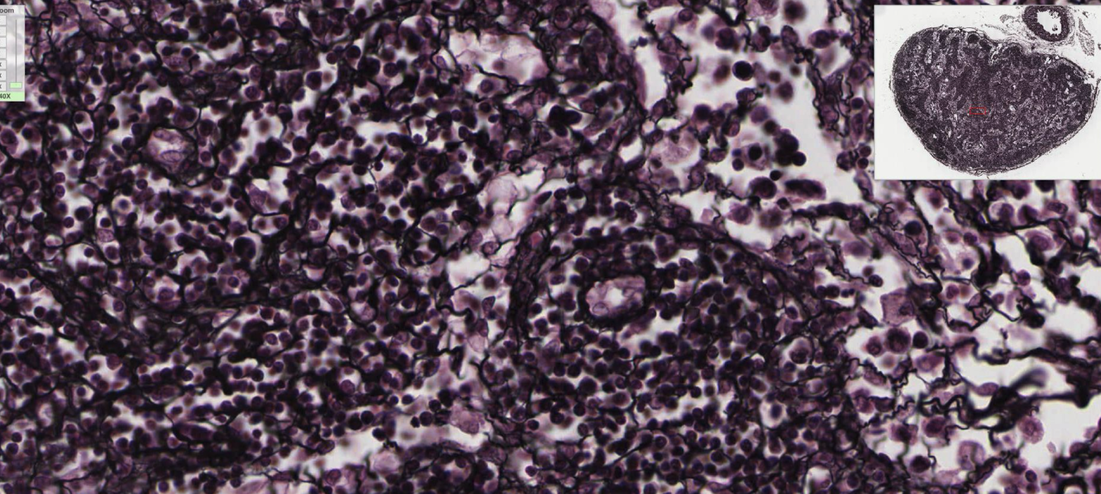
reticular fibers
fibroblast (right box), elastic fibers
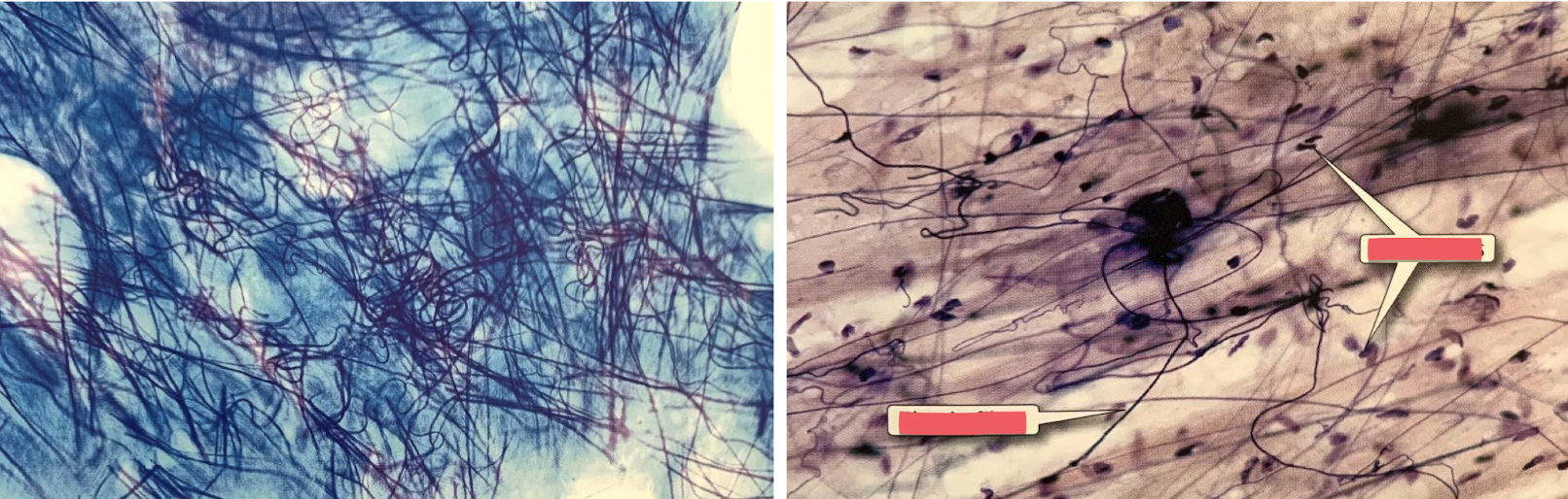
elastic fibers
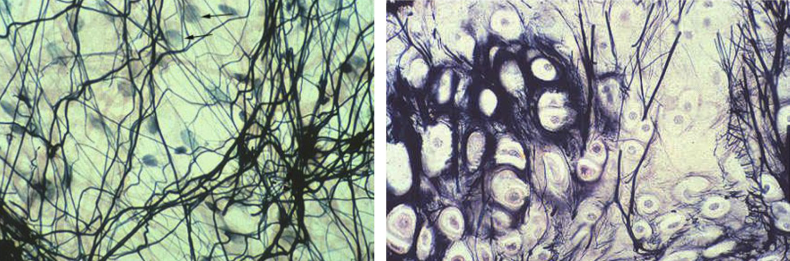
enamel (left box) internal elastic lamina (right box)
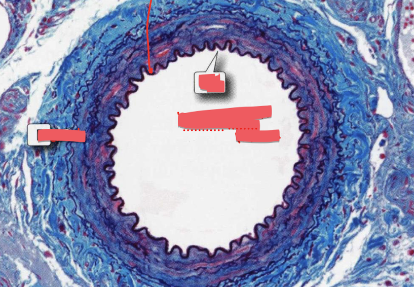
elastic (right box), collagen (left box)
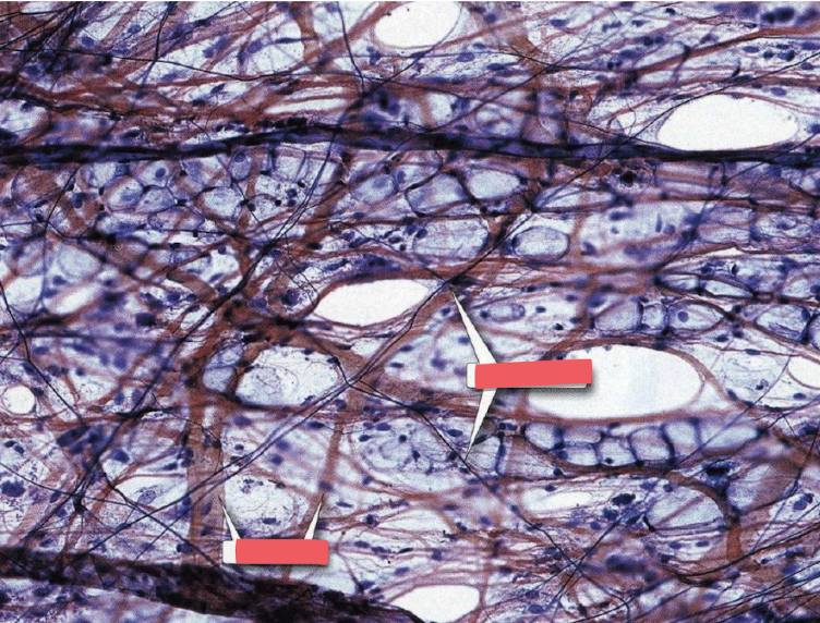
loose connective tissue
identify the type of connective tissue
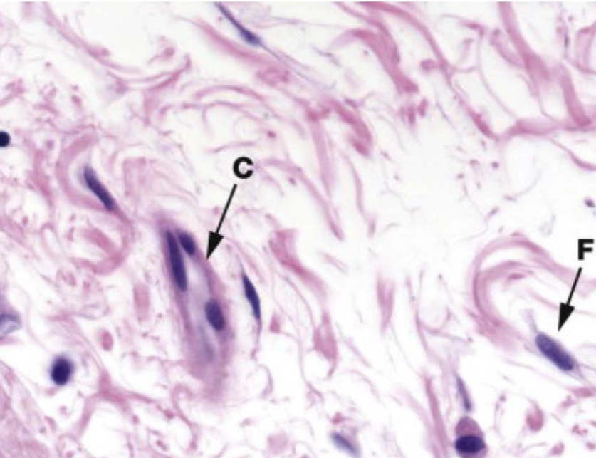
dense conective tissue
identify the type of connective tissue
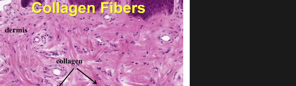
Loose connective tissue: adipose tissue
unilocular adipocyte (A)
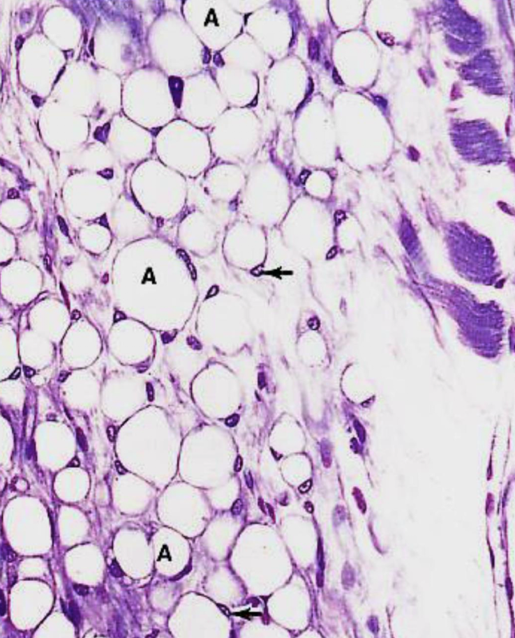
unilocular adipocyte
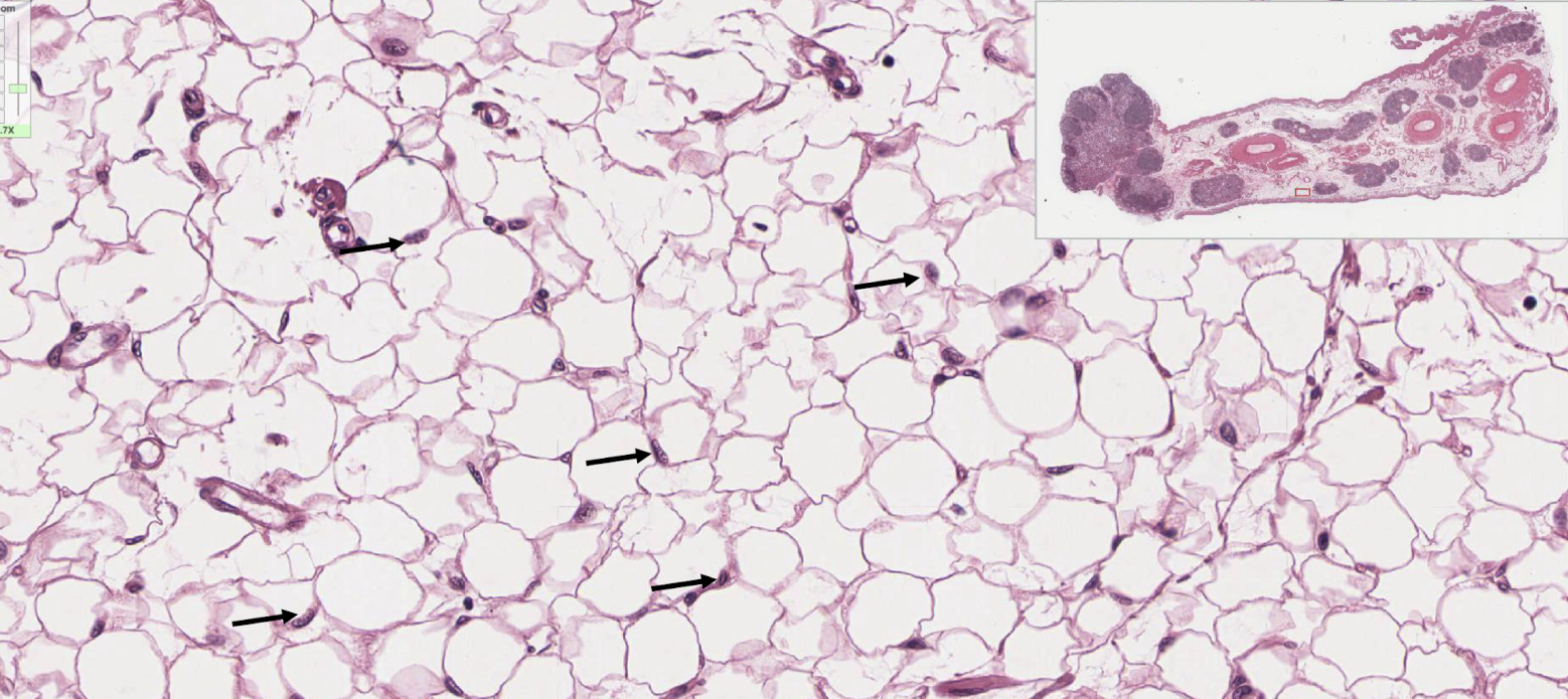
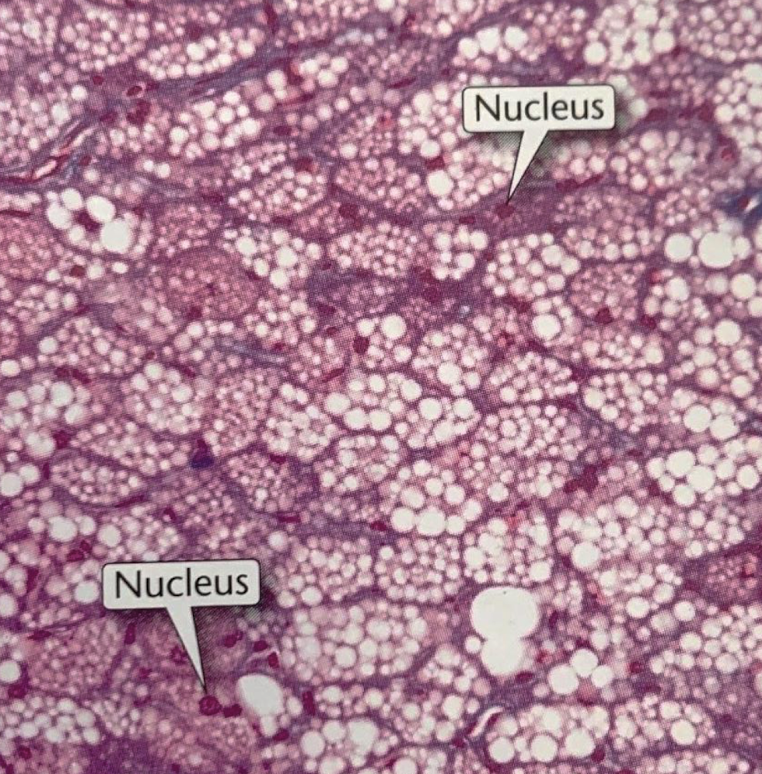
loose connective tissue: brown adipose tissue
loose connective tissue: reticular tissue
loose connective tissue: reticular tissue
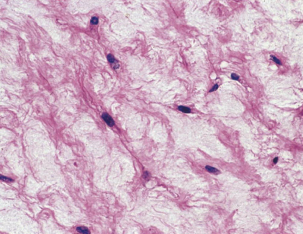
loose connective tissue: mucous connective tissue
loose connective tissue: mesentery