Lecture 6: Disorders of the Equine Foot
 Call Kai
 Learn
Learn Practice Test
 Spaced Repetition
 Match
Match Flashcards
 Knowt Play
1/70

There's no tags or description
Looks like no tags are added yet.
Name | Mastery | Learn | Test | Matching | Spaced | Call with Kai | Chat |
|---|
No analytics yet
Send a link to your students to track their progress
71 Terms
PE focusing on the foot
look for changes in color, odor, consistency of the hoof
clean the sole and examine the entire foot
flex and extend the foot
use hoof testers
observe the horse walking and trotting
hoof shape and balance
current state of trimming and shoeing
wear of hoof
what would make hoof cracks significant?
if it involves sensitive lamina vs insensitive
what would make a digital pulse significant?
increased or bounding
diagnostics for the foot
nerve blocks → localize the pain to the foot, PD or abaxial nerve block
radiographs → plain films, contrast studies
nuclear scintigraphy
CT
MRI
thrush =
infection of the frog
white line disease =
infection in the white line aka seedy toe
mixed infections
aerobic
fungal
anaerobic → fusobacterium necrophorum
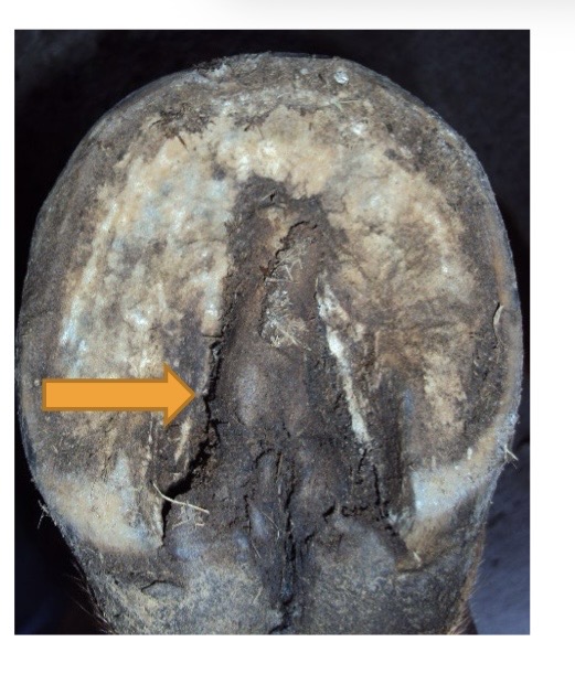
what is this?
thrush
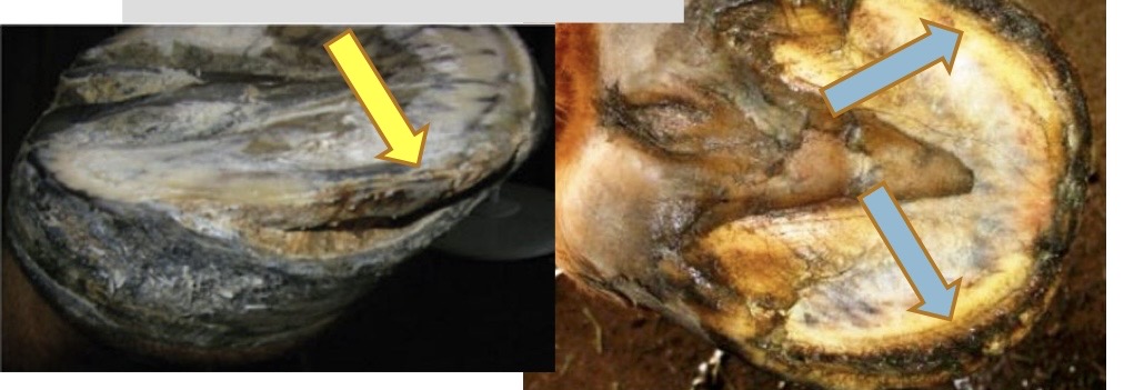
what is this?
white line disease
etiology of thrush and white line disease
moisture
poor hygiene
poor hoof care
reduced weight bearing
immunocompromised
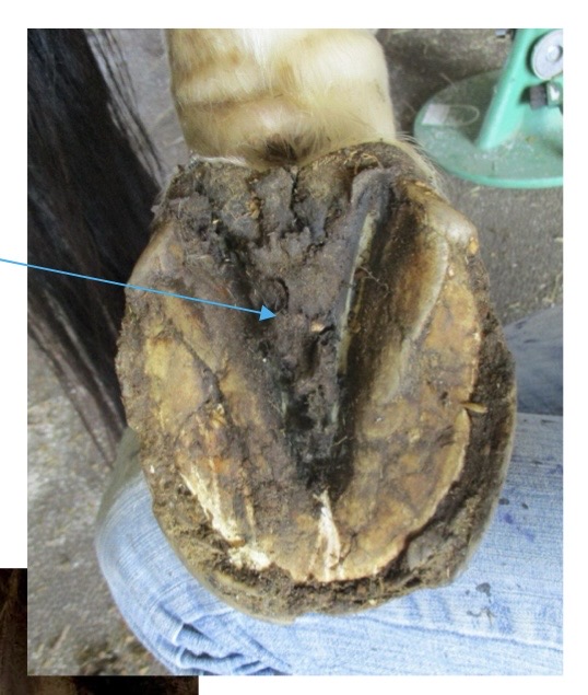
what is this?
thrush (the frog)
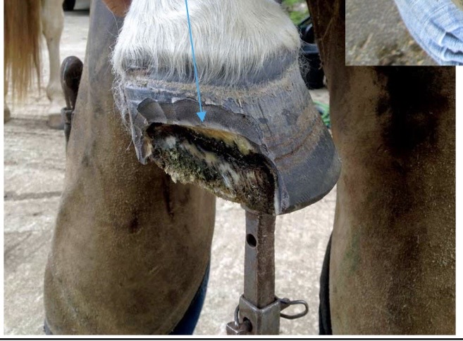
what is this?
white line disease
clinical signs of thrush/WLD
smell
hoof abnormalities
lame only with severe case
treatment of thrush/WLD
improve hoof care and stall hygiene
debride damaged tissue
copper naphthenate
iodine foot soak
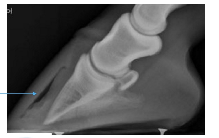
what is this showing?
gas with WLD
canker
common in draft horses
chronic inflammation and proliferation
hypertrophy of the sensitive lamina, degeneration of the superficial horn
looks like granulation tissue
cause is unknown
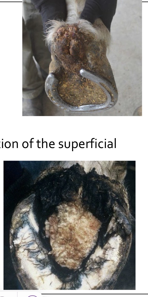
what are these showing?
canker
diagnosis of canker
± mild lameness
soft, greasy, friable, proliferative tissue over frog
characteristic smell → yeasty
can affect multiple hooves
treatment for canker
surgical debridement
local antibiotics (metronidazole)
liquid nitrogen
owner compliance → months of treatment
can’t always cure!
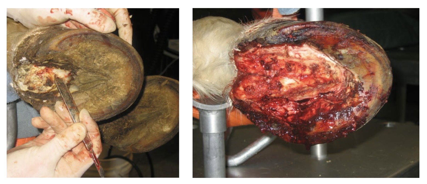
what is this?
canker
subsolar bruising etiology
thin-soled horses
wet conditions
rocky ground
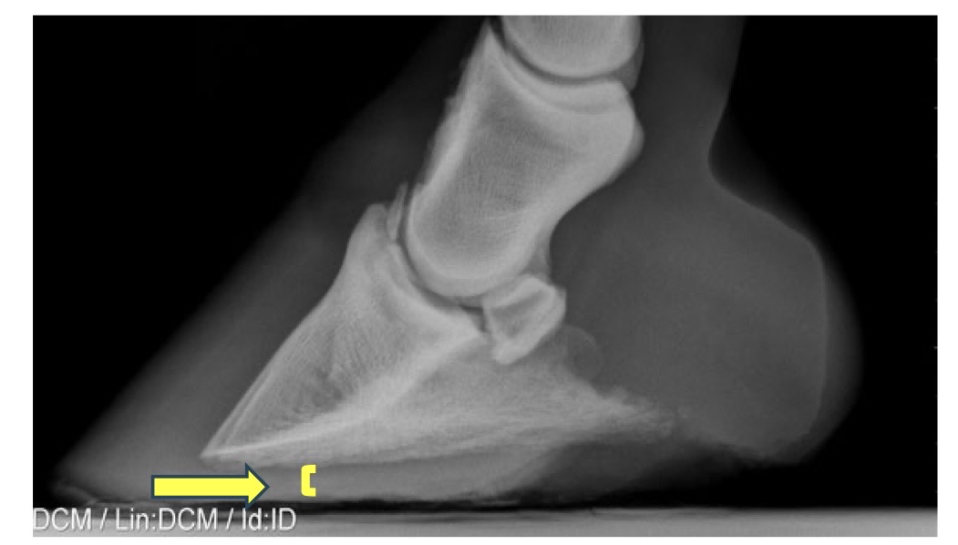
what is this?
subsolar bruising
corn
special area of sole bruise at angle of sole
usually from overdue shoeing
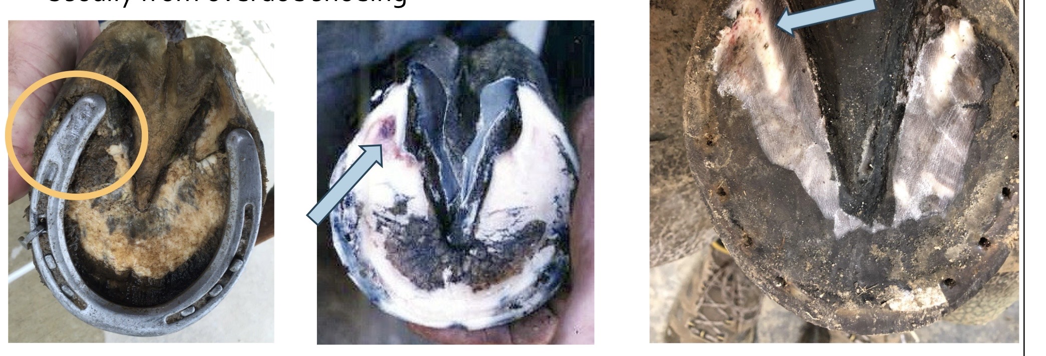
what are these?
corn
diagnosis of subsolar bruising
variable lameness
worse on rocky ground
positive to hoof testers → diffuse, squishy sole
treatment for subsolar bruising
poultices, shoes, keep out of mud
what is one of the most common causes of severe lameness in horses?
subsolar abscess
etiology of subsolar abscess
wet conditions
keratoma
sequel to a bad bruise
puncture wound
nail from shoe → quicking, pricked, close or blind
bacteria enter through failed hoof barrier
pus accumulates → pressure → pain
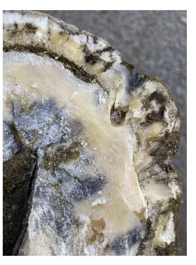
what is this?
subsolar abscess
subsolar abscess may break out at
the coronet or heel → reduced lameness, still want to establish ventral drainage if possible!
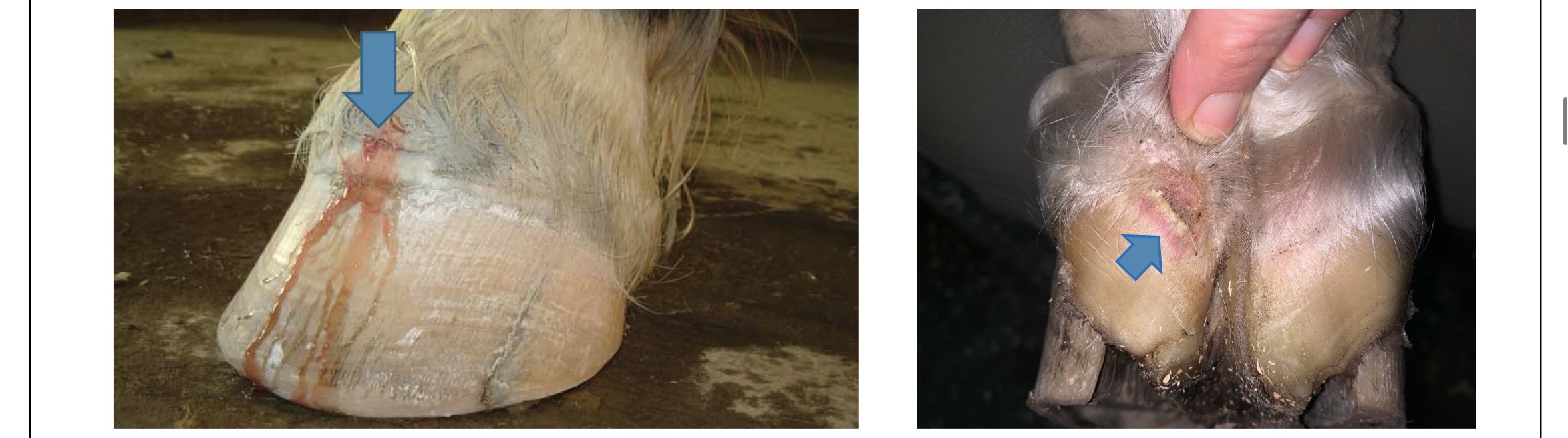
what is this?
subsolar abscess breaking out of coronet and heel
what is the goal of subsolar abscess?
draining
diagnosis of subsolar abscess
acute onset, severe lameness
increased digital pulse
possible distal limb swelling
squishy spot at coronet
+ hoof testers over area of abscess
gas pockets on radiographs
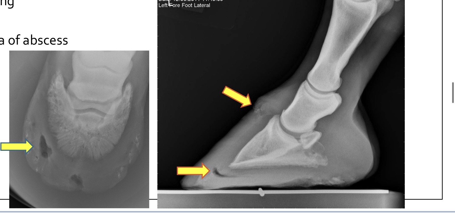
what are these showing?
subsolar abscess gas pockets
treatment of subsolar abscess
pain relief
tetanus toxoid vaccine
pare out abscess → ventral drainage
soak foot
poultice foot
protect foot
poulticing feet
reduce inflammation → hyperosmotics, DMSO, clay poultice, animalintex
harden hoof → hyperosmotics, tincture of iodine, keratex
distal hoof wall defects
failure of hoof strength
usually superficial → not a cause of lameness
painful if down to sensitive tissues
proximal hoof wall defects
trauma to the coronary band
usually painful
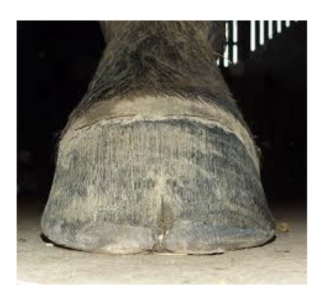
what is this?
distal hoof wall crack
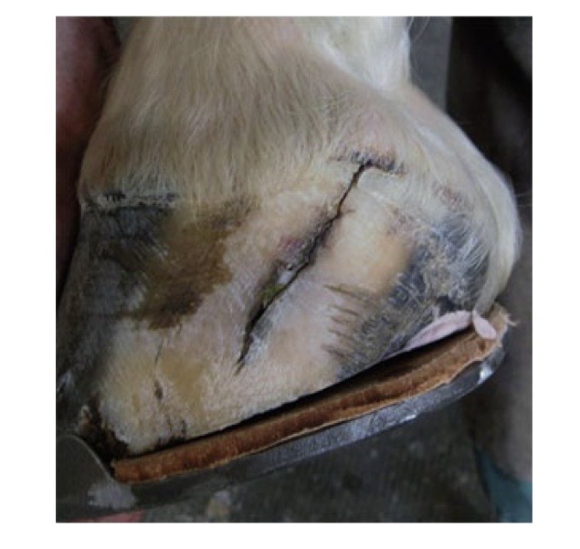
what is this?
proximal hoof wall crack
floating crack and widen crack
mostly at heel and quarters
reduce weight bearing and shear on crack
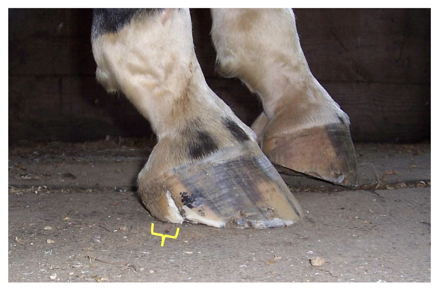
what is this?
floating crack
notch to halt propagation
mostly where floating isn’t feasible (aka the toe)
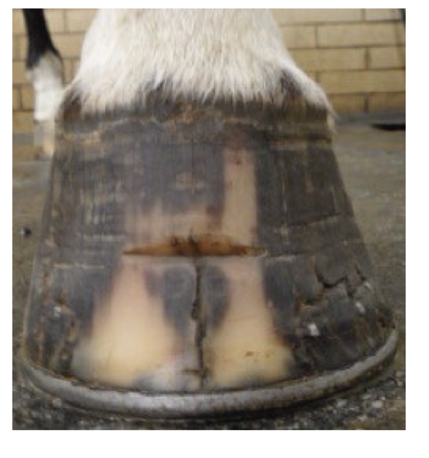
what is this?
notch to halt propagation
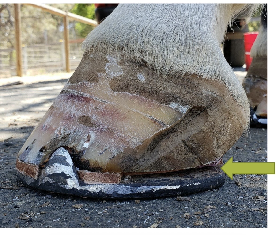
what is this?
heel crack
penetrating wounds to the sole
do not take objects out until after rads
if it does get pulled out before you take rads, try contrast radiography
if involves synovial structure, follow rules for diagnosis and treatment as for septic arthritis
treatment for penetrating wounds to the sole
early, aggressive treatment necessary
broad spectrum antimicrobials
local antimicrobials → intrasynovial, regional limb perfusion
NSAIDs
tetanus toxoid
clean and disinfect the sole and tract standing
if a penetrating wound involves a synovial structure =
refer!
surgical debridement under general anesthesia for penetrating wounds
street nail procedure
navicular bursoscopy
flexor tendon tenoscopy
coffin joint arthroscopy
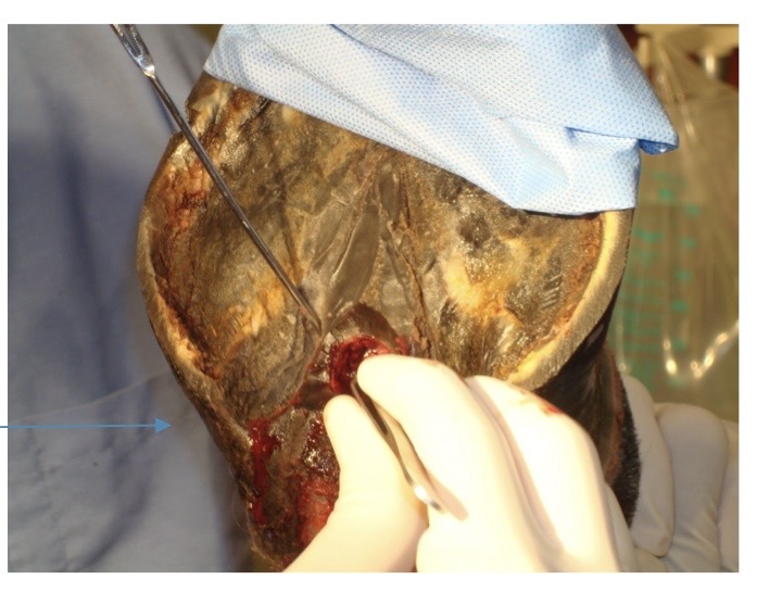
what is this?
street nail procedure
street nail procedure aftercare
keep clean → sterile bandages until granulation, hospital plate shoe
continued lavages as needed
antimicrobials and NSAIDs → several weeks
prognosis → guarded
features of the distal phalanx (P3)
acute, moderate to severe lameness → not usually fracture lame
owners may or may not know of the traumatic event
hoof tester sensitivity
increased digital pulse
diagnosis of P3 fractures
radiographs → if fracture line is not visible, re-radiograph in 7-10 days, often need obliques
± arthrocentesis of the coffin joint → helps differentiate between articular and non-articular fractures
synovial fluid → blood tinged with articular fractures
± CT/MRI
treatment for type I, II, and III P3 fractures
NSAIDs
stall rest for 2-4 months
stabilization of hoof capsule → foot cast, rim shoe
may never form a boney union
horse can be sound but radiographs still look the same
treatment for type III P3 fractures
some surgeons recommend internal fixation
may be advantageous with a step at the articular surface
lots of complications
sepsis!
treatment type IV P3 fractures
arthroscopic or arthrotomy removal
not all cause lameness
buttress foot
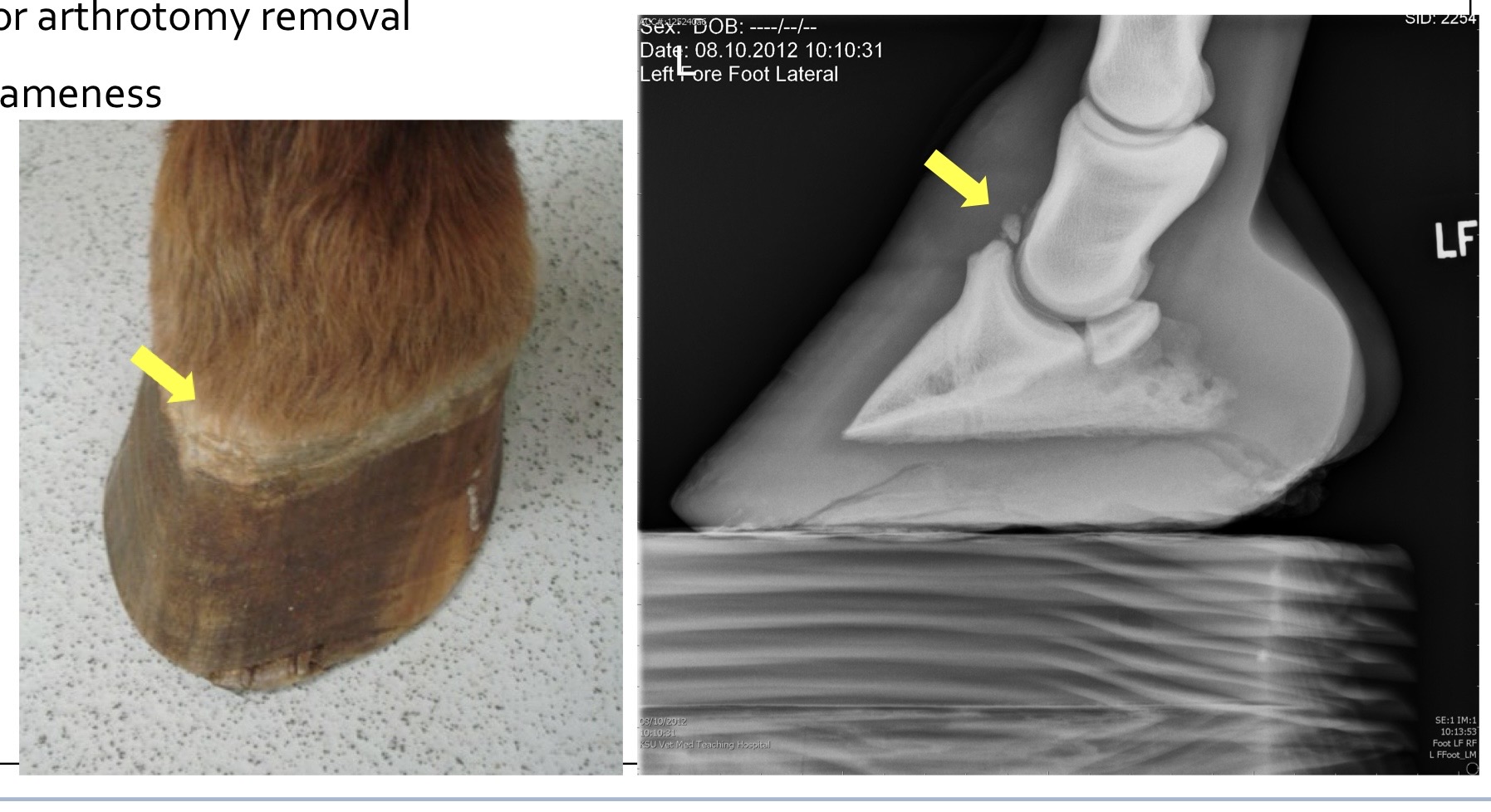
what are these showing?
buttress foot → type IV P3 fracture
treatment for type V P3 fracture
comminuted P3 fractures
severe lameness
poor prognosis for future soundness
euthanasia is recommended
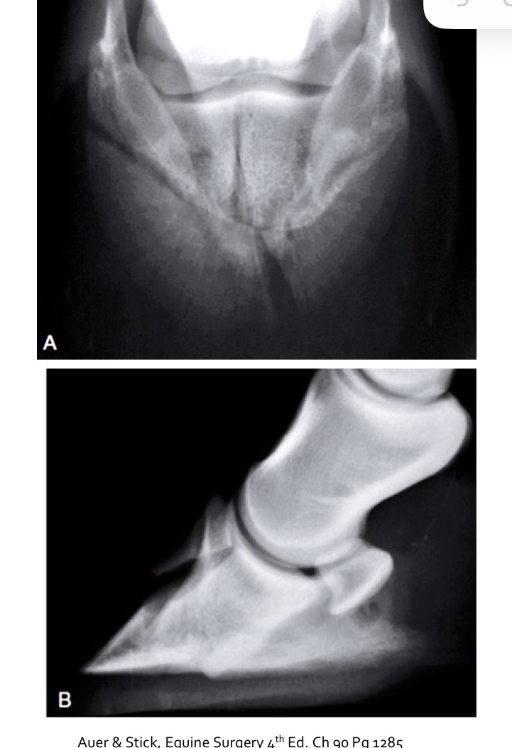
what is this showing?
type V P3 fracture
treatment for type VI P3 fracture
solar margin fractures
may occur because of chronic laminitis or trauma (pedal osteitis)
removal only if necessary if septic
most fractures heal, small fragments are resorbed
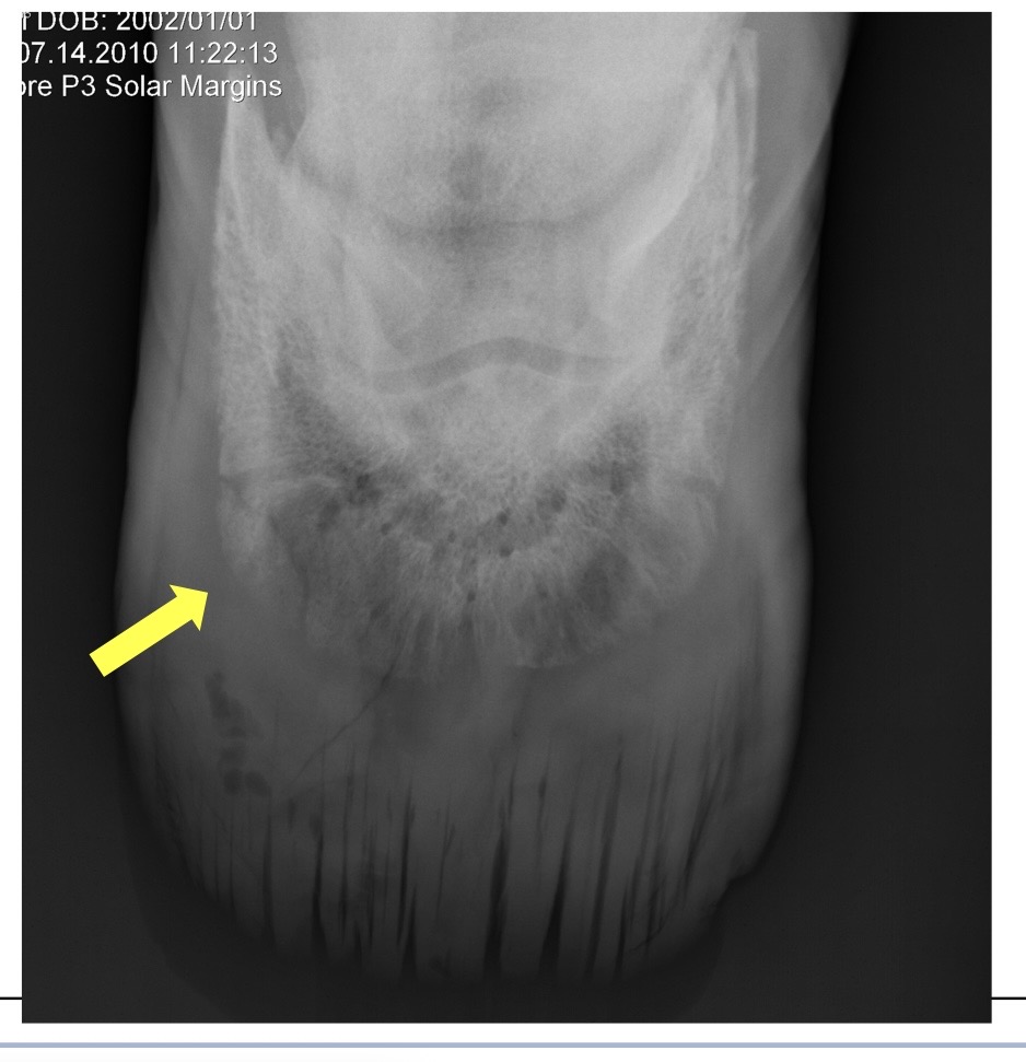
what is this?
type VI P3 fracture
prognosis for athletics after type I and II P3 fracture
good to fair
depends on amount of joint involvement for type 2
prognosis for athletics after type III P3 fracture
fair
depends on quality of reduction and subsequent coffin OA
prognosis for athletics after type IV P3 fracture
good
depends on amount of coffin OA
can be incidental finding
prognosis for athletics after type V P3 fracture
guarded for life
prognosis for athletics after type VI P3 fracture
good
keratoma
abnormal thickening of the hoof wall → likely secondary to inflammation or trauma to the hoof, presses on P3 and causes remodeling
clinical signs of keratoma
can cause distortion of P3
recurrent subsolar abscesses
treatment for keratoma
surgical removal of the abnormal tissue
better prognosis than conservative management ?
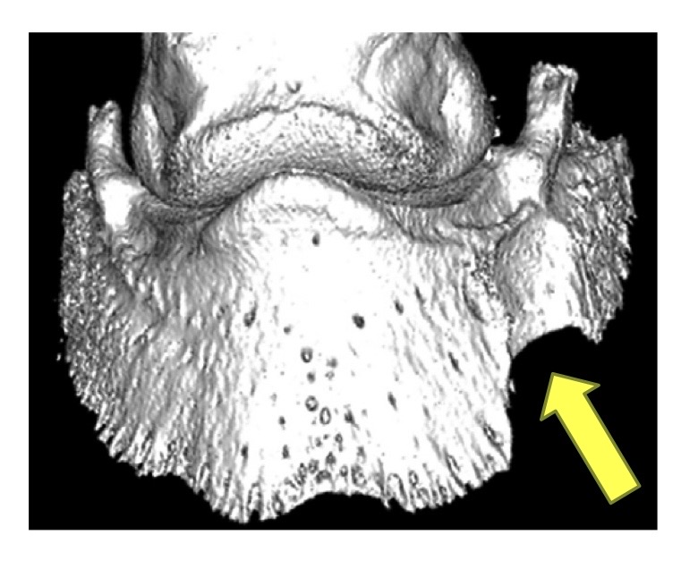
what is this?
keratoma
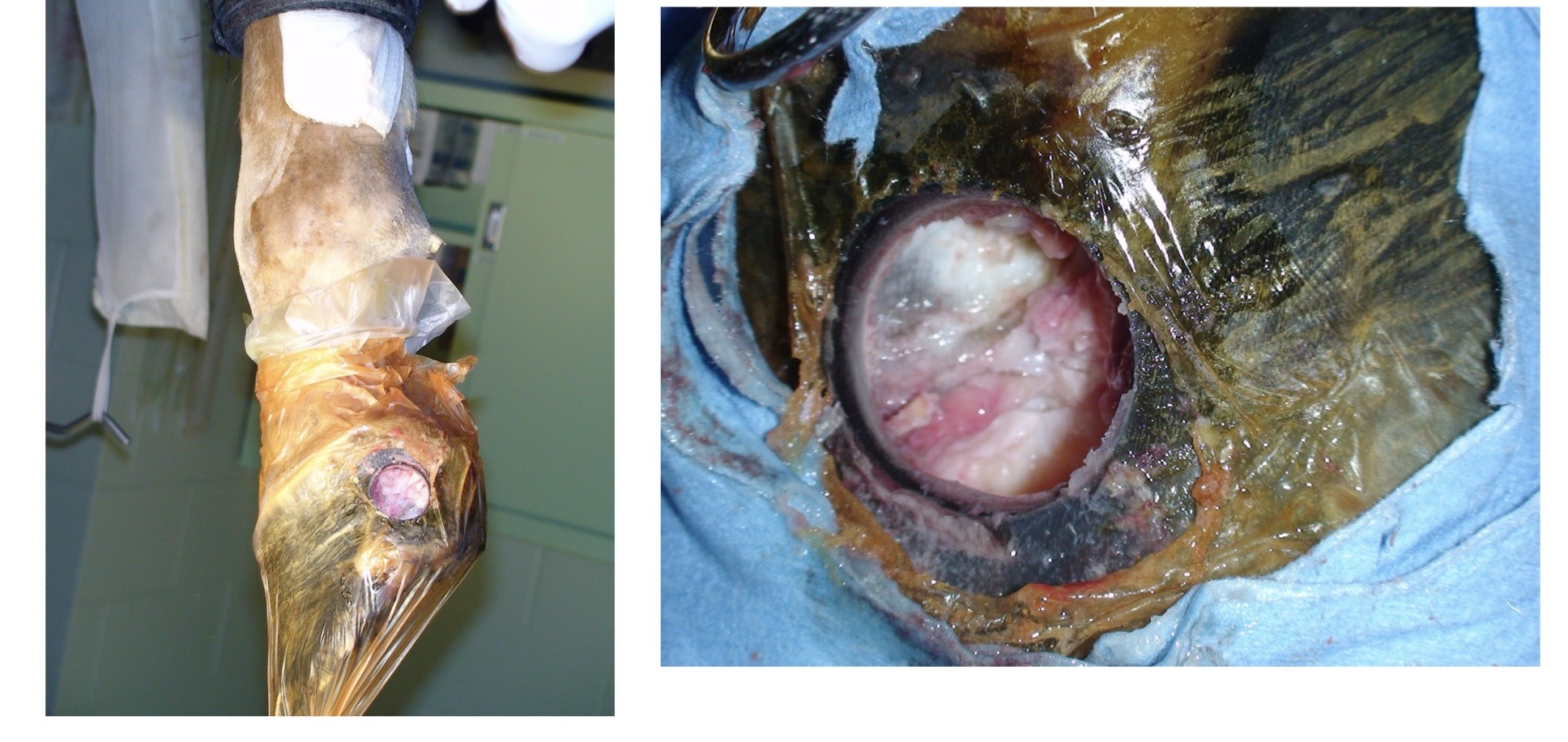
what is this?
keratoma