N325 Exam 2 (Musculoskeletal and Pain)
 Call Kai
 Learn
Learn Practice Test
 Spaced Repetition
 Match
Match Flashcards
 Knowt Play
1/60

There's no tags or description
Looks like no tags are added yet.
Name | Mastery | Learn | Test | Matching | Spaced | Call with Kai | Chat |
|---|
No analytics yet
Send a link to your students to track their progress
61 Terms
What is a sprain? Where do sprains most commonly occur?
INJURY OF LIGAMENT resulting from abnormal twisting or stretching at joints/spinal musculature (often from impact); most commonly occur at ankles, wrists, and knees
What are some signs and symptoms of a sprain?
pain, swelling, bruising, and limited ability to move joint
What is a strain? Where do strains most commonly occur?
overstretching of muscle, fascial sheath, or tendon; most commonly occur in larger muscle groups (lower back, neck, shoulder, and hamstring)
What are some signs and symptoms of a strain?
Pain, tenderness, swelling, bruising/discoloration, muscle weakness, stiffness, limited ROM
What are some clinical manifestations of both strains and sprains?
pain, edema, decreased function, ecchymosis, spasms, limited ROM
What is ecchymosis?
a discoloration of the skin resulting from bleeding within the body that moves to surface with gravity; differs from bruising as bruising occurs from local impact in which capillaries burst
How are sprains and strains diagnosed?
X-rays (cheaper) and MRI to rule out fractures/joint damage that require more complex or surgical interventions, ROM/mobility tests, assessment of amount of swelling and pain
What are some complications of sprains and strains?
unstable joint structure (especially relevant in ankle and knees b/c weight bearing), avulsion fracture, hemoarthrosis (bleeding into joint), long-term deficits (especially with more severe/repeated injuries; can eventually lead to issues with mobility), compartment syndrome (very rare with sprains and strains)
How can sprains and strains be prevented?
stretching, warming up, strength training, balance training, education on proper body mechanics/movements
How do you treat sprains and strains?
- Stopping offending activity
- RICE (rest, ice, compression, elevation) and POLICE (protection, optimal loading, ice, compression, elevation); elevate above level of heart to reduce edema and when icing, do 20 min on/20 min off cycle
- after 24-48 hrs rest + elevation, begin little movements
- pain relief
What is a repetitive strain injury (RSI)?
Catch-all term for symptoms and signs of sprains and strains caused by repetitive activities located in the neck, upper back, shoulder, arm, elbow, hand, wrist and fingers (e.g. Carpal Tunnel Syndrome)
Who is at risk of repetitive strain injury (RSI)?
- repetitive activities
- doing a high-intensity activity for a long time without rest
- poor posture or activities that involve working in an awkward position (e.g. car mechanic having to squeeze under car)
- Certain professions (e.g. poor body mechanics in nursing)
- Computer use
- Overtraining for sports
What are some interventions for those with repetitive strain injury (RSI)?
correct precipitating factor (don't do the thing), modifying behavior (e.g. changing the way you are holding hook while knitting), pain management (heat/cold application, NSAIDs), rest, PT, lifestyle modifications
*heat helps blood flow by dilating vessels, cold numbs pain
What is carpal tunnel syndrome? What is the etiology?
Carpal tunnel syndrome is a repetitive strain injury caused by compression of the median nerve. It is the most common neuropathy of the upper extremity
Who is more at risk of developing carpal tunnel syndrome?
female, cancer (puts body in inflammatory state and also treatments can lead to fluid shifts), ganglion cysts, RA, DM, PVD, repetitive wrist movements (e.g. typing)
What are some common assessment findings in patients with carpal tunnel syndromes?
weakness in the hand (decreased hand drip), burning pain in fingers, numbness (more numbness indicates nerve death), impaired sensation, Tinel's sign (holding hand up and tapping on median nerve with percussion hammer creates pain), Phalen's sign (creates pain in fingers - see image)
Memory tool: Tinels = Tapping nerve , Phalen's = Phalanges pointed downward
What are interventions for patients with carpal tunnel syndrome?
- Prevention is key
- Early interventions: stop activity or modify behavior, rest, hand splints (especially at night when not in use)
- Later interventions: NSAIDs, cortisone injection to reduce inflammation, PT, OT, more rest
- If lasting longer than 6 months: carpal tunnel release (snip tendon overlying median nerve)
What are some common causes of fractures?
trauma, pathological (e.g. cancer creating bony metastases that lead to fracture
How do you classify fractures?
Communication with the environment
- open (skin break)
- closed (no skin break)
Extent
- complete (two or more pieces of bone now)
- incomplete (still in one piece but damaged)
Displacement
- displaced (misalignment)
- non-displaced (bone pieces still properly aligned)
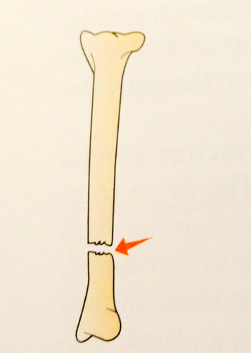
What does a transverse fracture look like?
complete fracture occurring straight across bone
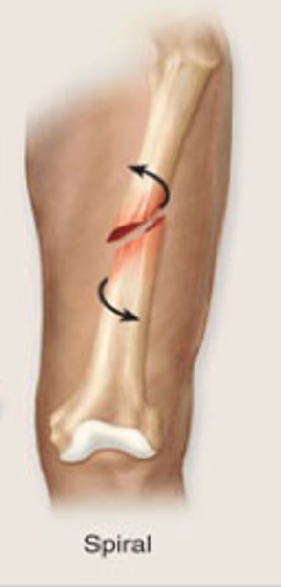
What does a spiral fracture look like?
ragged break occurs when excessive twisting forces are applied to a bone
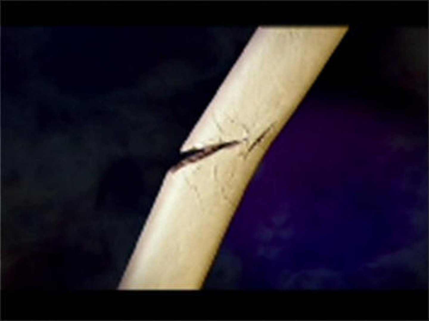
What does a greenstick fracture look like?
bending and incomplete break of a bone; most often seen in children
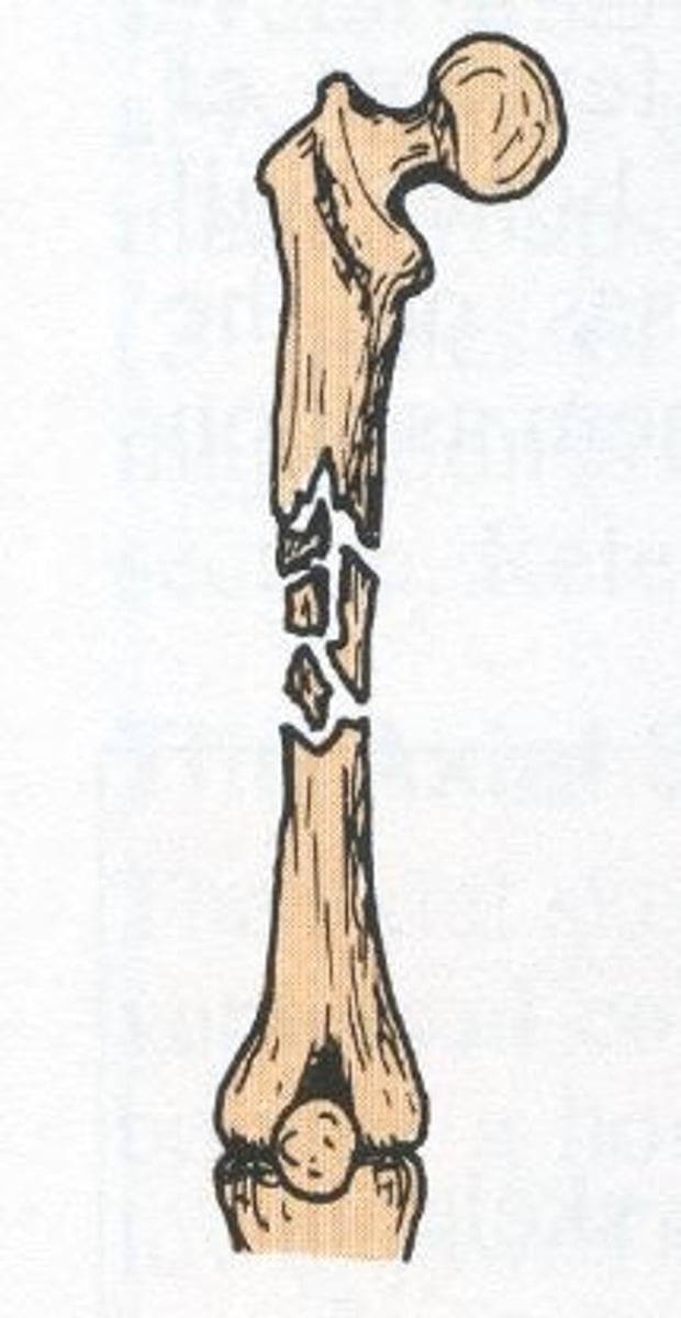
What does a comminuted fracture look like?
fracture in which the bone is splintered or crushed; breaks into many fragments
What does an oblique fracture look like?
occurs at an angle across the bone
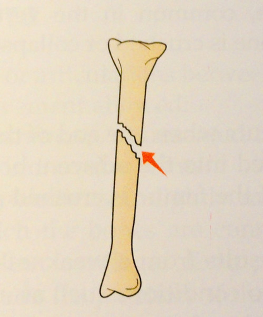
What does a pathologic fracture look like?
occurs when a weakened bone breaks under normal strain
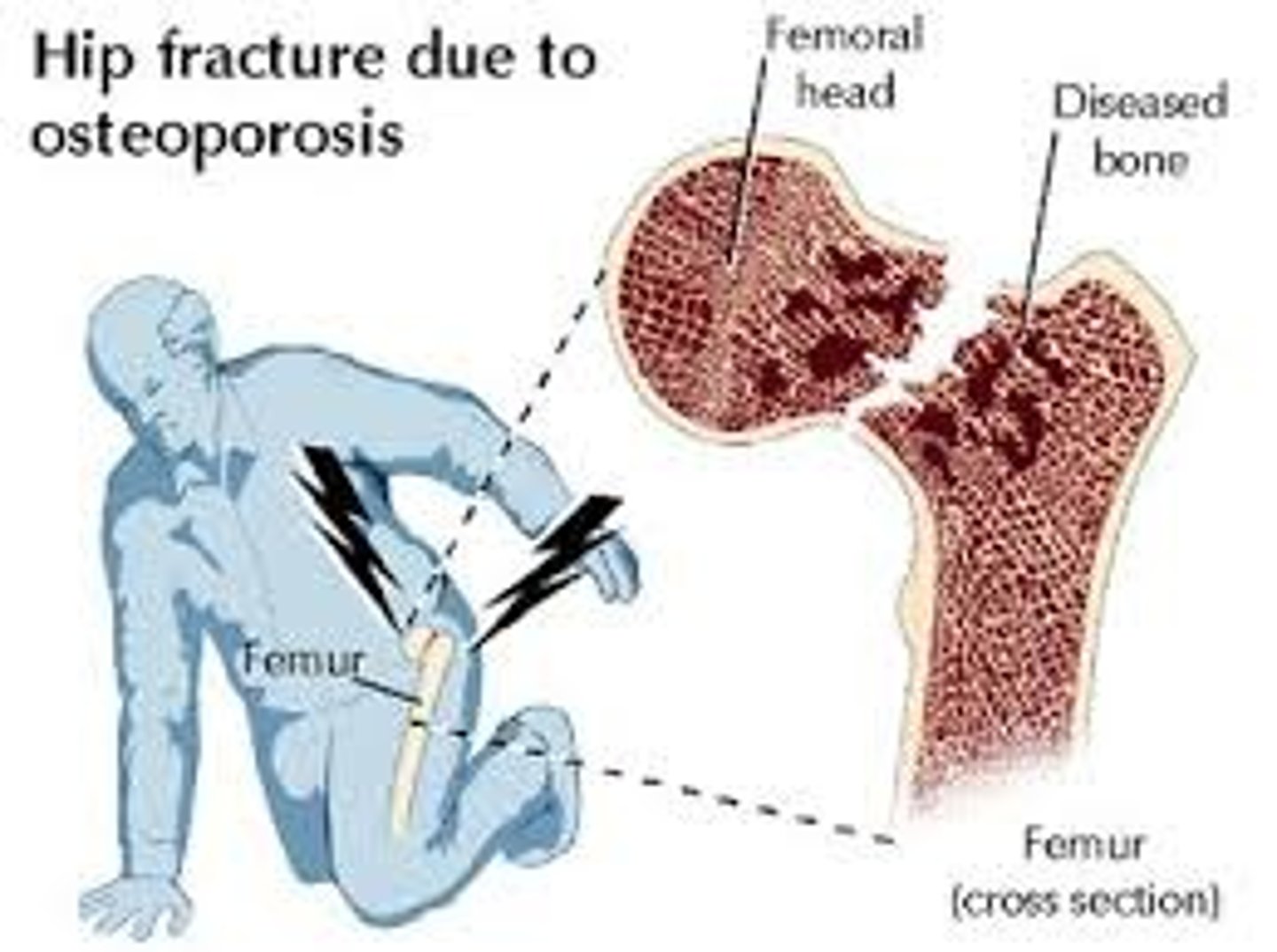
What does a stress fracture look like?
a small crack in the bone that often develops from chronic, excessive impact
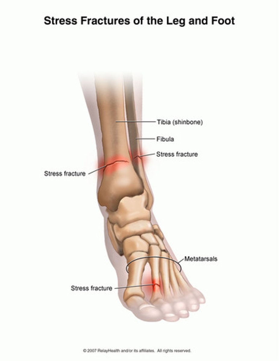
What are the clinical manifestations of a fracture?
edema, pain, spasm, deformity, ecchymosis, loss of function, crepitation
What are the causes and significance of edema in fractures?
edema is caused by disruption/penetration through skin/soft tissues and bleeding; too much edema at site of fracture can occlude circulation and lead to nerve damage + compartment syndrome
What are the causes and significance of pain in fractures?
pain is caused by trauma, spasm, pressure, movement of fracture; can lead to reduction in motion
What are the causes and significance of spasm in fractures?
caused by irritation and body trying to protect site, but can further displace a fracture
What are the causes and significance of deformities in fractures?
caused by abnormal positioning of bones; signals dysfunction and prevents proper union during healing of bone
What are the causes and significance of ecchymosis in fractures?
caused by extravasation of blood; usually seen distal to/below site of fracture
What are the causes and significance of loss of function in fractures?
caused by disruption in MS system; must be addressed so that function can be restored b/c of healing does not occur properly, will have future issues with mobility
What are the causes and significance of crepitation in fractures?
caused by crunching of bone fragments; sound signals possible nonunion and micromovement can lead to osteogenesis + the formation of new joints that should not be there
How should you assess a possible fracture that is first presented to you?
*compare affected side to contralateral side!!*
- vascular: assess skin color, skin temperature, pulses, capillary refill, and swelling
- neuro: movement, sensation, pain, numbess/tingling (can indicate nerve damage), reflexes
*make sure to assess distal to possible fracture as well!* e.g. if someone's upper arm is broken, do they still have movement and sensation in their fingers?
What are the main goals for patients with fractures?
realign bone fragments, immobilize for proper healing, and restore function through use of closed or open reduction methods
What does closed reduction of a fracture entail?
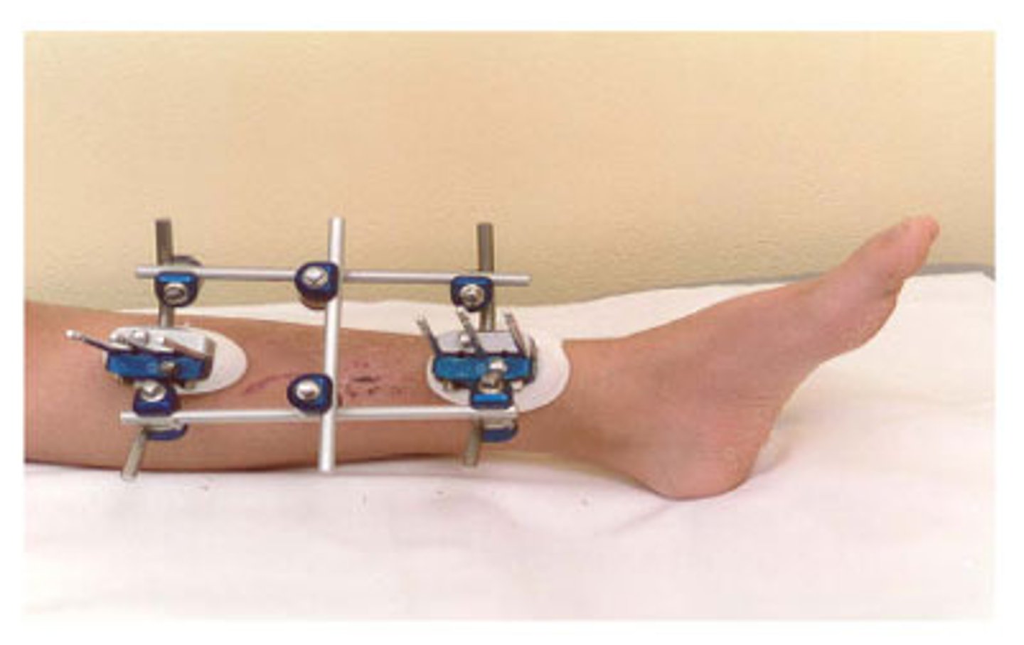
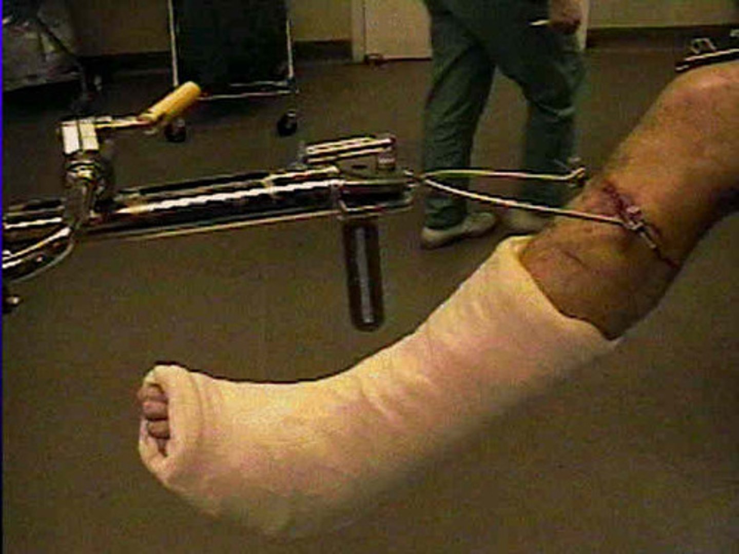
non-surgical, manual realignment of bones; may involve use of traction, casts, splints, external fixation (see photo), and braces
What does open reduction of a fracture entail? What are the disadvantages of this method?
surgical internal fixation (with use of things like pins, screws, and plates); puts patient at risk of infection, anesthesia complications, and healing complications with comorbid disease (pt with preexisting issues such as DM and heart problems will have more difficulties)
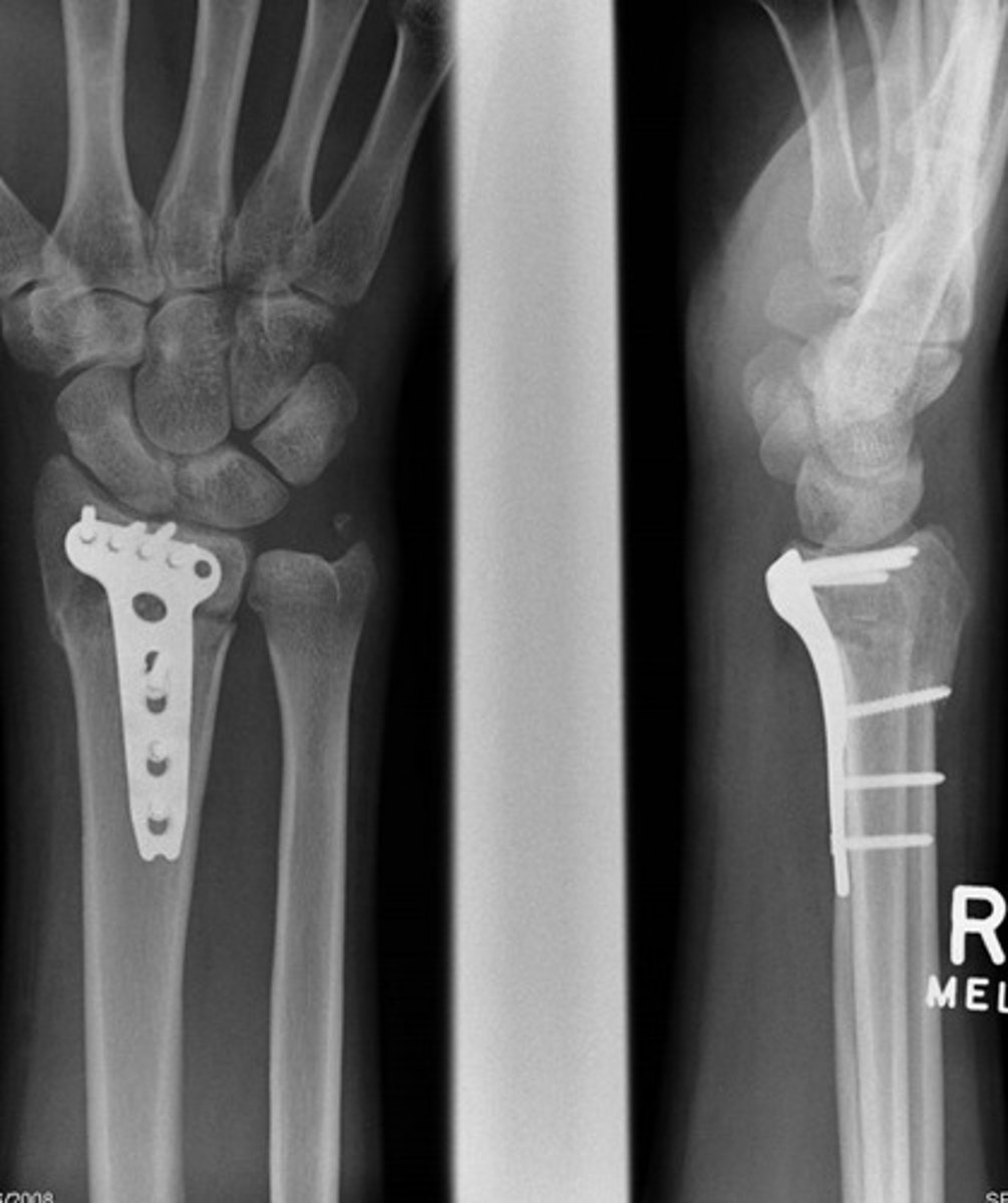
What are the stages of fracture healing?
First two weeks:
A. hematoma forms - increased blood flow to area brings essential components for healing to area of fracture
B. organization of components into a fibrous networks
C. osteoblasts mobilize, calcium deposits form; some remodeling occurs
After first two weeks:
D. callus formation - big chunk of bone made by osteoblasts
E. remodeling - osteoclasts break off excess bone
What is the purpose of casting fractures?
allow for ADLs while immobilizing fracture so that proper healing can occur
What are the different variations of casts? (names + structures they immobilize)
- short arm: stable wrist and fingers
- long arm: stable forearm/elbow, unstable wrist
- short leg: stable foot, ankle
- long leg: stable tibia, femur
- cylinder: stable knee or lower leg
- hip spica: stable pelvis and one or both legs
- body jacket: stable spine; fitted t-shirt underneath (it is a snug cast) and often hole cut out over stomach/GI system
What are some complications that can arise with the use of body jacket casts?
Can be difficult to breathe
Cast syndrome = small bowel obstruction
- s/s: abdominal pain and pressure, nausea & vomiting
- interventions: placement of NG tube, reapplication of cast (probably with abdominal hole cut out)
What should we educate patients to do when they have a cast for a fracture (cast care)?
- elevate for first 48 hours (above level of heart)
- watch for 6 P's related to compartment syndrome (pain, paresthesia, pallor, pulselessness, paralysis, poikilothermia)
- apply ice in PLASTIC BAG
- if gets wet, dry completely (with hair dryer on low)
- for itches, use hair dryer on cool + Benadryl
- move joints above/below cast
- don't get wet unless waterproof
What should we educate patients to NOT do when they have a cast for a fracture (cast care)?
- bear weight for 48 hours
- get cast wet (unless waterproof)
- insert items into cast or remove parts of it
When discharging patients with a cast, what education should we provide regarding meds, nutrition, and maintaining health while healing?
- proper use and SE of their Rx/OTC meds (e.g. muscle relaxants like flexeril for spasms, NSAIDs for swelling, opioids for pain, tetanus and diphtheria vaccinations & cephalosporin antibiotics for open fractures)
- proper nutrition to promote healing (increased vitamin C & D, protein, calcium, zinc, magnesium)
- options for mobility (e.g. teach use of crutches and scooters if indicated)
- no smoking (switch to other nicotine methods)! impedes bone healing
What is traction and its purpose? What are the different types?
-Traction is a pulling force applied to affected areas (counter-traction pulls in opposite direction); pressure is applied to distal end of fx to align bone w/ proximal end
-Purpose: to prevent/reduce pain and muscle spasms, immobilize area, reduce fracture while waiting for surgery, and treat pathology
-Types: Skin, Skeletal, and Buck's
What are the characteristics of skin traction?
▷Short term treatment
▷Tape, boots, splints
▷Maintains alignment but does not immobilize
▷Assists in reduction (possibly until surgery can be done)
▷Diminishes muscle spasms
▷Weights typically 5 or 10 lbs except pelvis
▷Example: Buck's traction
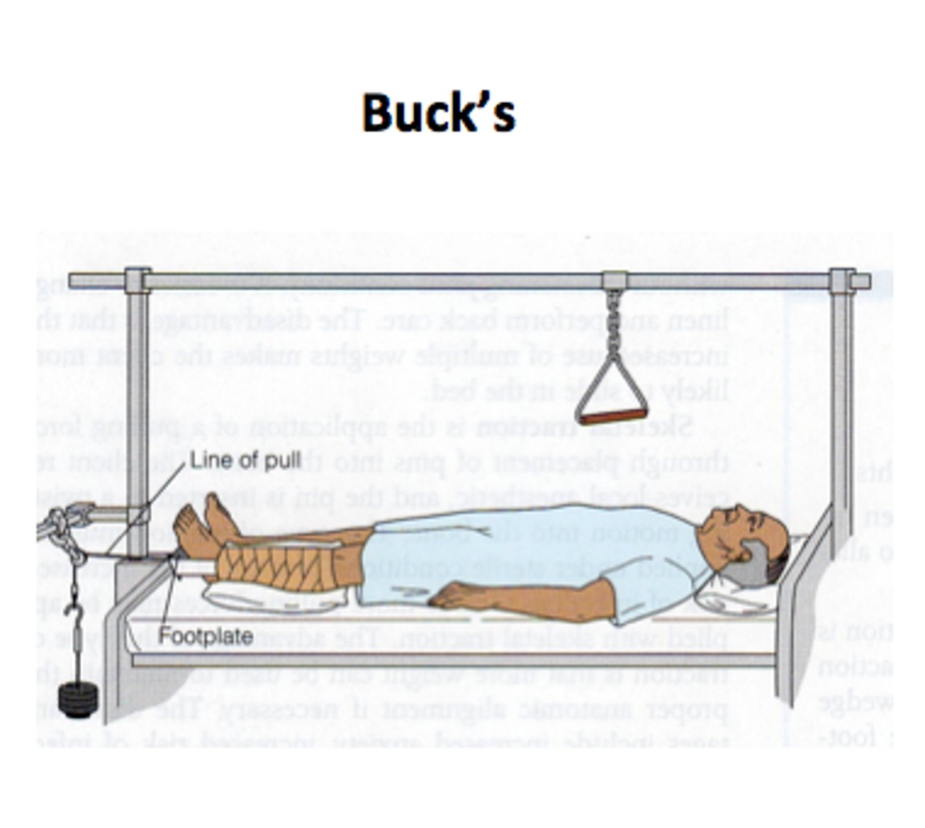
What are the characteristics of skeletal traction?
▷Longer treatment
▷Treats fx, contractures, hip dysplasia
▷Wire/pin inserted into bone
▷Immobilizes
▷Weights = 5 to 45lbs
▷Disadvantages: risk of infection, consequences of immobility (e.g. pressure sores)
What is a common example of skin traction and what is a nurse's role in caring for a patient with this type of traction?
▷Must keep traction forces on at all times (this might mean needing assistance when changing the position of a patient)
▷No weights touching floor
▷Weights must move freely through pulleys
What are some possible complications that can arise during the fracture healing process?
- infection (common if other comorbidities w/ circulation impairment like DM, malnourishment, or injury leading to fracture involved entrance of microorganisms, e.g. motor vehicle accident in which asphalt entered wound)
- union problems: nonunion (pseudoarthritis) and malunion (especially if immobilization was done incorrectly)
- compartment syndrome
- VTE
- myositis ossificans
- fat embolism
- osteomyelitis
- avascular necrosis
Who is most at risk for infection during fracture healing process and how do we treat these patients?
- patient has other comorbidities that impair healing, such as circulation issues (e.g. PVD) and DM (delays healing but also high blood sugar is food for bacteria)
- patient's injury leading to fracture involved entrance of microorganisms, e.g. motor vehicle accident in which asphalt entered wound
Treatment: Aggressive surgical debridement and IV AB 7 days to 6 weeks
What are some common causes of union problems in fracture healing?
poor blood supply, insufficient nutrients, insufficient immobilization, metabolic disorders (PVD, DM), smoking
What are the causes of compartment syndrome and what are the manifestations?
Etiology: trapped fluid compresses arterial flow and affects nerves too
- internal: swelling, hemorrhage, fluid
- external: casts or dressings too tight
Manifestations
- Pain (not just at site, but distal too; fracture in wrist would have pain in fingers with this syndrome)
- Paresthesia (numbness and tingling; again, distal to site is significant)
- Pallor (caused by decreased arterial flow)
- Paralysis (distal to site is significant)
- Pulselessness or diminished pulses (compare to contralateral side; caused by impeded arterial flow)
- Poikilothermia (coolness or decreased temp due to decreased arterial flow; again, compare to contralateral side)
What are some interventions for patients with compartment syndrome?
- do not elevate limb, keep at the limb of the heart
- no cold packs (will cause vasocontsriction, further decreasing circulation)
- remove or loosen bandages, split cast
- monitor BP, prevent hypotension
- O2 delivery (maximize saturation)
- urgent surgical consult for fasciotomy if internal swelling that cannot be alleviated by cast/dressing removal
What are common causes/risk factors of venous thromboembolism (VTE)? What are some signs and symptoms?
Causes (musculoskeletal)/risk factors
- Surgery
- Fx hip, pelvis, long bones
- Multiple trauma
- Immobility (necessary after surgery)
- Prior VTE
- Venous stasis
- Hypercoagulable state
- Endothelial injury
S/S
- Thigh/calf pain or tenderness
- Edema (more just enlargement)
- Warm to touch
- Red streaks or reddish discoloration
How do we diagnose and treat VTE?
Diagnosis: clotting factor lab, imaging like doppler ultrasound
Treatment: heparin/enoxaparin, early immobilization or fracture, early ambulation, compression stockings, SCDs
What are the causes and risk factors associated with the formation of dangerous fat embolisms?
Long bone break (femur, tibia, ribs, pelvis) and other pathologies can lead to release of bone marrow and then systemic fat globules distributed to tissues/organs
Causes/risk factors
- long bone break
- multiple fracture or crush injury
- hip/total joint replacement
- spinal fusion
- liposuction
- bone marrow transplant
What are the s/s of a fat embolism and when do s/s usually manifest?
Usually manifest 24-48 hrs after fracture.
- LOC changes, confusion, restlessness
- dyspnea, tachypnea, tachycardia
- fever
- petechiae
- progressive hypoxia
What are the interventions for fat embolism?
- Prevention is key; includes early stabilization of fracture, patient education about s/s of fat embolism at patient's discharge as well as how to monitor respirations and importance of TCDB + repositioning
- oxygen, IV fluids
- monitor respirations, TCDB, repositioning
- possible intubation and mechanical ventilation
What is osteomyelitis and what are some common causes/risk factors?
severe infection of the bone, bone marrow, and/or periosteum
causes/risk factors
- open fractures/wounds
- prosthetic devices
- other: periodontal disease, UTI, TB, infectious pathogens
- vascular insufficiency, immunocompromised pts
Which drug can block referred pain?
ketamine