neurophysiology - from cells to networks
 Call Kai
 Learn
Learn Practice Test
 Spaced Repetition
 Match
Match Flashcards
 Knowt Play
1/89

There's no tags or description
Looks like no tags are added yet.
Name | Mastery | Learn | Test | Matching | Spaced | Call with Kai | Chat |
|---|
No analytics yet
Send a link to your students to track their progress
90 Terms
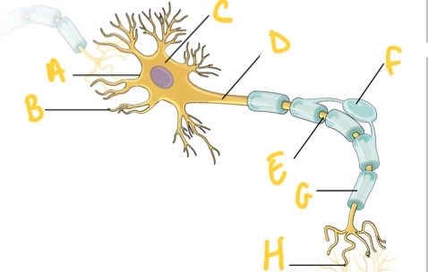
What is A?
Cell membrane
What is B?
Dendrites
What is C?
Cell body (soma)
What is D?
Axon
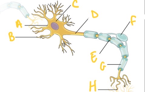
What is E?
Node of ranvier
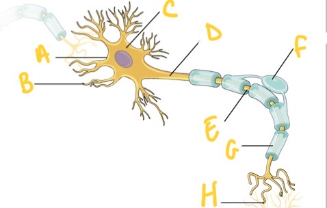
What is F?
Oligondrocyte
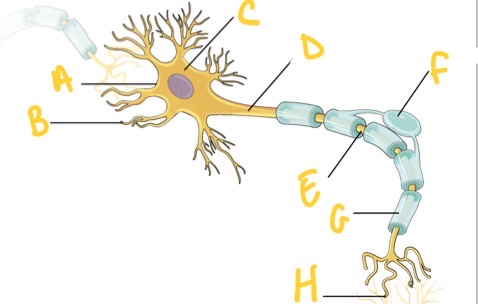
What is G?
Myelin sheath
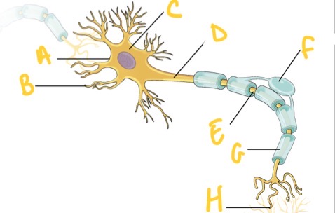
What is H?
Synapse
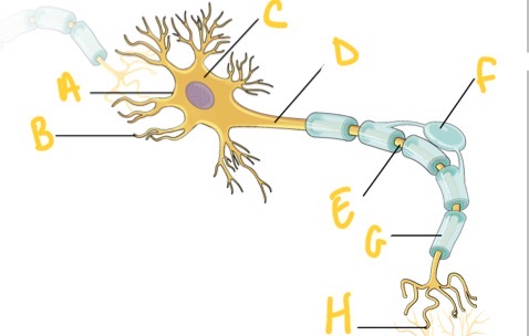
What type of neuronal cell is this ? And what is its function?
Neurone - communication
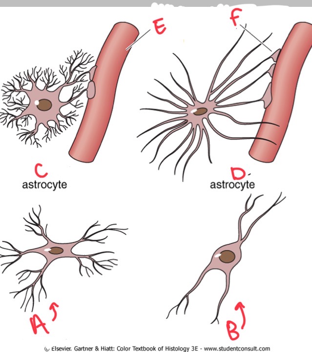
What is A?
Microglia
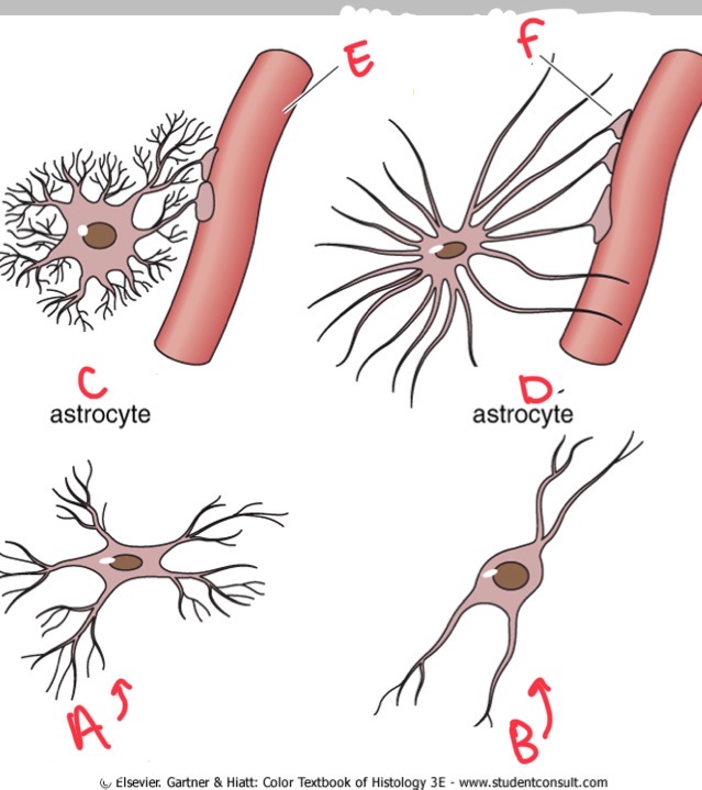
What is B?
Oligodendrocyte
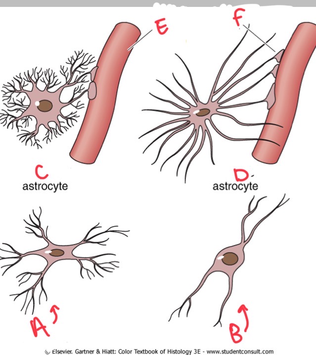
What is C?
Protoplasmic
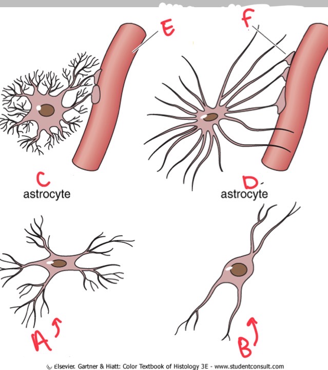
What is D?
Fibrous
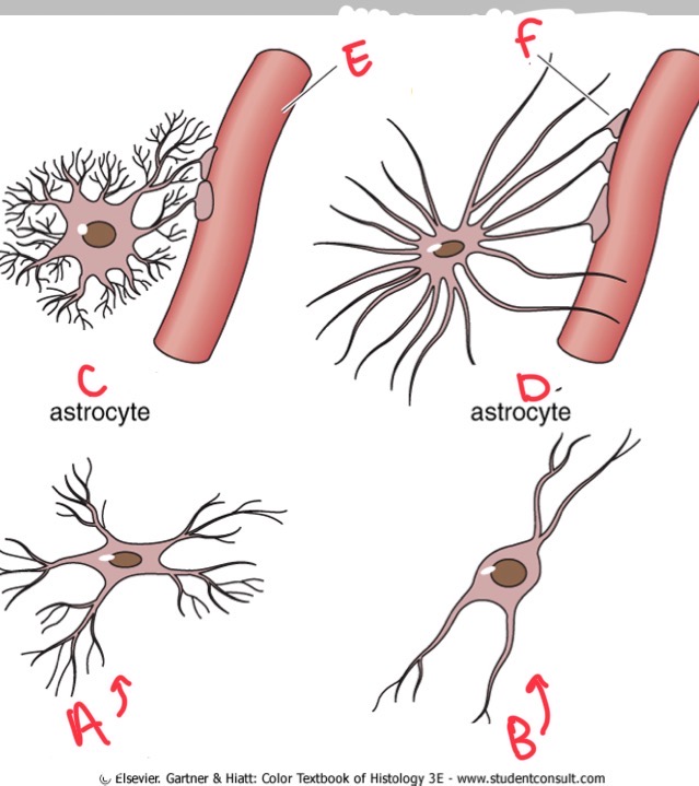
What is E?
Blood vessel
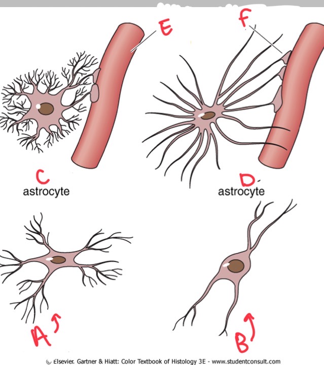
What is F?
Perivascular foot
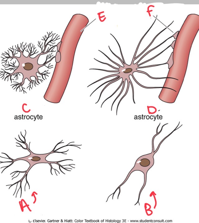
What neuronal cells are this? What is its role ?
Glia
Support functions, immune regulation
What are neurones ?
The basic unit of structure and function in the nervous system
What do neurones do?
Conduct impulses
process information
Sense environmental changes
Communicate changes to other neurons
Command body response
Why do neurones have high energy usage ?
Constant need for glucose and oxygen
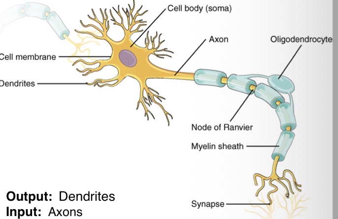
What does the cell body contain?
Contain nucleus and cellular activity
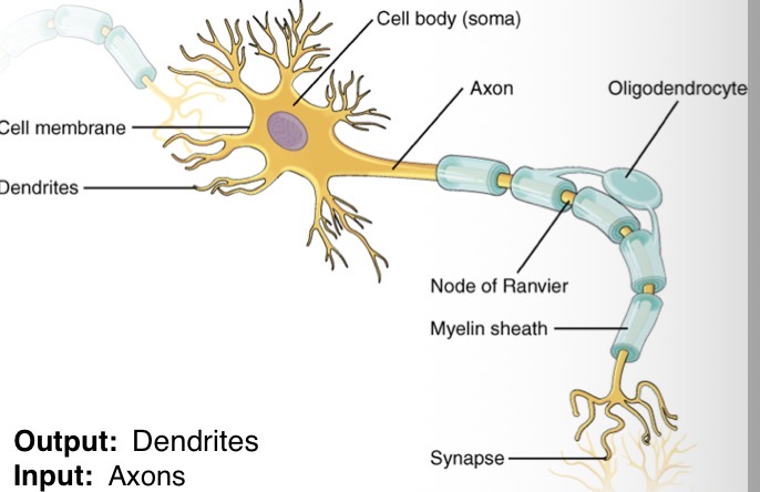
What are axons ?
Single extension of the neurones providing output
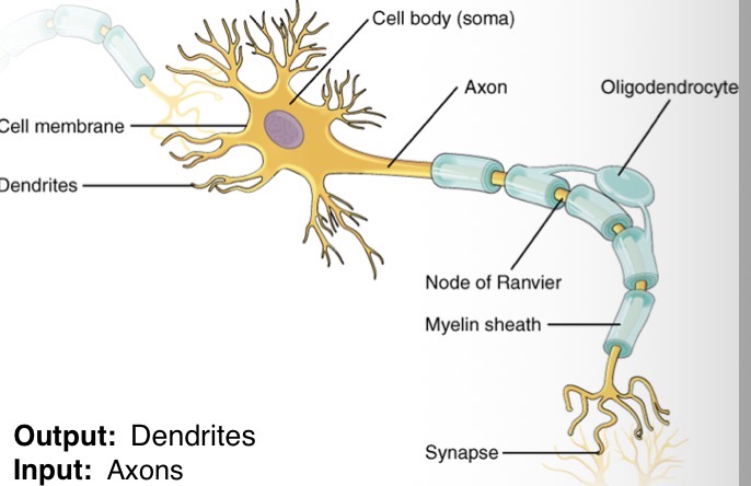
What are dendrites?
Branch like extensions that receive messages from other neurones
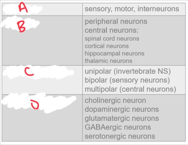
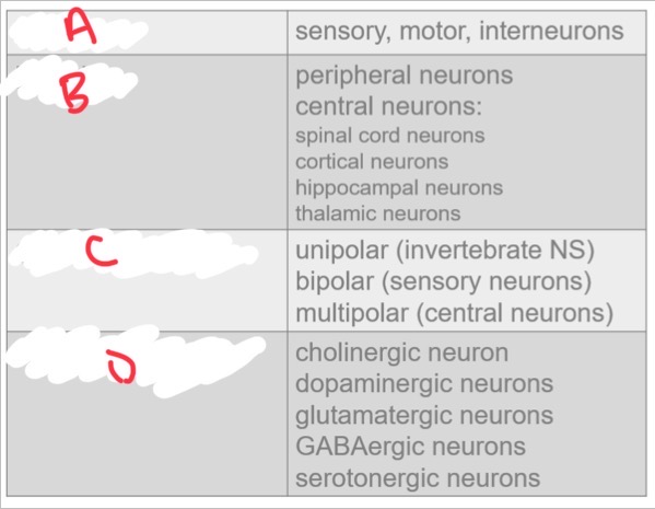
What is A?
Function
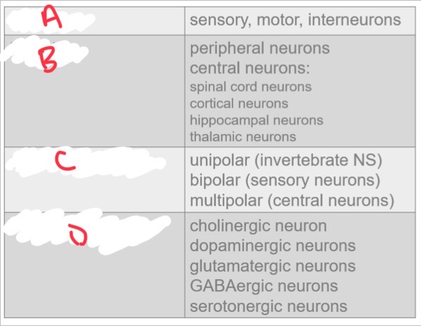
What is B?
Locality
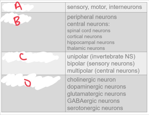
What is C?
Cell morphology
What is D?
Neurotransmitter
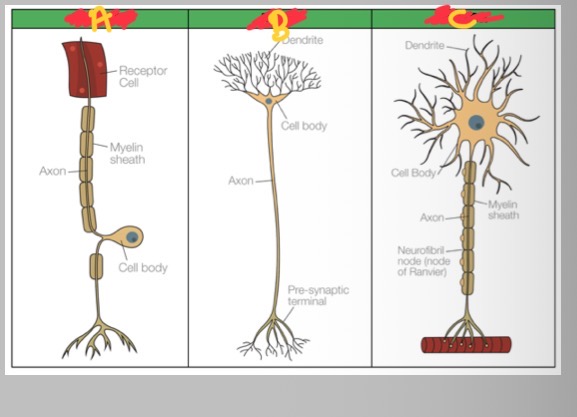
What is A?
Sensory
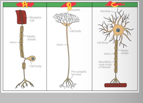
What is B?
Relay
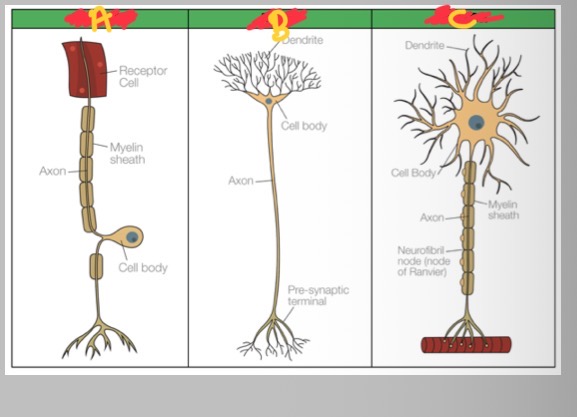
What is C?
Motor
What do motor neurones do?
Carry impulses away from the brain and spinal cord
What do sensory neurones do?
Carry impulses from inside/outside the body to brain/spinal cord
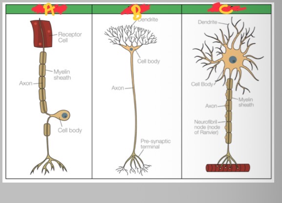
What are relay neurones ?
Process incoming impulses and pass them onto motor neurones
Each phospholipid has what?
phosphate “head”
Two fatty acid tails -
arranged back to back or form the belayer
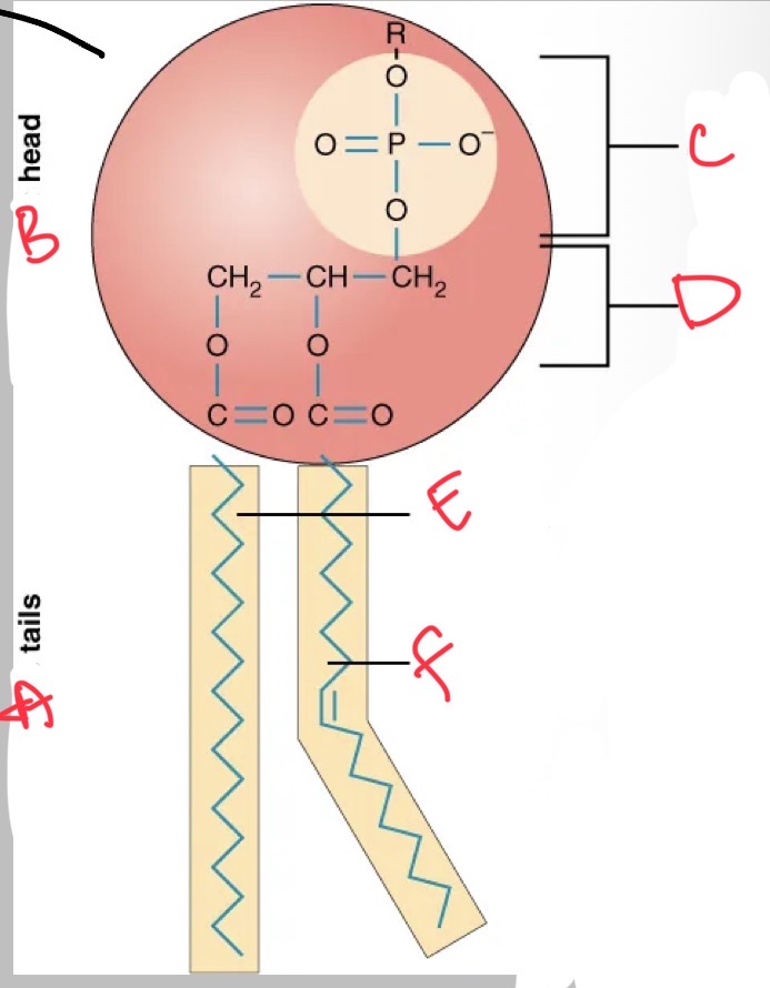
What is A?
Hydrophobic
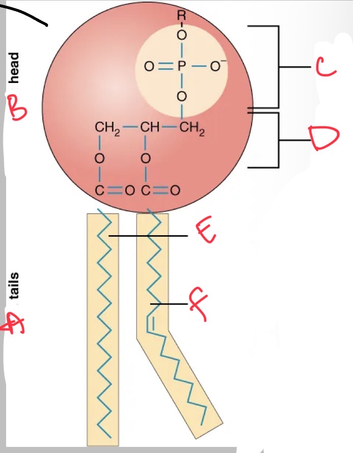
What is B?
Hydrophilic
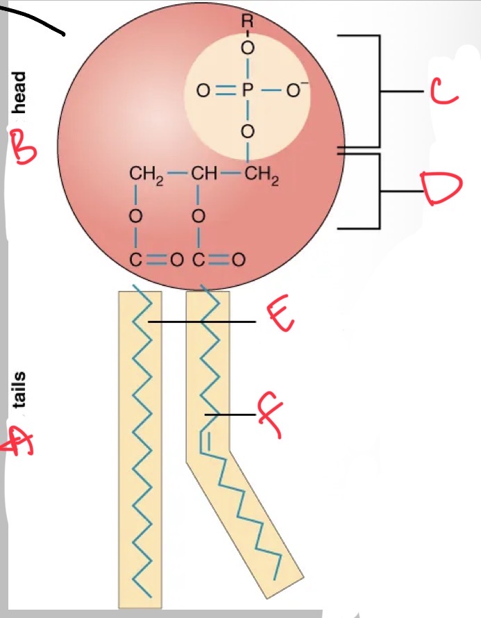
What is C?
Phosphate
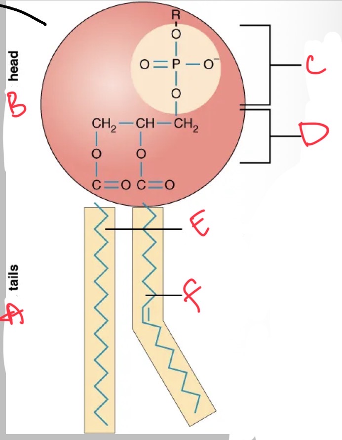
What is D?
Glycerol
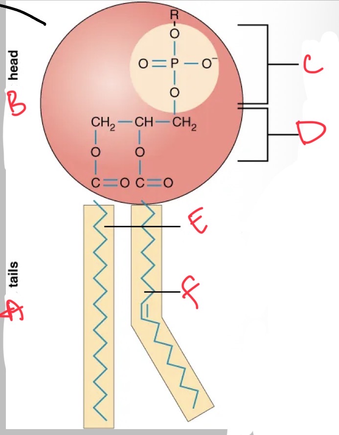
What is E?
Saturated fatty acid
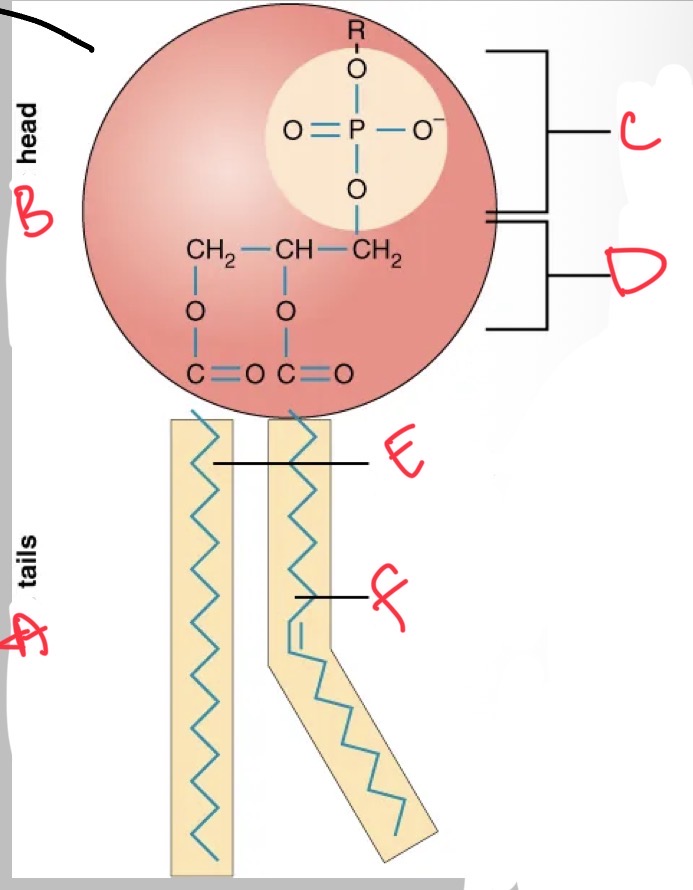
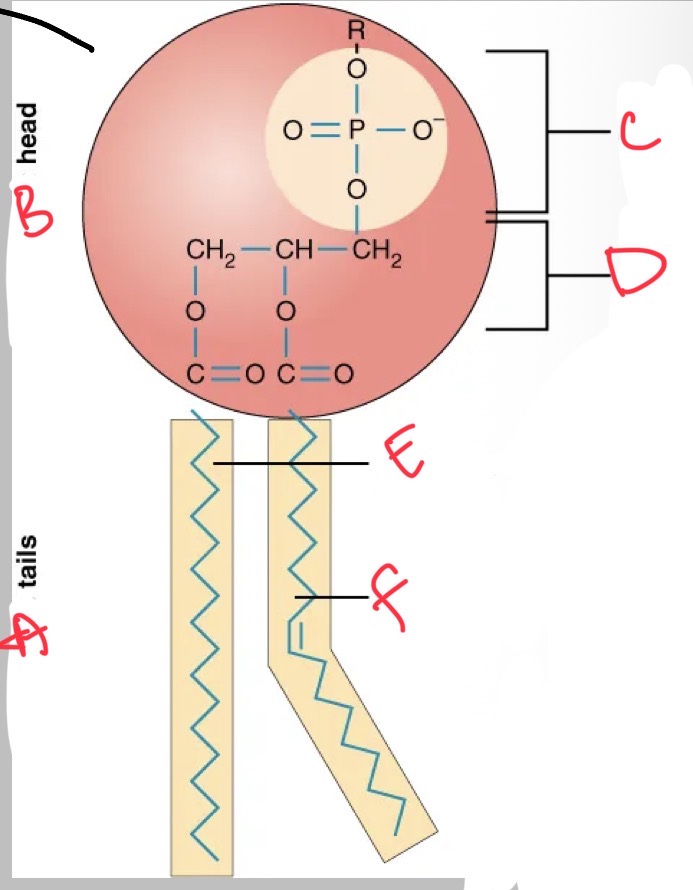
What is F?
Unsaturated fatty acid
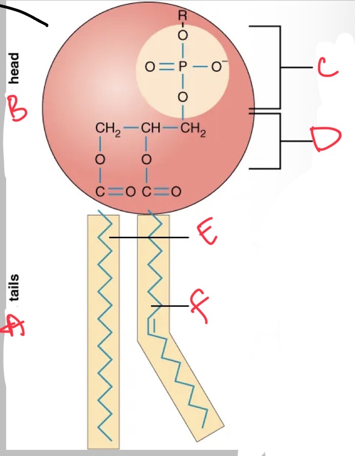
What are the properties of the phosphate head?
How does this form a bilayer?
negatively charged
Polar and hydrophilic - water loving
Attracted to: Extracellular fluid, intracellular fluid
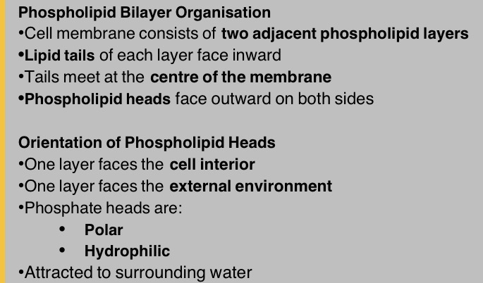
Faces outwards on both sides of the membrane
What are functions of the fatty acid tails? How does they form the bilayer?
uncharged and non polar
Hydrophobic - water fearing
Oriented inwards - away from water
Form the membranes hydrophobic core
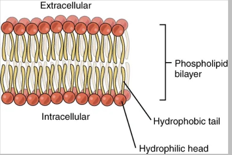
What are the fluid environments of the phospholipid bilayer?
Intracellular fluid (ICF) - fluid inside the cell
Extracellular fluid (ECF) - fluid outside the cell
Interstitial fluid (IF) - ECF not contained within blood vessels
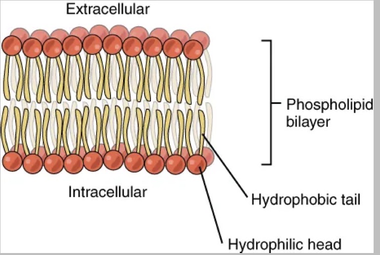
How is the phospholipid bilayer organised?
See below
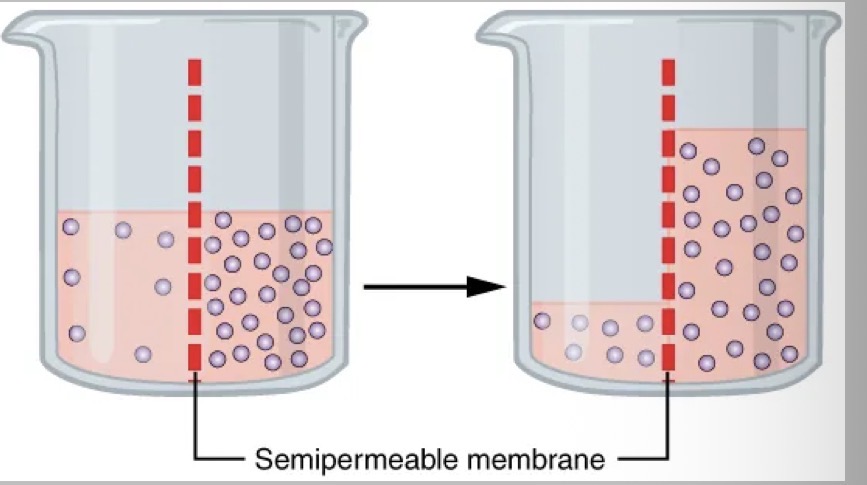
How does the neuronal membrane act as a semipermeable barrier?
Separates intracellular fluid (ICF) from Extracellular fluid (ECF)
Allows some substances to cross while restricting others
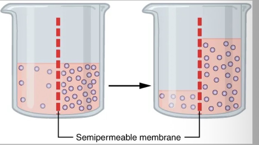
How are neurons selectively permeable ?
neuronal membranes are:
highly selective
Not freely permeable to most ions
Ion movement depends on:
specific ion channels
Membrane properties
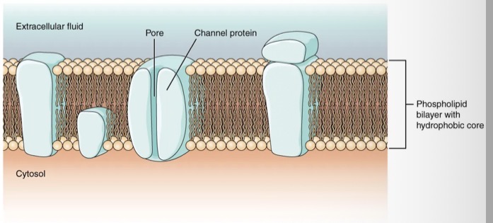
what are properties of channel proteins ?
span the membrane
Contain a central pore
Provide a hydrophobic pathway through the hydrophobic core
Allow specific ions to cross the membrane
How are ion channels selective ?
only certain ions can pass, Selectivity depends on:
Channel size
Charge lining the pore
Critical for controlling neuronal excitability
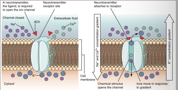
Explain the ligand gated ion channels
channel is closed at rest
Neurotransmitter (ligand) bind to receptor site
Binding opens the ion channel
Creates a hydrophilic pore through the membrane
Ions move down their electrochemical gradients
Na+ / Ca2+ enter, K+ exits
Ion movement changes membrane potential
Convert chemical signal → electrical response
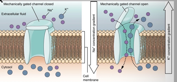
Explain how the mechanically gated ion channels enable sensory transduction
channel closed at rest
Opens in response to mechanical deformation (stretch, pressure etc)
Mechanical force causes conformational change in channel protein
Channel opening creates a hydrophilic pore
Ions move down their electrochemical gradients
Na+ enters, K+ exits
Ion flow alters membrane potential
Enables sensory transduction
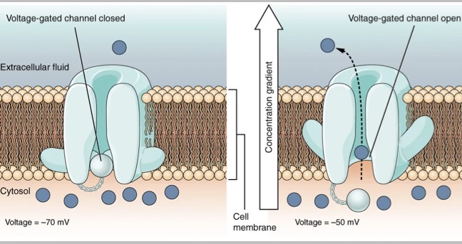
Explain how an action potential is generated and propagated
channel closed at resting membrane potential
Membrane depolarisation triggers channel opening
Opening occurs at threshold voltage (approx 50mv)
Voltage change causes conformational shift in channel protein
Channel opens → hydrophilic pore formed
Ions move down their electrochemical gradients
Rapid ion flux changes membrane potential
Essential for action potential generation and propagation
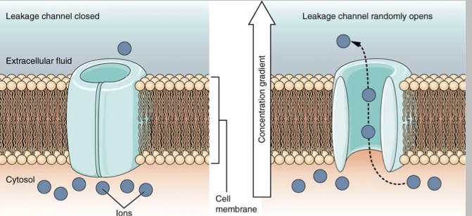
Explain why membrane potential is stable but never static
channels are not gated by stimuli
Randomly open and close at rest
Allow continuous passive ion movement
Ions move gradually down their concentration gradients
Most permeable to K+ (some Na+)
Net K+ efflux dominates
Establish and maintain the resting membrane potential
Membrane potential is stable but never static
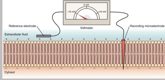
How can we measure resting membrane potential?
Measured using 2 electrodes:
reference electrode in Extracellular fluid
Recording micro electrode inside the neuron
Electrodes connected to a voltmeter
Voltmeter measures electrical potential difference
What is the resting membrane potential?
Voltage difference across the neuronal membrane
At rest what mV is the neuron?
-70mv
Inside of the neuron is what relative to outside?
Negative
What do the results of measuring the resting potential membrane reflect?
ion concentration gradients
Selective membrane permeability
Leak channel activity
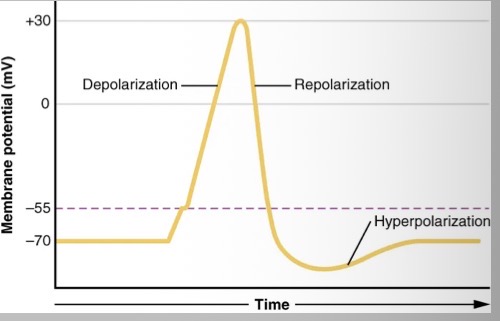
Explain the process of depolarisation into repolarisation into hyper polarisation then returning to rest
threshold reached at approx 55mv
voltage gated Na+ channels open
Rapid Na+ influx
Membrane potential rises to approx 30mv
Na+ channels inactivate
Voltage gated K+ channels open
K+ efflux restores negativity
K+ channels remain open briefly
Membrane potential becomes more negative than rest
K+ channels close
Leak channels and ion pumps re-establish resting potential
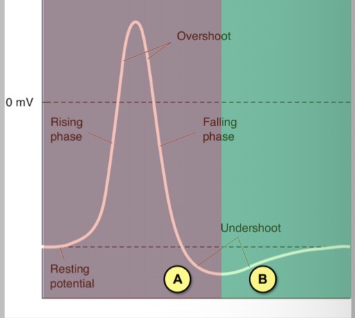
When does absolute refractory period occur ?
During rising phase, overshoot, early falling phase
What causes absolute refractory period and what does it ensure?
Caused as Voltage gated Na+ channels are inactivated
Ensures:
one way propagation of action potentials
Maximum firing frequency is limited
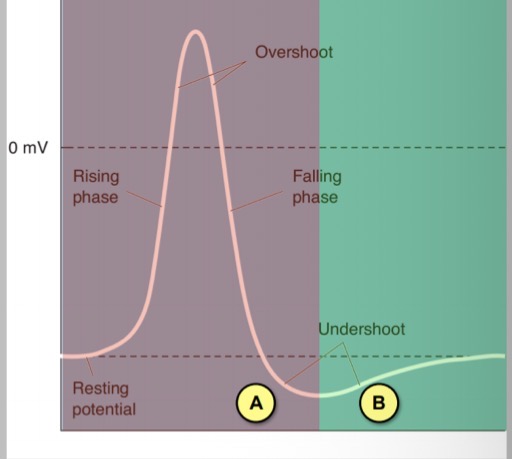
When does relative refractory period occur?
Late falling phase, undershoot (Hyper polarisation)
What is the cause of a relative refractory period ?
Stronger than normal stimulus is required
Caused by some Na+ channels being recovered
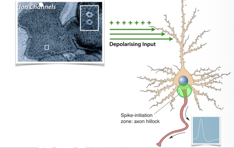
Why do action potentials start at the axon hillock?
Due to the high proportion of voltage gated Na+ and K+ channels there
This high proportion of voltage gated Na+ and K+ channels continues down the axon to the terminal
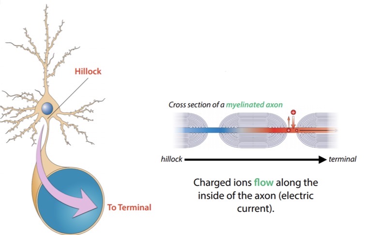
What happens to an action potentials after its initiated at the hillock?
Propagates in one direction towards the terminal
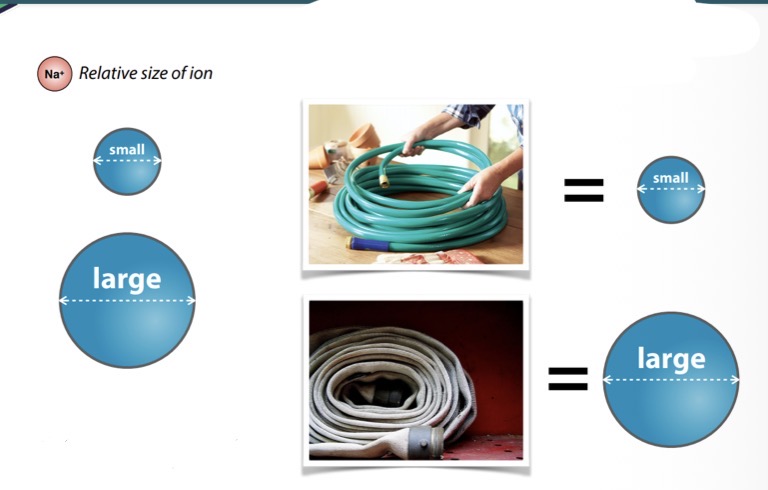
What does it mean when an axon has a large diameter?
Large diameter allows more ions to flow through them
Ions have a much harder time flowing through small diameter axons
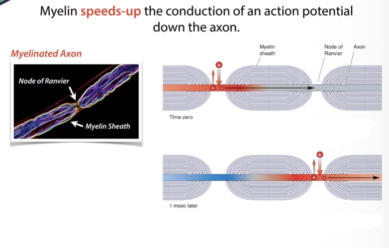
How does myelin speed up the conduction of an action potentials firing down the axon?
Action potentials are only generated at each node of ranvier
Action potentials ‘skip’ their way down the axon
Action potential regeneration at each node is called saltatory conduction, saltatory conduction in a myelinated axon is significantly faster
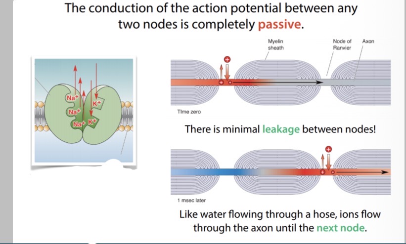
What are the benefits of myelination?
gives action potentials a speed boost
Gives Na+/K+ pumps a rest
What is synaptic transmission?
The process of information transfer between neurons
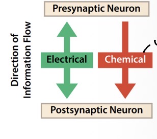
What is a chemical and electrical synapse?
Electrical synapse: information can pass between cels in either direction
Chemical synapse: information flow is unidirectional
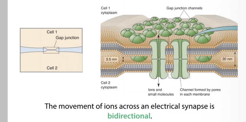
At an electrical synapse neurons are coupled together by what?
Gap junctions
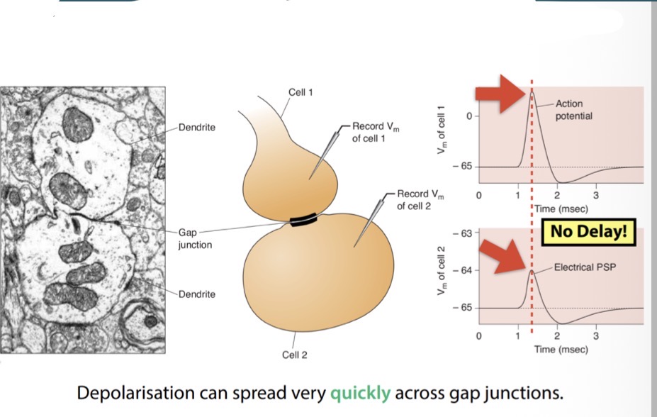
How can depolarisation spread quickly across gap junctions?
As there is no measurable delay in signal propagation across an electrical synapse
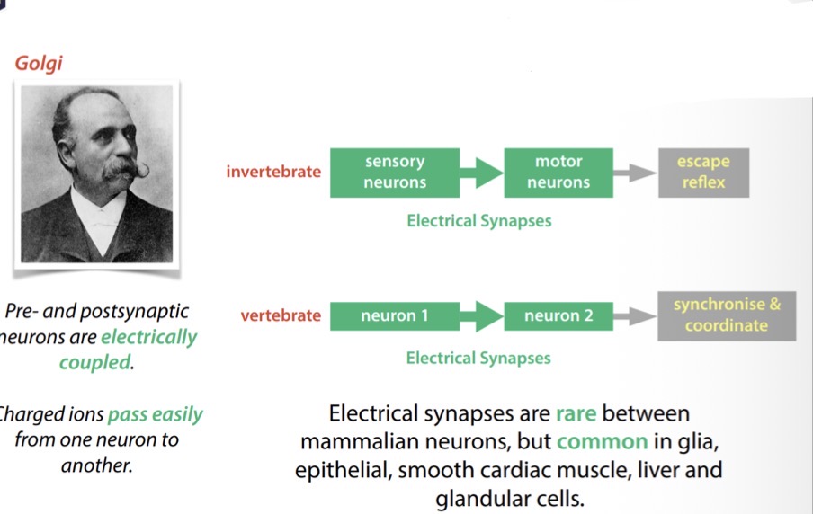
Electrical synapses mediate what?
Important reflexes including the coordination of heart muscle contractions
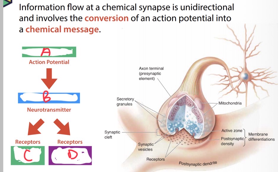
What are A, B, C and D?
A- electrical signal
B- chemical message
C- electrical signal
D- cell metabolism
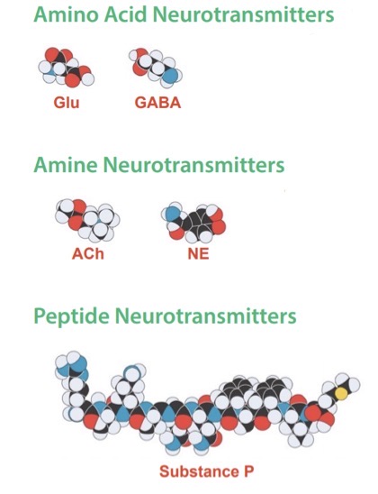
What size and speed are amino acid neurotransmitters?
small and fast acting
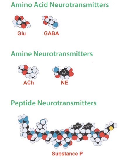
What size and speed are amine neurotransmitters?
intermediate size: both fast and modulatory actions
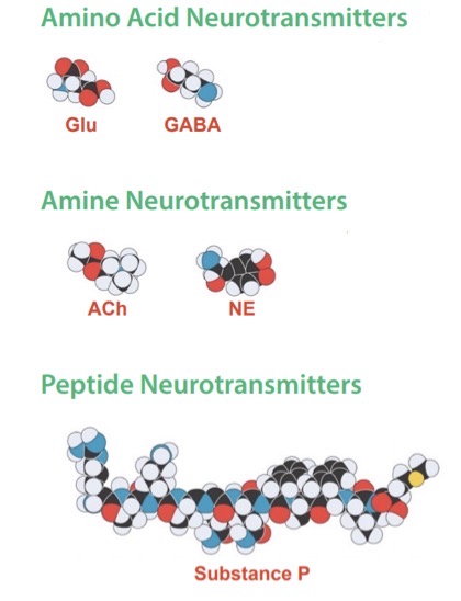
What size and speed are peptide neurotransmitters?
large size with slow and specialised actions
Where are small and intermediate sized neurotransmitters made ?
Directly in the axon terminal
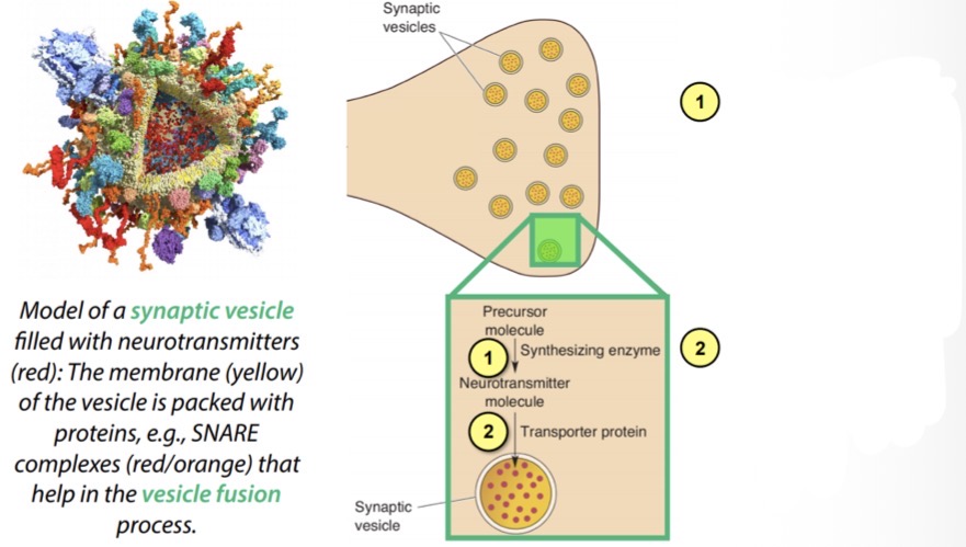
What is happening in this image?
1) precursor molecule interacts with an enzyme in the cytoskeleton at the terminal region and is converted inti a neurotransmitter
2) neurotransmitter is packaged and stored in synaptic vesicles
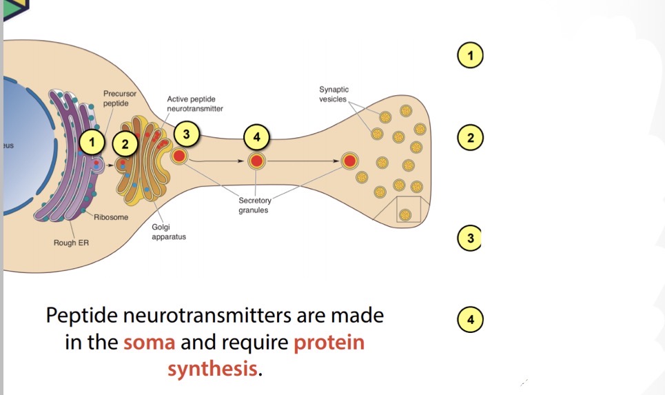
Describe what’s happening in this image
1) precursor peptide is synthesised in the rough ER
2) in the Golgi apparatus the new peptide is sorted, activated and packaged
3) secretory granules “bud” away from the Golgi apparatus
4) the granules are transported to the terminal via axoplasmic transport
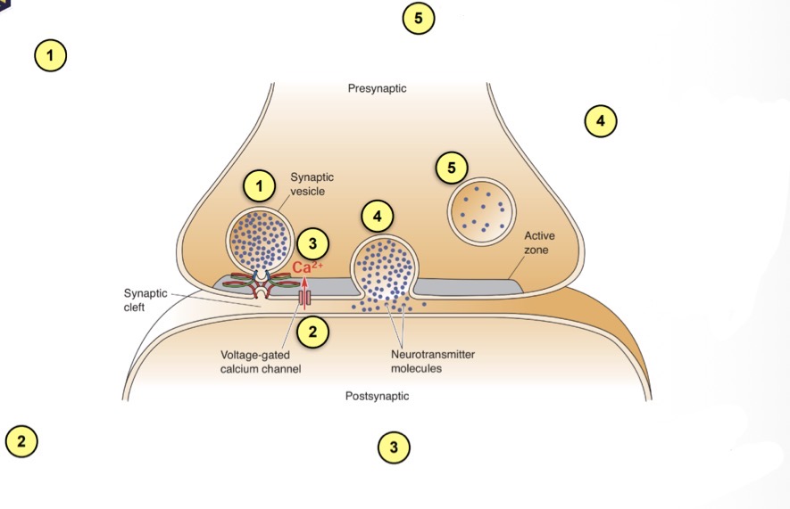
Describe how this process leads to endocytosis?
1) vesicle is docked along inside of presynaptic membrane
2) an action potentials depolarises terminal opening voltage-gated Ca2+ channels
3) Ca2+ rapidly floods into the terminal
4) Ca2+ interacts with snare proteins causing the vesicle to fuse with the presynaptic membrane and spill its contents (exocytosis)
5) the vesicle membrane is recycled (endocytosis)
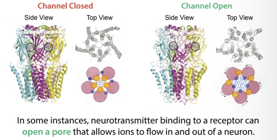
What happens to a receptor when a neurotransmitter binds to it ?
It affects the functioning of that receptor
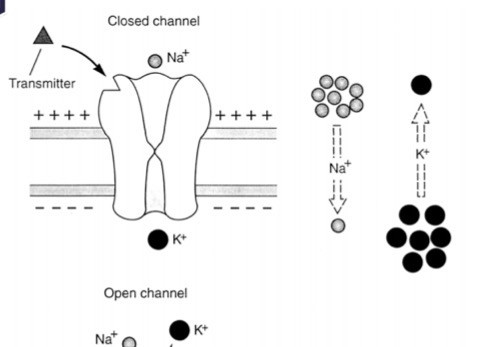
What happens when there is no neurotransmitter bound to the receptors ?
Then the ion channel remains closed and impermeable to ions
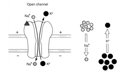
How are certain ions allowed to pass?
The neurotransmitter causes a change in the shape of the subunits that form the channel allowing certain ions to pass
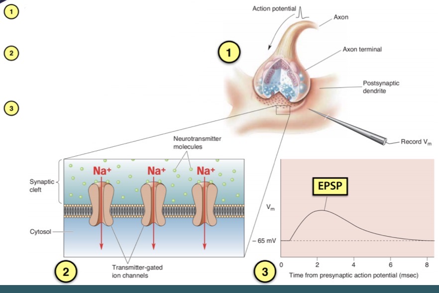
How is an excitatory postsynaptic potential formed (EPSP)
1) neurotransmitter binds to receptors which opens ion channels
2) the influx of Na+ into the postsynaptic cell will depolarise Vm towards threshold
3) this is called an excitatory postsynaptic potential
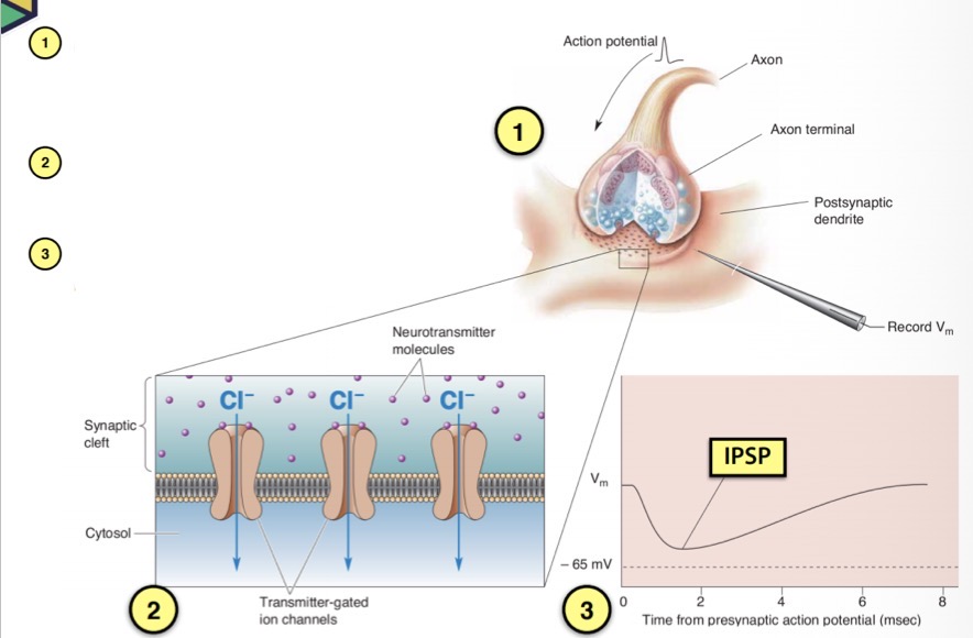
How is an inhibitory postsynaptic potential (IPSP) formed?
1) neurotransmitter binds to receptors which changes the conformation of the receptors and opens ion channels
2) this influx of Cl- into the postsynaptic cell will repolarise Vm towards rest
3) this is called an inhibitory postsynaptic potential
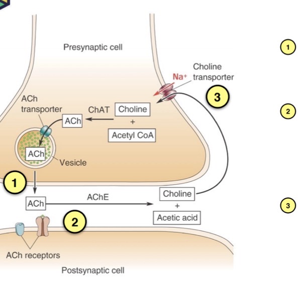
Describe what is happening in this image
1) acetylcholine (ACH) is released upon the arrival of an action potential
2) ach acts for a short time on postsynaptic receptors before it is degraded by acetylcholinesterase
3) choline is recycled and pumped back into the presynaptic terminal by the choline transporter
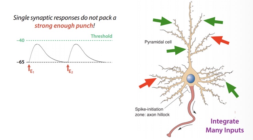
A single EPSP is usually too small to make a neuron fire an action potential, what is done about this?
EPSPs have to be summed together to bring a neuron to firing threshold
Activity across space and time is additive
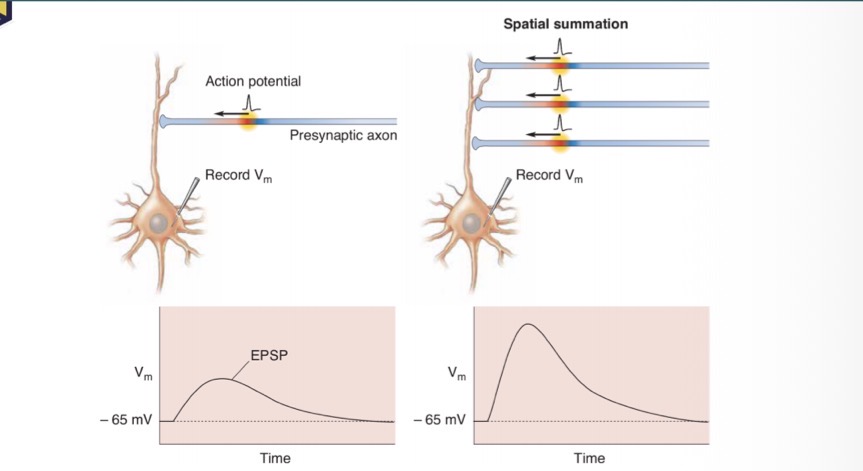
How does activation of a single synapse vs activation of ,ultiple inputs generate ?
Single synapse activation generates a small amplitude EPSP
Multiple input activation simultaneously generates a much larger postsynaptic response
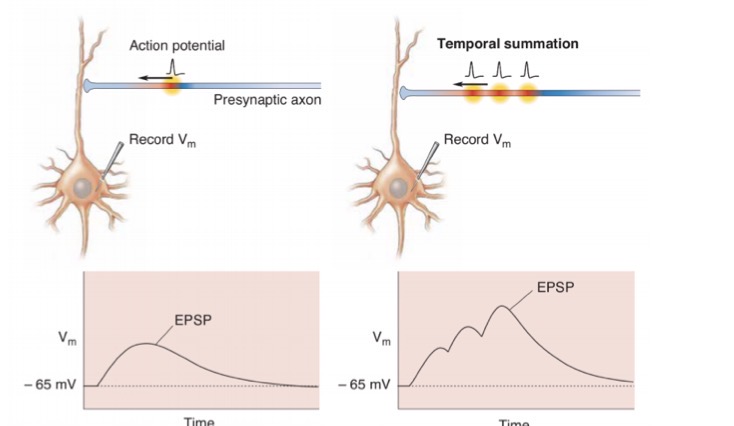
How does the image on the right show us how a much larger postsynaptic response can be generated?
Activating the same synapse in rapid succession
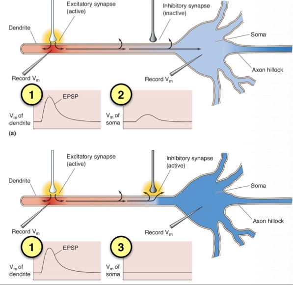
What is happening in this image ?
1) the EPSP recorded at the active synapse is large in amplitude
2) EPSP loses much of its “punch” with distance
3) a tonically active inhibitory synapse would drive Cl- inwards and K+ outwards as the EPSP approached