A&P II Lab: Quiz 1 - Visual Senses & Hearing and Equilibrium
 Call Kai
 Learn
Learn Practice Test
 Spaced Repetition
 Match
Match Flashcards
 Knowt Play
1/23

Name | Mastery | Learn | Test | Matching | Spaced | Call with Kai | Chat |
|---|
No analytics yet
Send a link to your students to track their progress
24 Terms
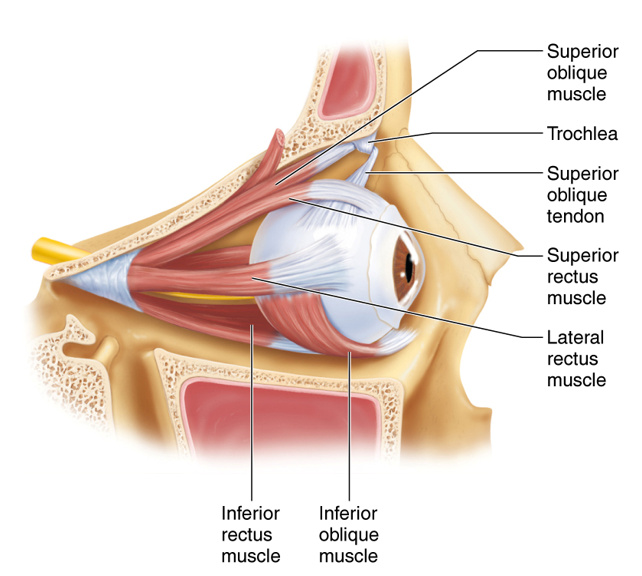
Rectus
Straight Muscles
Oblique
Slanting Muscles
Superior Oblique
Depresses, turns laterally
Inferior Oblique
Elevates, turns laterally
Superior Rectus
Elevates, turns medially
Inferior Rectus
Depresses, turns medially
Medial/Lateral Rectus
Turns eye medially/laterally
Trochlea
Pulley system
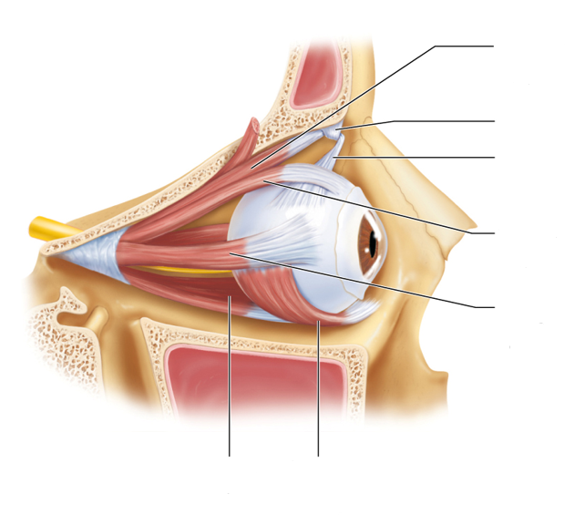
Eye Muscles Diagram
External/ Fibrous Layer
Sclera - White connective tissue of the eye
Cornea - Continuous with sclera; Transparent anterior bulge with no blood vessels
Middle/ Vascular Layer
Choroid - Blood vessel-rich dark membrane, produces nutrients and melanin
Ciliary Body - Choroid modification that encircles the lens
Ciliary Muscle - Smooth muscle in ciliary body
Ciliary Process - Radiating folds of ciliary muscle with capillaries
Ciliary Zonule - Suspensory Ligament; Halo of fine fibers extending from ciliary process around lens
Iris & Pupil - Eye pigment; 2 layers of smooth muscle: sphincter pupillae and dilator pupillae
Sphincter pupillae: Contracts to constrict pupil
Dilator pupillae: Contracts to dilate pupil
Inner Layer/ Retina
Pigmented Layer - Outer layer composed of a single layer of melanocytes (pigmented cells)
Neural Layer - Thicker layer composed of 3 neurons: 1. Photoreceptors (rods and cones), 2. Bipolar cells, 3. Ganglion cells
Macula Lutea - Part of retina responsible for sharp, detailed, central vision
Fovea Centralis - Center of macula lutea, responsible for Fovea vision (sharp, central vision)
Optic Disc - Location where ganglion cell axons exit the eye to form optic nerve
No light sensitive rods/cones to respond to light stimulus – blind spot
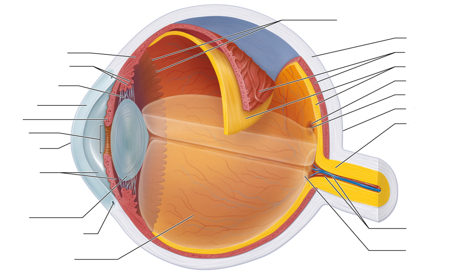
Internal Eye Anatomy
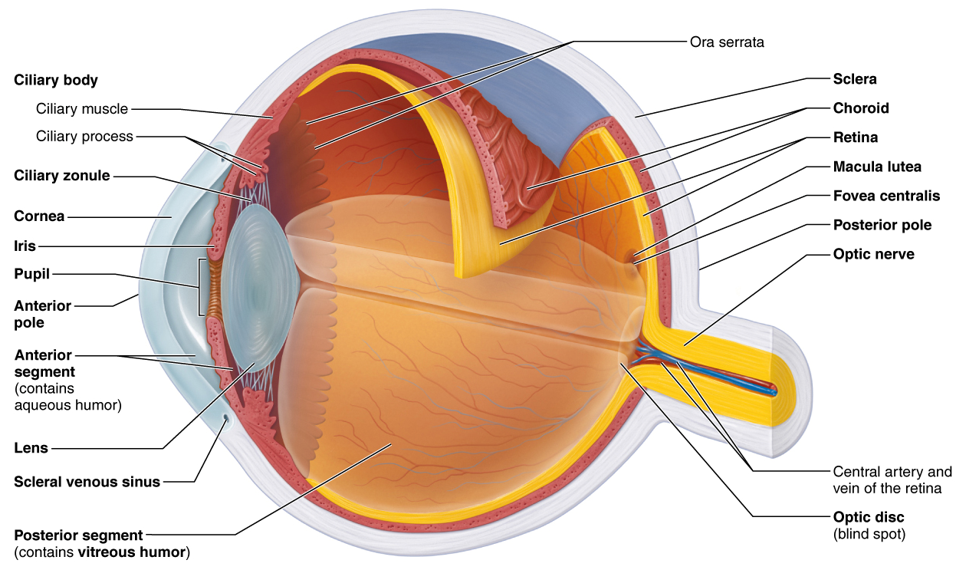
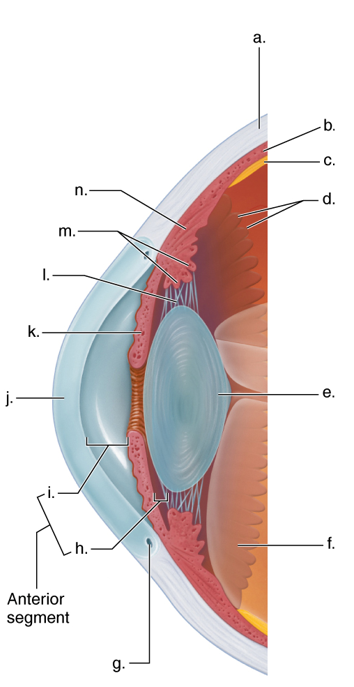
Internal Eye Anatomy 2
a. Sclera
b. Choroid
c. Retina
d. Ciliary processes
e. Lens
f. Vitreous humor
g. Aqueous humor
h. Iris
i. Cornea
j. Anterior chamber
k. Pupil
l. Suspensory ligaments (zonular fibers)
m. Ciliary muscle
n. Conjunctiva
Cones and Rods
Cones = Colors
Rods = Low Light Levels
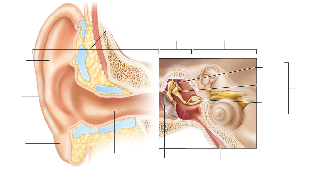
Ear Anatomy
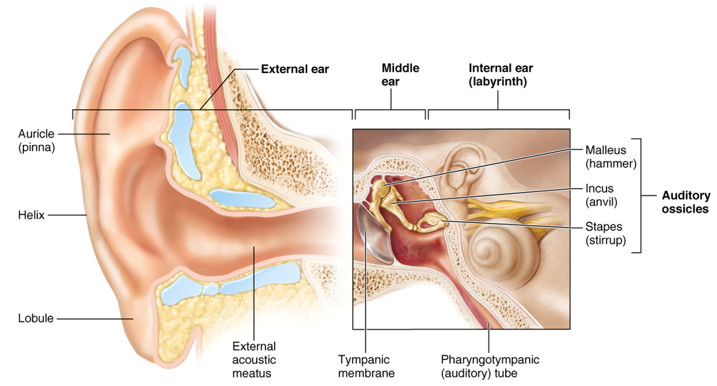
External Ear
Auricle (pinna) - Outer part of the ear; Elastic cartilage covered with skin
Collects and directs sound waves into the external acoustic meatus
Lobule (“earlobe”) - Part of auricle inferior to external acoustic meatus; Where ears are pierce
Helix - Between lobule and auricle
External acoustic meatus (“ear canal”) - Short, narrow canal carved into temporal bone lined with ceruminous glands
Transports sound waves from the auricle to the tympanic membrane
Tympanic Membrane (“ear drum”) - Thin membrane that separates the external ear from the middle ear
External Ear Simplified
Collect then transport sound waves:
Auricle → Lobule → External Acoustic Meatus → Tympanic Membrane
Middle Ear/Tympanic Cavity
Auditory Ossicles
Malleus (hammer) - Tiny “hammer”-like bone with its “handle” attached to the eardrum
Incus (anvil) - Tiny “anvil”-like bone that articulates with malleus and stapes
Stapes (stirrup) - Tiny “stirrup”-like bone with its “base” fitted inside the oval window
Oval Window - Oval-shaped membrane located deep to the stapes
Pharyngotympanic / Auditory Tube - Tube that connects the middle ear to the superior portion of the pharynx (throat)
Middle Ear Simplified
Transmit and amplify vibrations:
Ear Drum → Malleus → Incus → Stapes → Oval Window → Perilymph
Internal Ear
Cochlea
Membranous Labyrinth: Cochlea duct
Structure containing receptors: Spiral organ
Function of receptors: Hearing
Vestibule
Membranous Labyrinth: Utricle and saccule
Structure containing receptors: Maculae
Function of receptors: Equilibrium - Static equilibrium and linear acceleration of the head
Semicircular Canals
Membranous Labyrinth: Semicircular ducts
Structure containing receptors: Ampullae
Function of receptors: Equilibrium - Rotational acceleration of the head
Hair Cell Detection
Otoliths (ear stones) - Calcium / Ear crystals that serve as weights on top of the hair cells that detect the movement
If the crystal is knocked off the hair cell, the person will feel a sense of motion/spinning, dizziness – vertigo
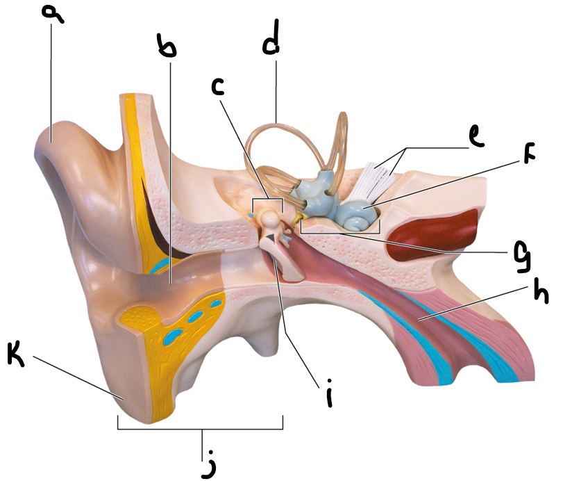
Ear Anatomy 2
a. Auricle (pinna)
b. External auditory meatus
c. Malleus (hammer)
d. Semicircular canals
e. Vestibulocochlear nerve
f. Cochlea
g. Middle ear cavity (tympanic cavity)
h. Auditory tube
i. Tympanic membrane (eardrum)
j. External ear
k. Lobule
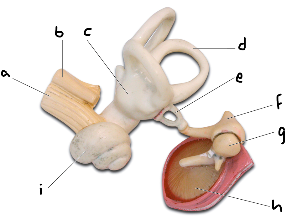
Internal Ear Anatomy
a. Cochlear nerve
b. Vestibular nerve
c. Semicircular canals
d. Semicircular canal
e. Incus (anvil)
f. Malleus (hammer)
g. Stapes (stirrup)
h. Tympanic membrane (eardrum)
i. Cochlea