Mild TBI & Neck Pain SG
 Call Kai
 Learn
Learn Practice Test
 Spaced Repetition
 Match
Match Flashcards
 Knowt Play
1/106

There's no tags or description
Looks like no tags are added yet.
Name | Mastery | Learn | Test | Matching | Spaced | Call with Kai | Chat |
|---|
No analytics yet
Send a link to your students to track their progress
107 Terms
SCM, scalenes, upper trapezius, cervical erector spinae, levator scapulae
Global muscles of cervical spine:
Longus colli, rectus capitis anterior/lateralis, longus capitis, multifidi
Deep segmental stabilizers of the spine:
- Origin: External occipital protuberance, nuchal ligament
- Insertion: Lateral 1/3 of clavicle
- Action: Upward rotation, retraction, and elevation of scapula.
- Innervation: CN XI (C3-C4 sensory)
Upper Trapezius

- Origin: Manubrium, clavicle
- Insertion: Mastoid process
- Action:
- Bilateral: Extends head, flexes neck, raises thorax
- Unilateral: Ipsilateral lateral flexion and contralateral rotation
- Innervation: CN XI (C2-3 sensory)
Sternocleidomastoid (SCM)

- Origin: Transverse processes of C1-C4
- Insertion: Superior angle of scapula
- Action: Elevates, downwardly rotates, retraction of scapula
- Innervation: Dorsal scapular nerve (C4, C5), Anterior rami (C3,C4)
- Circulatory: Dorsal scapular artery
Levator Scapula

- Origin:
- Anterior: Transverse processes C3-6
- Middle: Transverse process of C2-7
- Posterior: Transverse process of C4-6
- Insertion:
- Anterior and middle: 1st rib
- Posterior: 2nd rib
- Action:
- All: Flexes, laterally flexes neck, rotates neck opposite, elevates 1st rib
- Innervation:
- Anterior: Ventral rami C4-6
- Middle: Ventral rami C3-8
- Posterior: Ventral rami C6-8
- Circulatory: Branches of subclavian artery
Scalenes
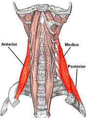
1. Longus colli
2. Longus capitis
3. Rectus capitis anterior
4. Rectus capitis lateralis
What are the anterior prevertebral muscles?

- Origin: Transverse processes C3-C5; Bodies of C5-T3
- Insertion: Anterior tubercle of atlas (C1), anterior vertebral bodies of C1-3, transverse processes of C3-C6
- Action:
- Bilateral: Flexes neck
- Unilateral: Contralateral rotation
- Innervation: Ventral Rami of cervical spinal nerves
Longus colli
- Origin: Transverse processes of C3-C6
- Insertion: Inferior surface occipital bone
- Action: Flexes neck
- Innervation: Ventral Rami of cervical spinal nerves
Longus capitis
- Origin: Anterior, lateral mass of atlas (C1)
- Insertion: Base of occipital bone anterior to foramen magnum
- Action: Flexes head, stabilizes A-O joint
- Innervation: Ventral Rami of cervical spinal nerves
Rectus capitis anterior
- Origin: Transverse process of atlas
- Insertion: Jugular process of the occipital bone
- Action: Lateral flexes head
- Innervation: Branches of loop between C1-C2
Rectus capitis lateralis
1. Rectus capitus posterior major
2. Rectus capitus posterior minor
3. Inferior oblique of the head
4. Superior oblique of the head
What are the suboccipital muscles?

- Origin: C2 spinous process
- Insertion: Inferior nuchal line
- Action:
- Unilateral: ipsilateral rotation of head
- Bilateral: Extends head at AO joint
- Innervation: Suboccipital N (C1)
Rectus capitus posterior major
- Origin: C1 posterior tubercle
- Insertion: Inferior nuchal line
- Action:
- Unilateral: ipsilateral rotation of head
- Bilateral: Extends head at AO joint
- Innervation: Suboccipital N (C1)
Rectus capitus posterior minor
- Origin: C2 spinous process
- Insertion: C1 transverse process
- Action: Ipsilateral rotates head
- Innervation: Suboccipital N (C1)
Inferior oblique of the head
- Origin: C1 transverse process
- Insertion: Occipital bone (B/w superior and inferior nuchal lines)
- Action: Extends, lateral flexes head
- Innervation: Suboccipital N (C1)
Superior oblique of the head
7
How many cervical vertebrae are there?
8; above except C8 exits between C7-T1
How many cervical spinal nerves?
Exit above or below same named vertebra? Except?
Atlanto-occipital
_________ joints: Condylar synovial joint that permits flexion and extension of the cranium. This motion is often noted when nodding the head to say "Yes"
Atlanto-axial
________ joints: Plane synovial joints that permit flexion, extension, lateral flexion, and rotation of the spin. The majority of rotation of the skull on the spinal column occurs here.
Intervertebral
________ joints: Formed by the superior and inferior surfaces of the vertebral bodies and the associated disk.
Zygapophyseal
________ joints: Formed by the right and left superior articular facets of one vertebra and the right and left inferior articular facets of an adjacent superior vertebra.
Intervertebral discs
________: Formed by a dense layer of collagen fibers and fibrocartilage called the annulus fibrosus as well as a flexible inner layer called the nucleus pulposus.

Posterior and superior
Cervical superior articular facets face?
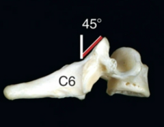
1. Capsular pattern restriction: Lateral flexion and rotation equally limited, then extension
2. Loose pack (Facet): Midway between flexion and extension
3. Closed pack (Facet): Extension
Cervical capsular pattern/closed pack/loose pack:
1. Flexion/extension
2. Side bending
3. Rotation
4. Anterior/posterior shear
5. Lateral shift
6. Compression/distraction
Six degrees of motion of the spine:
Upper cervical extension with lower cervical flexion
Protraction of c-spine:
Upper cervical flexion with lower cervical extension
Retraction of c-spine:
Alar ligament
______ ligament: From dens (C2) to occipital condyle; limit axial rotation of head and atlas relative to the dens.

Transverse ligament
______ ligament: Between the lateral masses of C1 and functions to provide posterior support to the dens (forming a collar with the anterior arch of C1)

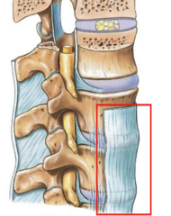
Posterior longitudinal ligament
_________ ligament: Limits flexion, reinforces posterior disc.

Anterior longitudinal ligament
_________ ligament: Limits extension of the spine and reinforces the anterior portion of the intervertebral disks and vertebrae.
1. Passive (bones/ligaments resist motion)
2. Active (muscles respond dynamically)
3. Neural control (nervous system coordinates muscle activation)
Spinal stability is supported by three subsystems:
A. Anterior cerebral artery
B. Internal carotid artery
C. Posterior communicating artery
D. Posterior cerebral artery
E. Vertebral artery
F. Basilar artery
G. Arterial circle
H. Middle cerebral artery
I. Anterior communicating artery
Circle of Willis

1. Roots (C5-T1)
2. Trunks (superior, middle, inferior)
3. Divisions (anterior, posterior)
4. Cords (lateral, medial, posterior)
5. Branches (Musculocutaneous, axillary, median, radial, ulnar)
Brachial plexus

1. Dorsal scapular (rhomboids, levator scapulae)
2. Long thoracic (Serratus anterior)
Brachial plexus: Nerves/muscles from the rami of the plexus
1. Nerve to subclavius (subclavius)
2. Suprascapular (infraspinatus/supraspinatus)
Brachial plexus: From the trunks of the plexus
1. Lateral pectoral (pectoralis major - clavicular head)
2. Musculocutaneous (coracobrachialis, biceps brachii, brachialis)
3. Lateral root of the median (flexor muscle in forearm, except flexor carpi ulnaris, and five muscles in the hand)
Brachial plexus: From the lateral cord of the plexus
1. Medial pectoral (pectoralis major, pectoralis minor)
2. Ulnar (flexor carpi ulnaris, flexor digitorum profundus, most small muscles of hand)
3. Medial root of the media (flexor muscles in the forearm, except flexor carpi ulnaris, and five muscles of the hand)
Brachial plexus: From the medial cord of the plexus
1. Upper subscapular (subscapularis)
2. Thoracodorsal (latissimus dorsi)
3.Lower subscapular (subscapularis, teres major)
4. Axillary (deltoid, teres minor)
5. Radial (brachioradialis, triceps, supinator, wrist extensors, anconeus)
Brachial plexus: From the posterior cord of the plexus
"Oh Oh Oh to touch and feel very good velvet, ah heaven"
"Some say marry money but my brother say big brains matter most"
CN mnemonics
Olfactory
CN I: ________
Classification: Sensory
Function: Smell
Test: Identify familiar odors
Optic
CN II: ________
Classification: Sensory
Function: Sight
Test: Test visual fields
Oculomotor
CN III: ________
Classification: Motor
Function: Eye Movement: Look R, L, up, down, and parasympathetic motor
Test: Upward, downward, and medial gaze/reaction to light - smooth pursuit
Trochlear
CN IV: ________
Classification: Motor
Function: Eye muscle: Superior oblique
Test: Downward and inward gaze
Trigeminal
CN V: ________
Classification: Both
Function: Face sensation and mastication
Test: Face sensation. corneal reflex, clench teeth
Abducens
CN VII: ________
Classification: motor
Function: Eye muscle: lateral rectus muscle
Test: Lateral gaze
Facial
CN VII: ________
Classification: Both
Function: Taste (anterior tongue), facial expression muscles, and parasympathetic motor
Test: Closes eye, smile, puff cheeks, familiar tastes
Vestibulocochlear
CN VIII: ________
Classification: Sensory
Function: Hearing and balance
Test: Hearing test/eyes closed
Glossopharyngeal
CN IX: ________
Classification: Both
Function: Taste and parasympathetic motor
Test: Gag reflex/ability to swallow
Vagus
CN X: ________
Classification: Both
Function: Taste, fore/midgut sensory and parasympathetic motor
Test: Gag reflex, ability to swallow, say "Ahhh"
Accesory
CN XI: ________
Classification: Motor
Function: Trapezius & SCM
Test: Resisted shoulder shrug
Hypoglossal
CN XII: ________
Classification: Motor
Function: Muscle of tongue
Test: Tongue protrusion (if inured, tongue deviates toward injured side)
1. C5, C6 Musculocutaneous
2. C5, C6 Radial
3. C6, C7 Radial
Match the following:
1. Biceps
2. Brachioradialis
3. Triceps
C6, C7 Radial
C5, C6 Musculocutaneous
C5, C6 Radial
Meninges
____________: Consist of three layers of connective tissue covering the brain and spinal cord. They provide protection from contusion and infection. There are blood vessels and cerebrospinal fluid within them.
Dura mater, arachnoid mater, pia mater
Put in order from superficial to deep:
- Pia mater
- Dura mater
- Arachnoid mater
Blood-brain barrier
___________: Consists of the meninges, protective glial cells, and capillary beds of the brain. It is responsible for exchange of nutrients between the CNS and the vascular system. Provides protection for the CNS by restricting certain molecules from crossing the barrier while others are able to do freely.
Cerebral spinal fluid
_________: Clear, fluid-like substance that cushions the brain and spinal cord from injury and provides shock absorption, circulates nutrients and chemicals filtered from the blood, and removes waste products.
Causes structural and biochemical changes:
- Stretching of axons
- Damage to cell membranes
- Dysregulated ion flow
- Increased glutamate release
This disrupts normal neurological function and produce concussion symptoms
What is occurring physiologically during a concussion?
1. CNS/brain
2. Vestibular system
3. Oculomotor/visual
4. Cervical musculoskeletal system
5. Autonomic/exertional system
6. Cognitive/emotional systems
What tissues or body systems are involved in a concussion?
- Blunt force or jolt to the head or neck
- Direct blow to the head, face, neck
- Force elsewhere on the body transmitted to the head
- Common mechanisms: falls (main), sports collision, and blast injuries
- Causes brain to rapidly twist or bounce inside of skull
What causes a concussion?
- Symptoms may occur immediately
- LOC may occur but is not required
- PTA/altered mental status may occur (Antero/retrograde)
- Recovery can occur as quickly as 7-10 days
- >14 days = protracted recovery
- Some develop persistent symptoms lasting months or longer
- Progress from relative rest for the first 24-48 hours and gradually return to daily activity/exercise based on symptoms
What causes a concussion to progress/resolve?
1. Cognitive/fatigue: Fogginess, concentration/memory problems fatigue
2. Vestibular: Dizziness, nausea, imbalance, motion sensitivity
3. Ocular: Blurred/double vision, difficulty focusing/reading
4. Migraine/headache: Headache, nausea, light/noise sensitivity
5. Anxiety/mood: Anxiety, depression, irritability, emotional changes
6. Motor/balance: Impaired balance, coordination, dual-tasking
7. Autonomic/exertional: Symptoms w/ PA and heart rate
8. Cervical: Neck pain, decreased ROM, cervicogenic
9. Sleep: difficulty sleeping or altered pattern
What impairments develop because of a concussion?
Anyone sustaining sufficient head/body trauma
Common populations:
- Athletes
- Military personnel
- Individuals who fall
Who develops a concussion?
1. Sports participation
2. Previous concussion
3. Female sex - risk factor
4. Concussion risk is greater during games
4. Collision/contact exposure
5. Military personnel
What characteristics or demographics are common in concussions?
1. Risk factors
- Previous concussion
- Sports
- Female sex
2. Medical history
- Cognitive/fatigue: Prior cognitive deficits or sleep disturbance
- Vestibular: History of motion sickness
- Ocular: Oculomotor impairment (strabismus)
- Migraine: Personal or family history
- Anxiety/mood: Hx of mental health disorder
3. Lifestyle/environmental
- Sports, continuing to participate after injury
- High physical/cognitive demands
- Poor sleep
- Visually stimulating environment
What are the predisposing risk factors for concussions?
Typical MOI:
- Direct blow to head, face, or neck
- Jolt/impulsive force transmission to head
- Fall (main cause)
Common patient history:
- Headache, dizziness, feeling dazed/foggy, confusion, balance problems, nausea, visual disturbance, difficulty concentrating/remembering, LOC may or may not occur, PTA may occur
MOI of concussions
Chief complaint: Reports of headache, dizziness, fogginess/fatigue, difficulty concentrating, balance problems, visual problems, neck pain, nausea, light/noise sensitivity.
Subjective findings during concussion: Chief complaints
- Symptoms vary between patients
- May have a few symptoms at rest but symptoms appear with activity
- Symptoms can increase with physical, visual, vestibular, or cognitive demands
- May become more symptomatic with dynamic movement and crowded environments
Subjective findings during concussion: Symptom behavior
Aggravating:
- Exercise/PA
- Head movement
- Reading/screens
- Bright lights/noise
- Busy/crowded environments
- Cognitive activity
- School/work
- Poor sleep
Easing:
- Relative rest
- Decreasing provocative stimulation/activity
- Pacing activities
(24-48 hours of relative rest followed by gradual return to daily activity)
Subjective findings during concussion: Aggravating and easing factors
School/work, reading/computer use, exercise/sports, walking/balance, driving, community activity, dual-tasking, return to previous PA
Subjective findings during concussion: Functional limitations
- What happened? MOI
- Did you lose consciousness? Length?
- Any post-traumatic amnesia?
- What symptoms occurred immediately after?
- Previous concussion?
- History of headaches/migraines?
- mental health or sleep history?
Subjective findings during concussion: Questions to ask
May observe:
- Appears dazed, foggy, or slowed
- Gait/balance difficulty
- Difficulty concentrating or following commands
- Symptoms may increase as examination becomes more provocative
- Normal at rest, but increase symptoms with activity
Objectives findings during concussion: Observation
May have decreased or painful cervical ROM with C-spine involvement
Objectives findings during concussion: ROM findings
With cervical involvement, may have:
- Decreased strength
- Decreased endurance
- Pain with resisted testing
Objectives findings during concussion: Strength
Testing may be normal but screen:
- Cranial nerves
- Sensation/motor function
- UMN
- Cognition/mental status
911 or seek ED if: One pupil larger than other, drowsiness or difficulty waking up, unrelenting or worsening headache, loss of sensation or strength on one side of body, slurred speech, nausea with vomiting, seizures, LOC, unusual behavior/agitated.
Objectives findings during concussion: Neurological
May demonstrate:
- Increased postural sway
- Increased balance errors
- Impaired static/dynamic balance
- Difficulty with gait
- Difficulty with dual-tasking/multitasking
Balance outcome measures include: BESS, SOT, HiMAT, DGI, FGA, and BEST
Objectives findings during concussion: Balance/Postural impairments
May have difficulty with:
- Exercise/sports
- School/work
- Reading/computer use
- Busy/crowded environments
- Busy/crowded environments
- Head movements
- Dual-task
- Return to PA
Objectives findings during concussion: Functional limitations
1. Prognosis
- Generally favorable
- Varies depending on symptom severity/systems involved
2. Typical recovery timeline
- Recovery can occur as quick as 7-10 days, protracted recovery is >14 days
- Some experience symptoms for months or longer
3. Factors with delayed healing
- Immediate LOC
- Post-traumatic amnesia
- Continuing sport after injury
- Prolonged headache
- Fatigue/fogginess/memory problems
- >3 initial symptoms
- Visual memory/processing speed deficits
- Premorbid headaches
- Previous concussion
- Worsening symptoms after injury
- Emotional symptoms on day of injury
- Female sex
Clinical Course - Concussion
1. Activity/work/school:
- Gradually increase physical and cognitive activity
- Use pacing strategies and accommodations as needed
- Progress based on symptom response and irritability
2. Exercise:
- Begin symptom-guided aerobic exercise when appropriate
3. Sport
- Team based decision
- Must be symptoms free at rest and tests including (VOMS, BCTT, phased return to sport, IMPAACT, practice, school demands)
- Impact test is compatible to baseline score
- If protracted recovery, consider ramp up period due to deconditioning
Return to activity/work/sport expectations: Concussion
1. Manual therapy to C-spine/T-spine
2. ROM exercises/ SNAGs
3. Strengthening - i.e., DNF, endurance training
4. Re-integrate cervico-ocular reflex with joint position error training (move head on stationary body, move body on stationary head position)
Recommended interventions: Cervical
1. Treat what you see
2. Recall treatments for saccades, smooth pursuit, convergence, etc.
Refer to vision specialist when necessary:
- Neuro-opthalmologist
- Neurobehavioral optometrist
Recommended interventions: Oculomotor
1. BPPV (Canalith repositioning)
2. Motion sensitivity (desensitization techniques)
3. VOR (vestibular rehab program)
4. Need to integrate into functional activities/sports specific activities
Recommended interventions: Vestibular
How: Stroke lateral plantar foot from heel toward forefoot
Normal response: Toes flex/down
Abnormal: Great toes extend with fanning of other toes
Interpretation: Positive = possible UMN/CNS pathology
Examination techniques concussion: UMN Babinski

How: Flick distal phalanx of middle finger
Normal: No thumb/index flexion
Abnormal: Thumb/index flexion
Interpretation: Positive = possible UMN/CNS pathology
Examination techniques concussion: UMN Hoffmann's

How: Rapidly dorsiflex ankle and maintain stretch
Normal: No sustained rhythmic contractions
Abnormal: Sustained/repetitive beats
Interpretation: Positive = Suggest UMN involvement
Examination techniques concussion: UMN Clonus

How: Tap brachioradialis tendon
Normal: Brachioradialis contraction/elbow flexion
Abnormal: Reduced response with finger flexion
Interpretation: Positive = cervical spine cord/UMN involvement
Examination techniques concussion: UMN Inverted supinator sign

1. Any high-risk factors?
- Age >=65
- Dangerous mechanism
- Paresthesia in extremities
2. Is there a low-risk factor allowing safe ROM testing?
- Simple rear-end MVC
- Able to assume sitting position in ER
- Ambulatory at any time
- Delayed onset neck pain
- No midline cervical tenderness
3. Can patient actively rotate neck 45 left and right?
- Yes - radiography not indicated
- No - radiography indicated
Examination techniques concussion: Canadian C-spine rules

How: Patient keeps head still and follows target with eyes
Normal: Smooth tracking
Abnormal: jerky/poor tracking or symptom provocation
Interpretation: Oculomotor involvement
Examination techniques concussion: Smooth pursuit
How: Patient rapidly looks between two targets horizontal and then vertical
Normal: Quick, accurate eye movements w/o symptoms
Abnormal: Poor accuracy/slowed movements or symptoms
Interpretation: Oculomotor involvement
Examination techniques concussion: Horizontal & vertical saccades
How: Slowly move target toward patient's nose and measure
Normal:
Examination techniques concussion: Convergence
How: Patient looks at stationary target while moving head horizontal and vertical (180 bpm)
Normal: Maintains fixation w/o symptoms
Abnormal: Difficulty maintaining target or symptom provocation
Interpretation: Vestibular/VOR impairment
Examination techniques concussion: Horizontal and vertical VOR
How: Patient focuses on thumb while rotating head, trunk, and arms together
Normal: Minimal/no symptoms
Abnormal: Increased dizziness, headache, nausea, or fogginess
Interpretation: Suggests visual-motion/vestibular sensitivity
Examination techniques concussion: Visual motion sensitivity

How: Stabilize C2 and move the head/atlas into side bend
Normal: Firm restriction
Abnormal: Excessive movement compared with opposite side or soft end-feel
Interpretation: Alar ligament instability
Examination techniques concussion: Alar ligament test

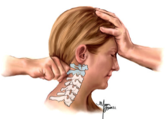
How: Patient seated with slight cervical flexion. Stabilize C2 and apply a posterior force through forehead
Normal: No excessive translation, clunk, or symptom change
Abnormal: Excessive movement, clunk, reduction of neurological symptoms
Interpretation: Transverse ligament/atlantoaxial instability
Examination techniques concussion: Sharp-Purser test
Symptoms: Noncontinuous, unilateral neck pain W/O side shift and associated headache (referred). Headache is precipitated by or aggravated by neck movements or sustained positions/postures.
Demographic: Mean age 42.9
Impairments: Positive CFRT, headache produced with provocation of upper cervical segments, limited cervical ROM, restricted upper cervical segmental mobility, strength, endurance, and coordination deficits of neck and scapulothoracic muscles.
Cause: Impairments in C-spine (upper), stemming from trigeminocervical nucleus
Neck pain with headaches: Symptoms, demographic, and impairments
- Female: male - 50/50
- Unilateral without side shift
- Occipital to frontoparietal and orbital
- Chronic, episodic
- Moderate-severe
- 1 hour to weeks
- Non-throbbing, and non lancinating, pain usually starts in the neck
- Triggers, neck movement, and postures, limited ROM, pressure over C0-3
Cluster: Decreased AROM of cervical extension, palpably painful somewhere from OA to C3/4 joint dysfunction, deep cervical flexor strength impairments w/ CCFT
Differential diagnosis of headaches: Cervicogenic headaches
- Female: male - 75:25
- 60% unilateral with side shift
- Frontal, periorbital, temporal
- 1-4 per month
- Moderate/severe
- 4-72 hours
- Throbbing, pulsating
- Triggers, multiple, neck movement not typical
- Associated symptoms nausea, vomiting, visual changes, phonophobia, photophobia
Differential diagnosis of headaches: Migraine
- Female: male 60:40
- Diffuse bilateral
- 1-30 per month
- Mild/moderate
- Days to weeks
- Dull
- Multiple triggers, neck movement not typical
- Associated symptoms, occasionally decreased appetite, phonophobia, photophobia
Differential diagnosis of headaches: Tension-type headache
Symptoms: Unilateral neck pain, neck stiffness, onset of symptoms often linked to a recent unguarded/awkward movement, may report recurrent bouts of pain, associated UE pain may be present.
Demographic: Spondylosis related to increased age/OA (typically 40-50)
Impairments: Limited c-spine AROM, neck pain at end AROM & PROM, hypomobile segmental mobility of C-spine/thoracic, cervical and scapulothoracic strength deficits.
Neck pain with mobility deficits (Cervicalgia, pain in thoracic spine, spondylosis): Symptoms, demographic, and impairments