Lec 2 ANSC323
 Call Kai
 Learn
Learn Practice Test
 Spaced Repetition
 Match
Match Flashcards
 Knowt Play
1/45

There's no tags or description
Looks like no tags are added yet.
Name | Mastery | Learn | Test | Matching | Spaced | Call with Kai | Chat |
|---|
No analytics yet
Send a link to your students to track their progress
46 Terms
brain facts: what does brain run on? weight? grey/white %? oxygen info?
-brain runs on electricity., this is how signals are conducted
•The brain has no pain receptors.
•Average human brain weighs 3 pounds
•By age 6, human brain is full size
•40% = gray matter 60% = white matter
•Brain uses 20% of the oxygen supply and 20% of glucose
•Lack of oxygen for 3 to 5 minutes causes
brain cells to die
neurons vs neuroglial,
Glial (Neuroglial) cells do not conduct nerve impulses,but support, nourish, and protect the neurons. Glial cells are far more numerous than neurons and, unlike neurons, are capable of mitosis. 50-150 billion glial cells
Neurons = conducting cells of the nervous system. A typical neuron consists of a cell body, containing the nucleus and the surrounding cytoplasm; dendrites, axon-which terminates in twiglike branches and may have branches projecting along its course ~90 billion neuron cells
what are 2 main systems of nervous system and their subdivisions?
Central Nervous System (CNS) is the command center, consisting of the brain and spinal cord. responsible for processing and interpreting sensory information, and it formulates responses that are sent out to the body
Peripheral Nervous System (PNS) consists of all the cranial and spinal nerves that branch off from the CNS to the rest of the body. includes all the nerves that branch out from the brain and spinal cord to the rest of the body. It serves as a communication relay between the CNS and the limbs and organs. The PNS itself is divided into two components: the somatic nervous system and the autonomic nervous system.
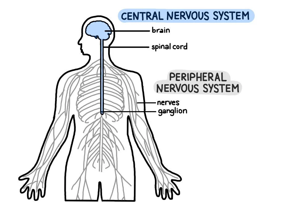
efferent vs afferent neurons
Afferent neurons carry sensory information toward the central nervous system, while efferent neurons carry motor commands away from it
Afferent = Arrives at the brain (or Ascends).
Efferent = Exits the brain.
SAME: Sensory Afferent, Motor Efferent.
somatics vs autonomic, what type of neurons are they, which nervous system?
BOTH EFFERENTNEURONS
Somatic nervous system: responsible for voluntary actions, such as movement and interaction with the environment. It controls skeletal muscles and mediates voluntary motor activities, allowing us to perform tasks like walking or picking up objects.
Autonomic nervous system, responsible for involuntary bodily functions, controls activities such as heart rate, digestion, and respiratory rate.
further divided into:
the sympathetic and parasympathetic nervous systems, which work in tandem to balance bodily functions.
sympathetic nervous system prepares the body for stress-related activities,
parasympathetic nervous system promotes rest and digestion.

what do somatic motor neurons control?
somatic motor neurons control skeletal muscles
what do autonomic motor neurons control? what are subdivisions? what are 3 key target tissues?
send signal to internal organs, control all the involuntary, automatic functions of body, regulate the activities of internal organs, glands, and blood vessels without you having to consciously think about it.
SYMPATHETIC: (Fight or Flight)
Prepares your body for action, stress, or emergencies.
Increases heart rate, dilates airways for more oxygen, dilates pupils, and pauses digestion.
PARASYMPATHETIC:(Rest and Digest)
Calms the body down and handles everyday maintenance.
Actions: Lowens heart rate, constricts airways, stimulates digestion, and allows the body to conserve and store energy.
Key tissues = cardiac muscle, smooth muscle, exocrine glands/cells, some endocrines and some adipose
what is enteric nervous system? what is it controlled by?
= independent network of millions of neurons embedded directly within the walls of your gastrointestinal (GI) tract.
sensory neurons stimulate it
autonomic neurons communicate w it
it controls smooth muscle, exocrine cells/glands, endrocine cell/glands
CAN OPPORATE ON THEIR OWN OR CONTROLLED BY ANS
exocrine vs endocrines
Exocrine glands secrete their substances through ducts onto an internal or external body surface, while endocrine glands secrete their hormones directly into the bloodstream without any ducts
what are some types of sensory receptors?
Receptors of vision; Receptors of hearing; Receptors of
balance; Receptors of taste; Receptors of smell; Receptors
on the skin (mechanoreceptors, thermoreceptors, or
nociceptors). Viscera organs contain nociceptors that
activate following inflammation and tissue damage.
3 main catergories of nervous sys. are?
Central nervous system CNS: brain and spinal cord
Peripheral nervous system
– Sensory (afferent) neurons
– Efferent neurons: somatic motor and autonomic divisions
▪ Autonomic divided into sympathetic and parasympathetic branches
Enteric nervous system
– Network of neurons in the walls of the digestive tract
– Controlled by autonomic nervous system but is able to function
autonomously
what are three target cells from action potential of neurons and what is target cell? does targets get touched by neurons?
a target cell= simply the destination cell that receives a chemical or electrical message from a neuron.
Neurons do not touch their targets directly; instead, they send an electrical signal (the action potential) down their axon, which triggers the release of chemical messengers (neurotransmitters) across a tiny gap (the synapse). Whichever cell is sitting on the receiving end of that gap is the target cell.
1. Neuron-to-Neuron Synapse= neuron communicating with another neuron.
The Target Cell: A postsynaptic neuron.
What happens: The first neuron excites or inhibits the target neuron, deciding whether the target neuron will fire its own action potential to keep passing the message along.
2. Neuromuscular Junction (Neuron + Muscle)= motor neuron communicating with a muscle fiber
The Target Cell: A skeletal, smooth, or cardiac muscle cell.
What happens: The neuron releases the neurotransmitter acetylcholine. The muscle target cell receives it and instantly responds by contracting (moving).
3. Neuroglandular Junction (Neuron + Gland)= neuron communicating with a gland cell.
The Target Cell: A glandular epithelial cell (an exocrine or endocrine cell).
What happens: The neuron signals the target gland cell to either secrete something (like releasing sweat from sweat glands or adrenaline from the adrenal gland) or stop secreting.

NEURON ANATOMY: what contains sodium channels, what has high lipid content, white vs grey sections? neurons in CNS vs PNS?

what is function of dendrites, cell body, axon hillock, presynaptic axon,myelin sheaths
dendrites= input signals
cell body= integration of signals
axon hillock= features a very high density of voltage-gated ion channels, gathers all incoming signals and can trigger action pot.
myelin sheaths= Insulate the axon to drastically speed up signal transmission.
presynaptic axon terminal= communicates w post-synaptic target cell, has synaptic vesicles packed with neurotransmitters (like dopamine, serotonin, or acetylcholine).
how do myelin sheaths work? what are they made of ? Schwann cells vs oligodendrocytes
This fatty layer (made by Schwann cells or oligodendrocytes) wraps around the axon like insulation on an electrical wire. It prevents the electrical charge from leaking out, forcing the signal to rapidly "hop" between uninsulated gaps called Nodes of Ranvier= saltatory conduction.
Schwann Cells: Found exclusively in the PNS
Oligodendrocytes: Found exclusively in the CNS
2. Structural Capacity
Schwann Cells: One Schwann cell can wrap around only a single segment of one axon. It takes thousands of individual Schwann cells lined up end-to-end to insulate a single long peripheral nerve.
Oligodendrocytes: One oligodendrocyte features multiple tentacle-like extensions that can wrap myelin around up to 50 different axon segments simultaneously.
FUNCTIONAL CATEGORIES OF NEURONS: pseudounipolar vs bipolar vs anaxonic vs multipolar. which are commonly found for sensory, interneurons of CNS and efferent?


efferent vs afferent neuron communication from PNS to CNS to effector organ

sensory neurons are equivalent afferent or efferent?
afferent
what are functions of glial cells? common names?
= the supportive, non-neuronal cells of the nervous system. While neurons act as the communication wires passing electrical messages, glial cells act as the ultimate support crew. They do not fire action potentials, but neurons cannot survive or function without them.
= neuroglia, glia
structural Support: hold neurons physically in place like a cellular scaffold.
Insulation:produce the fatty myelin sheath
Nutrient Supply: deliver oxygen and nutrients (like glucose) from blood vessels directly to neurons.
Cleanup & Defense: destroy pathogens and dispose dead or damaged neurons.
Chemical Regulation: absorb excess neurotransmitters and balance potassium levels around neurons.
PNS vs CNS glial cell/neuroglia, which from myelin sheaths?
CNS
Astrocytes (The Caretakers): Star-shaped cells that anchor neurons to their blood supply. form the blood-brain barrier (BBB)
Oligodendrocytes (The CNS Insulators): Wrap around CNS axons to form the protective myelin sheath.
Microglia (The Immune Force): The specialized immune cells of the brain. They act like macrophages
Ependymal Cells (The Fluid Producers): Line the fluid-filled cavities (ventricles) of the brain and central canal of the spinal cord. feature hair-like cilia that beat rhythmically to create and circulate cerebrospinal fluid
PNS
Schwann Cells (The PNS Insulators): Wrap around a single segment of a peripheral axon to form myelin sheath.
Satellite Cells: Flat cells that wrap around and cushion the cell bodies of peripheral neurons. They regulate the chemical environment and protect the neuron body

tract vs nerve
A tract is a collection of nerve fibers (axons) in the CNS
A nerve is a collection of nerve fibers (axons) in the PNS
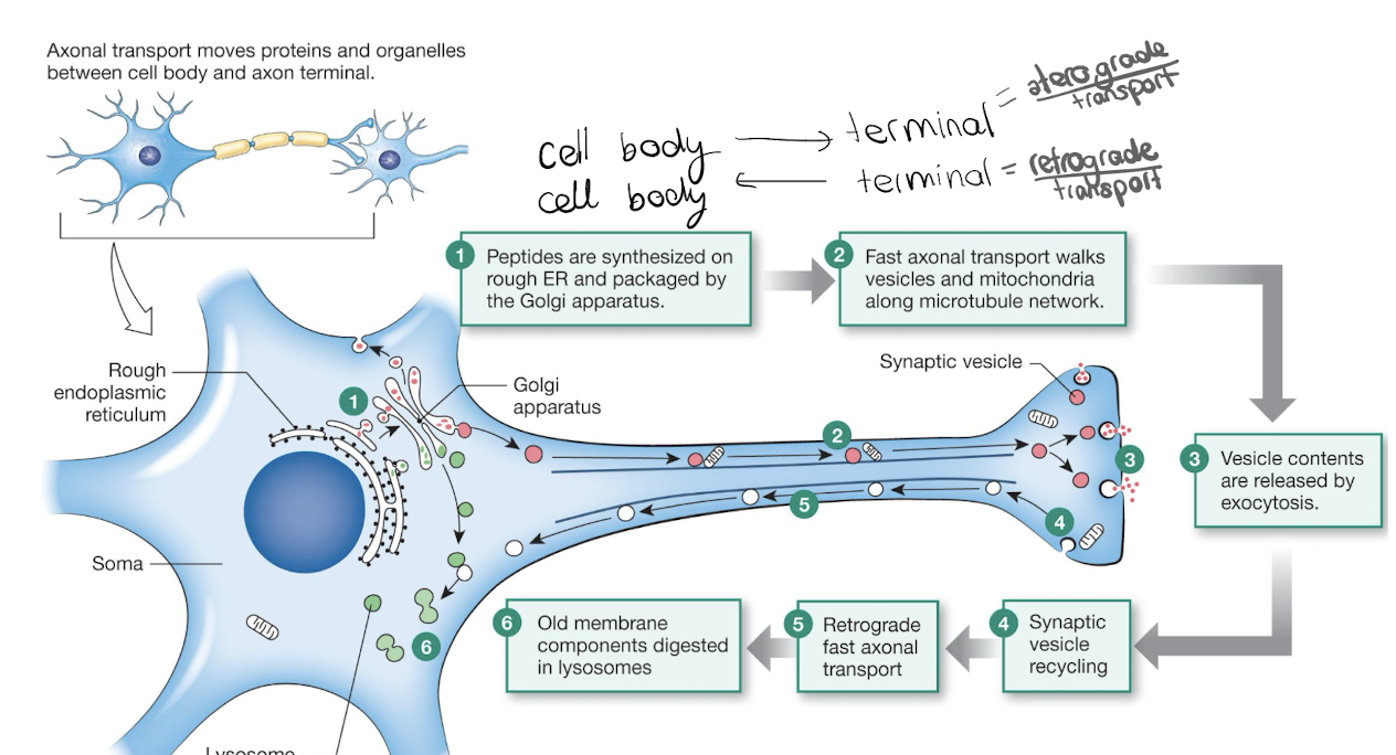
slow vs fast axonal transport
both used to move proteins, organelles, and chemical messengers across the long distances between the axon body and the axon terminal
Fast axonal transport
– Moves organelles at rates of up to 400 mm/day
relies on a "railway system" made of microtubules. Specialized motor proteins act as trains, consuming ATP
Directions: It operates in both directions:
Anterograde (Forward): Moves cargo from the cell body toward the axon terminal. Driven by the motor protein kinesin.
Retrograde (Backward): Moves cargo from the axon terminal back to the cell body. Driven by the motor protein dynein.
Slow axonal transport
– Moves material by axoplasmic (cytoplasmic) flow at 0.2–2.5 mm/day
what are three cytoplasmic protein fibers? what is their role
Microfilaments
– Actin fibers
Intermediate filaments
– Keratin
– Neurofilament
Microtubules
– Tubulin
= Associated accessory proteins, Involved in fast transport and structural support and cell movement with motor proteins
fast axonal transport steps
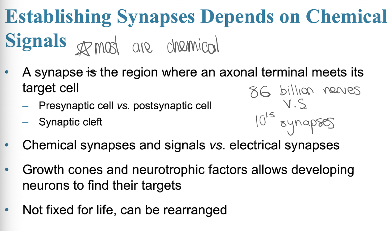
what do most synapses depend on to be established? nerve to synapse ratio?
what is neutrophic factor
specialized type of protein that acts like a growth hormone for the nervous system,
= reason why retrograde fast axonal transport exists in neurons BC when a presynaptic axon successfully reaches its target cell (like a muscle or another neuron) and establishes a good synapse, the target cell secretes a tiny reward package of neurotrophic factors
how is myelin formed in PNS cells? what are these cells? what are nodes of ranvier?

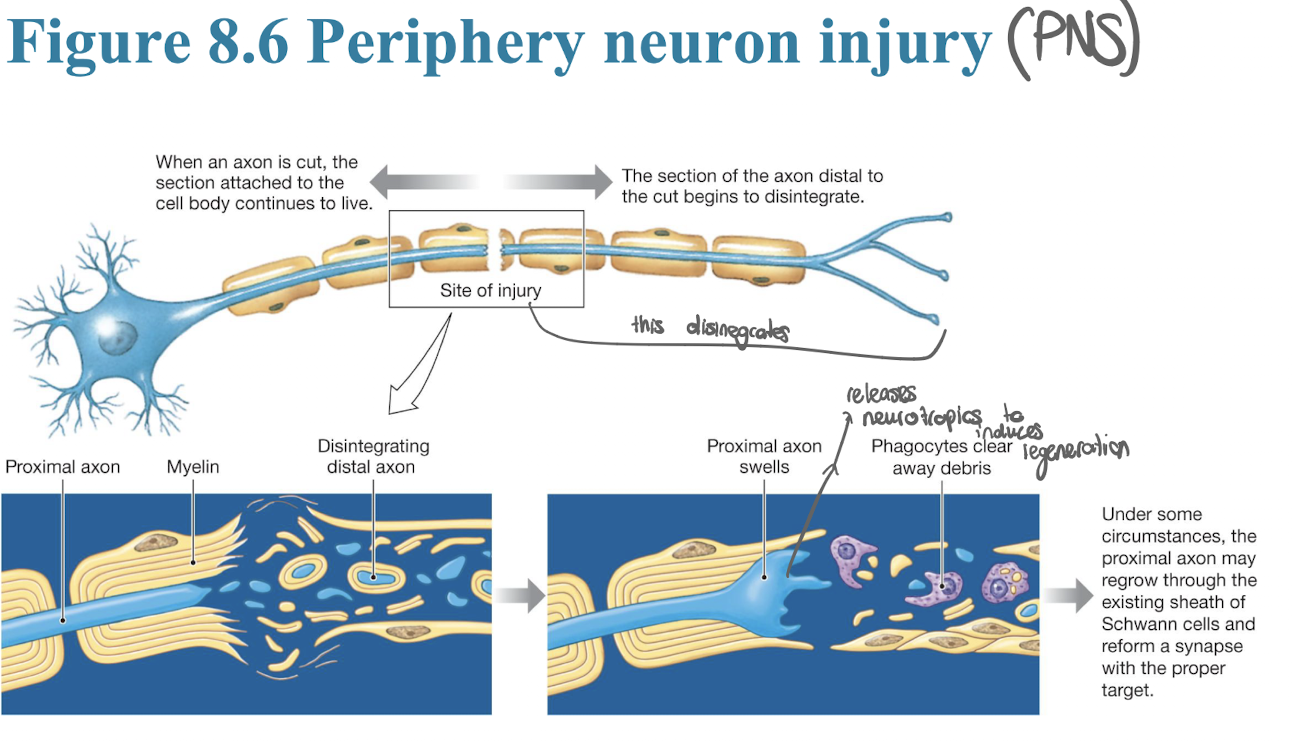
can damages neurons be repaired? what are options if cell body vs axon are damaged?
• If the cell body dies, the neuron dies
• If axon is severed, then cell body and attached segment survives; severed portion degenerates
– If its damaged motor neuron, then target muscle results in permanent paralysis
– If sensory neuron, then experience loss of sensation from innervated area
• Regeneration may occur in PNS; less likely in CNS
CHANNEL PROTEINS: what are open channels vs gated channels, what are channels filled with/. what are sub-categories of gated Chanels. what are the 4 Na+ channels
4 types of Na+ channels:
there is always a high conc. of Sodium outside the cell and low concentration inside, whenever any of these four doors open, Sodium will rush into the cell
Na+ leak: Always open; allows a slow, constant trickle of sodium inside.
Na+ voltage: Opens only when an electrical action potential arrives.
Na+ chemical: Opens only when a neurotransmitter lands on it.
Na+ mechanical: Opens only when the cell is physically stretched or squeezed.

what are normal ion conc. and equilibrium potentials (and where do they leak) of k+, Na+, Cl-, Ca2+
normally more +’ve outside of cell

what is membrane potential difference, resting potential
Membrane Potential: the electrical charge inside the cell compared to the outside.
Resting potential: When a neuron is completely quiet and not sending signals, it sits at its resting potential.

what are 3 states of polerization
DEPOLARIZATION: he cell becomes less negative (closer to zero or positive), Positive Na+doors open, and sodium rushes in the cell.
HYPERPOLARIZATION: cell becomes more negative than its normal resting state, positive K+ leaves the cell, or negative Cl- enters.
REPOLERIZATION: process of resetting the neuron's electrical charge back to a negative state after it fires.—→ voltage-gated Na+ channels close and lock shut, voltage-gated K+ channels open wide, K+ ions rapidly rush out of the cell= inside loses positive charge and plunges back into negatives
graph of depolarization


what are two basic types of electrical signals and what are they used for?
graded potentials= variable strengths, used for short distance
action potentials = very brief large depolarizations, rapid signals over long distances
what controls ion permeability of neuron? what is key about threhsold voltage rates?
gated ion channels prinarily control them , theyre names for primary ion that passes through them.
includes mechanically gated, chemically gated, voltage-gated
threshold volatges vary from one channel type to another, both activation and inactivation rates vary
GRADED POTENTIAL VS ACTION POTENTIAL; where they happen, How They Are Controlled,
1. Where They Happen
Graded Potential: Stays local in the dendrites and cell body (the receiving area).
Action Potential: Travels all the way down the long axon to the terminal.
2. How They Are Controlled (Channels & Ions)
Graded Potential: Uses any gate (chemical, mechanical, or voltage). It can use many ions like Na+, k+m Ca2+
Action Potential: Uses only voltage-gated channels. It relies strictly on Na+ and K+
3. Signal Strength & Rules
Graded Potential: Variable strength, bigger stimulus creates a bigger ripple. Multiple small ripples can add together (sum) to become stronger. It can go up (depolarize) or down (hyperpolarize).
Action Potential: Always the same size (All-or-None). cannot be added together because of a mandatory rest break (refractory period). It only goes up (depolarizes).
4. How They Start
Graded Potential: Starts from outside stimuli (like a touch or a neurotransmitter). It has no minimum requirement to start.
Action Potential: Started by a graded potential. If the graded potential ripples are strong enough to hit the threshold voltage, the action potential explodes into life.
what does graded pot vs action pot. look like when gently vs firmly tapping skin example?
Gentle Tap (Graded Potential Only)
barely touch your skin, you only open a few mechanically gated ion channels at the very tip of the neuron (the dendrites).
The Ripple: A tiny amount of sodium enters, creating a weak graded potential.
The Result: The signal is too weak. As it travels from your fingertip toward the cell body, it fades away completely before reaching the axon trigger zone. You might not even consciously notice the touch because no action potential was fired.
Tapping Repeatedly or Harder (Summation)
tap the exact same spot rapidly, or press down just a little bit harder.
Adding Up: Each individual tap creates its own small ripple. Because the taps are happening close together, the electrical ripples overlap and add together (summation).
Building Strength: The signal grows stronger and crawls closer to the axon hillock (the trigger zone).
The Firm Poke (Action Potential Fired)
press down firmly.
Hitting the Threshold: The combined graded potentials are now massive. They hit the axon hillock with enough electrical power to cross the magic threshold line (-55 mV).
The Explosion: The fire alarm is pulled. A wave of voltage-gated channels snaps open one after another all the way down the axon. This action potential travels at lightning speed up your arm into your spinal cord and brain.
The Result: Your brain registers the signal, and you think, "Ah, someone is poking my arm."
what is local current flow, how do graded potentials lose strength (2)?
local current flow= the movement of ions through the cytoplasm inside a neuron, away from the exact spot where they first entered= the physical mechanism that spreads a wave of depolarization along a membrane
movement of +’ve to negative
LOCAL CURRENT FLOW DOESNT LAST LONG BC:

what is conductance vs resistance in this case?
1. Resistance= how much the membrane blocks or opposes the flow of ions.
High Resistance: When a neuron is at rest, most of its gated ion channels are tightly closed. The phospholipid bilayer acts like a solid wall, making it incredibly difficult for ions to cross.
Low Resistance: When channels open, the barrier is broken down, and resistance drops significantly.
2. Conductance= how easily ions can flow through the membrane.
Low Conductance: When channels are closed, conductance is near zero because there is no path for the ions to travel.
High Conductance: When specific ion channels open, the conductance for that specific ion spikes dramatically.
subthreshold graded pot. vs suprathreshols graded pot.
subthreshold graded potential=stimulus that is too weak to reach the threshold voltag, does NOT initiate action potential
suprathreshold graded potential= stimulus that is strong enough to meet or exceed the threshold voltage.

DESCRIBE STEPS OF ACTION POTENTIAL: when does action potential begin? what occurs during rising phase? what happens at peak?


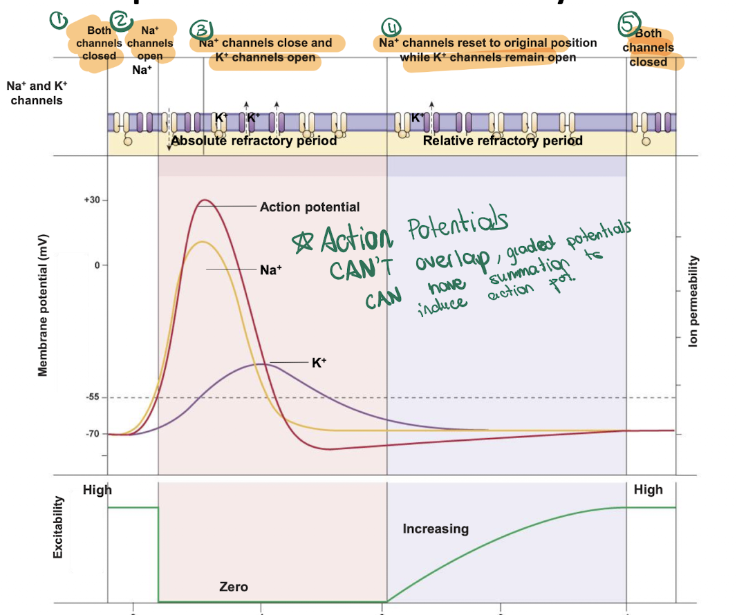
what are refractory periods ?
= ensures that nerve signals only travel in one direction (forward) and limits how fast a neuron can fire.
can action potentials go backwards?
no, but gradded potentials can have summation to INDUCE action pot.

where do synapses occur? where could a neuron terminate ?

what are 3 types of cell junctions relevant?

EPSP vs IPSP, what doe they do to threshold
When neurotransmitters bind to receptors on the target cell (dendrites or cell body), they alter the membrane voltage by creating a graded potential. This change is either an Excitatory Postsynaptic Potential (EPSP) or an Inhibitory Postsynaptic Potential (IPSP).
EPSP moves the neuron’s voltage closer to the threshold required to fire an action potential.
IPSP moves the neuron’s voltage farther away from the threshold, making it harder for the neuron to fire.
think EPSP green light, IPSP red light