week 8 - skeletal part 2
1/338
There's no tags or description
Looks like no tags are added yet.
Name | Mastery | Learn | Test | Matching | Spaced | Call with Kai | Chat |
|---|
No analytics yet
Send a link to your students to track their progress
339 Terms
What is osteopenia?
generalized breakdown of bones
bone loss that is greater than normal but not severe enough to be classified as osteoporosis.
results from an imbalance between bone formation and bone breakdown
characterized by decreased mineralization of bone
What is the normal purpose of the balance between bone formation and bone breakdown?
The balance between bone formation and bone breakdown helps replace damaged bone and maintain the amount and density of bone.
How does osteopenia contribute to disease?
contribute to the pathology of all metabolic bone diseases and predisposes individuals to fractures, particularly of the hip and vertebrae.
how to see osteopenia
special x-ray called DEXA scan
measures how much x-ray is absorbed by the bone
blood test for osteopneia
serum for
elevated alkaline phosphatase = abnormal bone turnover (increased bone turnover)
abnormal Ca+/P- levels also
predisposition of osteopenia
fractures (#) of hip and vertebra
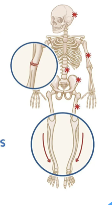
Define osteoporosis.
chronic, progressive metabolic bone disease
low bone mass
structural deterioration of bone tissue
lead to increased bone fragility, increase risk of # (fractures - hip/wrist/spine)
characterized by porous bone and decreased bone mass
result = increased fracture risk.
Why is osteoporosis described as "porous bone"?
causes loss of bone density and deterioration of bone structure, creating porous and fragile bones that are more susceptible to fractures.
why is osteoporosis more common in women?
low Ca+ intake
less bone mass (smaller frame)
bone reabsorption at an earlier age in women accelerated in menopause
breastfeeding = use skeletal reserve endless Ca+ intake adequate
longevity (women live longer than men)
What is trabecular bone tissue? - osteoporosis
spongy and porous bone material composed of hard and soft tissue components.
long bones + vertebrae (shock absorbers)
RBCs
quick bone remodelling
highly sensitive to hormonal changes (women and loss of estrogen in menopause)
see perforation in women (loss of connectivity and complete trabeculae)
men = thinning
hormonal decline more gradual
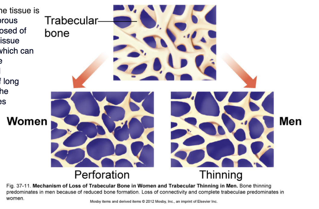
Where is trabecular bone located?
epiphyses and metaphyses of long bones and within vertebral bodies.
osteoporosis screening
predispositions
rheumatoid arthritis
steroid use (3+mths past year)
age
DEXA scan
What screening test is used to diagnose osteoporosis?
Dual-energy X-ray absorptiometry (DEXA scan).
What does a DEXA scan measure?
A DEXA scan measures bone density using a T-score.
Why are DEXA scans repeated over time?
evaluate changes in bone density over time and assess the effectiveness of treatments such as osteoporosis therapy.
What happens to bone absorption in osteoporosis?
Bone absorption (resorption) increases, resulting in progressive loss of bone mass.
risk factors
this leads to complications of SPINE and HIP
risk factors for osteoporosis
age
post-menopause women
physical inactivity (need regular weight bearing activity)
inadequate Ca+ and Vit D
CKD/endo disorders (hyperparathyroid)
What spinal complications can occur with osteoporosis?
vertebral compression fractures
bending, lifting, minor fall, minimal trauma
no pain, fracture unnoticed = experience repeated vertebral fractures = loss of height and kyphosis
Vertebral fractures
kyphosis
scoliosis - further worsens if already have
What hip complication commonly occurs with osteoporosis?
Hip fractures.
pain
reduced mobility
loss of independence
prolonged hospitalization
increase risk of mortality (older adults/comorbidities)
What is osteomalacia?
softening of bones due to inadequate mineralization of bone tissue.
osteomalacia = adults
rickets = children
inadeuate Ca+/P- into bone matrix = soft/decreae bone growth
What is osteoid in osteomalacia?
Osteoid refers to soft, inadequately mineralized bone.
What is the major cause of osteomalacia?
Conditions that result in vitamin D deficiency.
bone deformity as well
inadequate diet
malabsorption issues
disorders affected Vit D metabolism (poor intestinal/renal tubular disease)
osteomalacia patho
decrease Vit D = no mineralization of bone + decrease intestinal uptake of Ca+
decrease serum Ca+ = increase PTH
increase PTH secretion = …
increase osteoclast = increase serum Ca+ (from bone to blood)
increased Ca+ reabsorption by kidney
decrease PO4 = decrease mineralization
What is rickets?
disease of childhood characterized by weak and soft bones.
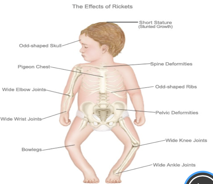
What causes rickets?
Extreme and prolonged vitamin D deficiency
genetic abnormalities
X-linked hypophosphatemic rickets
genetic causes Vit D deficiency
What complications can result from rickets?
Pain
poor growth
skeletal deformities.
What is Paget's disease of bone?
increased bone remodeling with excessive bone resorption and formation.
single or several bones
INITIALLY = NO S/S
rare = develop into a lesion (osteosarcoma)
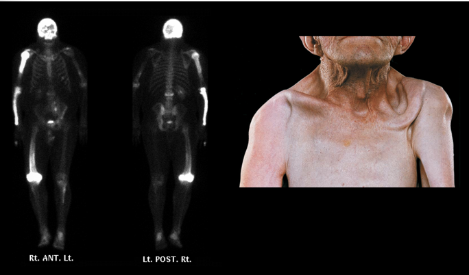
What happens to bones in Paget's disease?
Bones enlarge, more porous and become softer, and structurally abnormal.
disorganized manner
bone deformity + patho fractures
Which bones are commonly affected by Paget's disease?
Skull
vertebrae
sacrum
sternum
pelvis
femur.
What medication is commonly used to treat Paget's disease?
Bisphosphonates.
What is the "cotton wool patch" seen in Paget's disease?
A radiographic finding representing thickened, disorganized trabeculae.
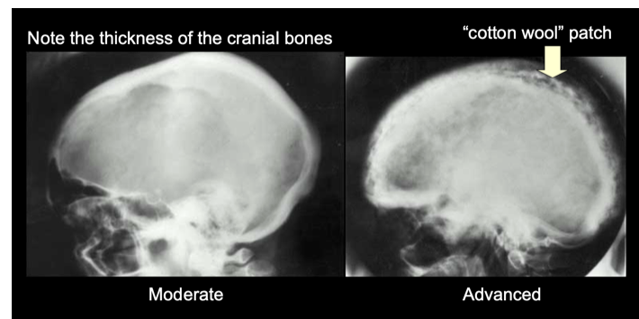
How can Paget's disease affect the brain?
Brain compression can cause altered cognitive function and dementia.
How can Paget's disease affect cranial nerves?
Compression of cranial nerves can cause sensory and motor changes.
What is osteomyelitis?
bacterial infection of bone.
What organism most commonly causes osteomyelitis?
Staphylococcus aureus.
What is endogenous osteomyelitis?
develops when infection spreads to bone through the bloodstream.
skin or dental infection to the bone
What is exogenous osteomyelitis?
direct contamination of bone from trauma, surgery, or adjacent infection.
Describe the pathophysiology of osteomyelitis.
Infection causes inflammation
blood vessel thrombosis
exudate accumulation within canaliculi (bone canals increase amount of pressure = disrupts blood supply = necrosis + sequestrum (hallmark of chronic infection)
Haversian canals
Volkmann canals.
How does exudate damage bone in osteomyelitis?
causes periosteal separation, disrupts blood supply, and leads to necrosis of underlying bone.
What is a sequestrum? - osteomyelitis
devascularized fragment of dead bone formed during osteomyelitis.
osteomyelitis complciations
weakens bone
spreads to joints
delayed wound healing
delayed healing of fractures
nonunion bone
bone destruction/infection
septicemia
chronic osteomyelitis
How does osteomyelitis affect bone strength?
It weakens bone and increases fracture risk.
inflammation and necrosis is the cause
What joint complication can result from osteomyelitis?
Spread of infection to joints.
lead to septic arthritis
How does osteomyelitis affect wound healing?
impaired blood flow of infected bone…
delayed wound healing
delayed fracture healing = nonunion
Failure of a fractured bone to heal properly
What severe systemic complication can occur with osteomyelitis?
Septicemia.
What chronic complication can develop after osteomyelitis?
Chronic osteomyelitis.
What is the primary treatment for osteomyelitis?
Intravenous antibiotics.
prolonged IV abx
start abx in hospital
get peripheral central access device or PIC line to continue abx IV at home for wsk
long b/c need to fully eradicate bacteria in poorly vascularized bone tissue
What is osteonecrosis?
bone death caused by ischemia due to inadequate blood supply.
avascular necrosis
needs constant Oz/nutrients from blood
no blood = bone cells die = weakne + collapse of bone
What are causes of osteonecrosis?
Bone fractures
thrombosis
embolism
vessel injury
increased intraosseous pressure causing compartment syndrome
pressure builds in bone = vessels + nerve + tissues compromise, accelerate ischemia
steroid therapy
alters lipid metabolism = fat accumulation in bone marrow blood vessels
ALL = decrease BF to bone
What are bone tumors?
Abnormal growths of tissue within bone that may be benign or malignant.
bone tumours
precursors from mesoderm
type of cells = determine type of tumour
fibroblast
osteoblast = bone-forming cells
osteogenic tumours x 3
chondroblast = cartilage precursor
fibroblast = collagen-producing cell
fibro
reticulum
various blood cells precursors in bone marrow
myelogenous tumor
giant cell tumour
ewing sarcoma - malignant tumour in children/adolescent
benign bone tumours
condroma
osteochondroma/exostosis
giant cell tumours/osteoclastoma
What is a chondroma? - benign bone tumours
A benign bone tumor composed of cartilage.
small bones of HANDS + FEET
location = class
endo = medullary of bone
surface
Where are chondromas most commonly found?
Small bones of the hands and feet.
What is an osteochondroma (exostosis)?
The most common primary benign bone tumor caused by overgrowth of cartilage near a growth plate (ends of bone)
popcorn appearance (cartilage calcifies irregular way)
Where are osteochondromas commonly found?
Long bones of the leg, pelvis, and scapula.
What are the clinical manifestations of osteochondroma?
A painless, hard, immobile mass.
What is a giant cell tumor (osteoclastoma)?
A bone tumour composed of mononuclear cells and osteoclast-type giant cells.
behaves like malignant tumour but is usually benign
grey zone tumours (benign but can progress in aggressive clinical behaviour)
Where are giant cell tumors commonly found?
Near joints at the ends of bones, especially the distal femur, proximal tibia, and distal radius.
What percentage of giant cell tumors are benign?
Approximately 90%.
What symptoms occur with giant cell tumors?
Localized swelling, pain, and altered joint function.
malignant bone tumours
osteosarcoma
chondrosarcoma
Ewing sarcoma
What are common signs and symptoms of malignant bone tumors?
Localized pain and swelling.
What is osteosarcoma? - malignant bone tumour
An aggressive malignant bone tumor arising from bone-forming osteoblasts.
What is the most common primary malignant bone tumor?
Osteosarcoma.
terry fox
children and adolescents
period of rapid growth (puberty)
Where is osteosarcoma commonly found? - malignant bone tumour
Metaphyses of long bones
distal femur
proximal tibia
pelvis
presentation of osteosarcoma - malignant bone tumour
localized pain + swelling
sometime palpable mass
1st sign = patho fracture since growing tumour weakened bone
danger of osteosarcoma - malignant bone tumour
highly aggressive
mestatize to the lungs
treatment = pre- surgery chemo (shrink tumour) followed by surgical recession of tumour
What is chondrosarcoma? - malignant bone tumour
A malignant tumor originating from cartilage.
most common in MIDDLE age + OLDER adult
pelvis, ribs, shoulder, prox femur
What is Ewing sarcoma? - malignant bone tumour
A malignant tumour that develops in the medullary cavity of long bones.
PEAD cancer (adolescents/YA) - rapid growth period
Where is Ewing sarcoma commonly found? - malignant bone tumour
Femur, humerus, pelvis, and chest wall.
long bones
Ewing sarcoma presentation - malignant bone tumour
initially resembles infection + local pain/swelling
warmth over the affected area
occupational visible mass
SYSTEMIC
What distinguishes Ewing sarcoma from many other bone tumors? - malignant bone tumour
It often produces systemic symptoms in advanced disease.
fever
fatigue
weight loss
blood = leukocytosis anemia (advanced disease progression)
variations in growth/development of bones
as children grow, bones remodel
What are angular and torsional deformities?
Variations in growth and development that affect alignment and rotational positioning of bones.
What is in-toeing?
Inward turning of the feet during walking.
What is out-toeing?
Outward turning of the feet during walking.
What is femoral torsion and tibial torsion?
Abnormal twisting of the femur that may be internal or external.
Abnormal twisting of the tibia that may be internal or external.
causes in and out toeing
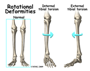
sitting and torsion
preferred sitting position
w sitting kneeling = frequent = enforce femoral torsion + intoeing
cross-legged position = enhance external torsion (outioesing gait)
encourage balance of positions (muscle develop)
outlook on femoral torsion and tibial torsion?
resolve naturally as kid more active + grows
angular changes of legs
can be normal infant/young toddler
preschool age - can be normal, resolve no treatment
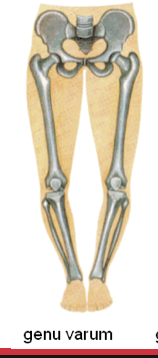
bowlegs (varum)
knock-knees (valgum)
need to know when normal and not to refer to a specialist
persistent
severe
asymmetrical
progressive deformity
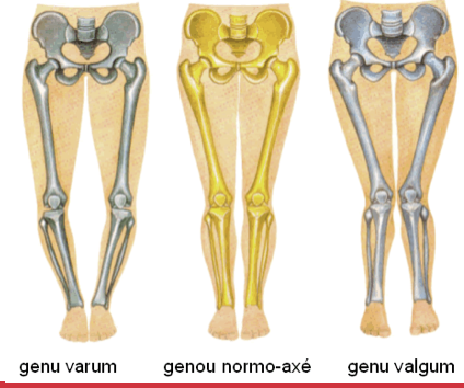
What is genu varum?
Bowlegs, where the knees remain apart when standing with feet together.
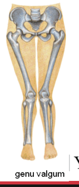
What is genu valgum?
Knock-knees, where the knees touch while the ankles remain apart.
congenital disorders
osteogenesis imperfecta
developmental dysplasia of the hip
clug footing
What is osteogenesis imperfecta?
ongenital disorder also known as brittle bone disease that results from a mutation in the collagen gene.
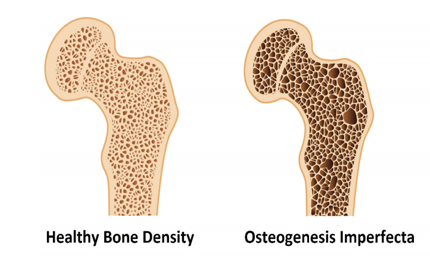
How does osteogenesis imperfecta affect bones?
The collagen gene mutation causes fragile bones that fracture easily and can lead to skeletal deformities.
What are the major consequences of osteogenesis imperfecta? - congenital disorders
Bone deformities and recurrent fractures due to bone fragility.
children presents with multiple fractures after minimal trauma
family hx of OI (osteogenesis imperfecta)
What is developmental dysplasia of the hip (DDH)?
A congenital disorder in which the hip joint develops abnormally, resulting in instability or dislocation of the hip.
What physical assessment finding may indicate developmental dysplasia of the hip?
Asymmetrical thigh folds or gluteal folds.
What are the Barlow and Ortolani tests used for?
Screening newborns and infants for developmental dysplasia of the hip.
also ultrasound
What does a positive Barlow test suggest?
Hip instability where the femoral head can be dislocated from the acetabulum.
What does a positive Ortolani test suggest?
A dislocated hip that can be reduced back into the acetabulum.
What is the primary treatment for developmental dysplasia of the hip?
Pavlik harness.
1st line treatment
wears harness 20+ hrs per day
hold hips in flex/abduction position = stable femur head sitting in acetabulum
let caregiver know routine follow-up + advise not to change straps
monitor for complications = femoral nerve pulse
decrease leg movement
reduce profusion to toes
How does a Pavlik harness treat developmental dysplasia of the hip?
It maintains the hips in flexion and abduction to promote proper alignment and development of the hip joint.
What is congenital clubfoot?
A congenital deformity in which one or both feet are abnormally positioned at birth.
one or both feed
common
dx before born sometimes or during NB assessment
What are common treatments for congenital clubfoot?
Serial casting and bracing.
stretching cast → bracing
akele tendon release surgery before bracing stage
What is scoliosis?
abnormal curvature of the spine in the coronal plane that is typically S-shaped or C-shaped and occurs in three dimensions.
rotation of vertebrae
3Dm
symmetry of trunk and shoulder
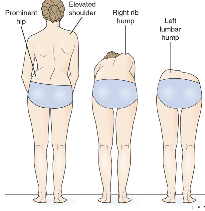
How is the spinal curve typically described in scoliosis?
An S-shaped or C-shaped curve.
What are possible causes of scoliosis?
idiopathic
periods of rapid growth
hereditary disorders
dystonia.
affect muscle control
from degenerative in older adults - alt spine alignment + stability
Degeneration of spinal discs
arthritis
osteoporosis
visual findings of scoliosis
elevated shoulder
rib hump with opposite side lumbar hump
prominent hip