quiz 3 key terms - methods in neuroscience
 Call Kai
 Learn
Learn Practice Test
 Spaced Repetition
 Match
Match Flashcards
 Knowt Play
1/63

There's no tags or description
Looks like no tags are added yet.
Name | Mastery | Learn | Test | Matching | Spaced | Call with Kai | Chat |
|---|
No analytics yet
Send a link to your students to track their progress
64 Terms
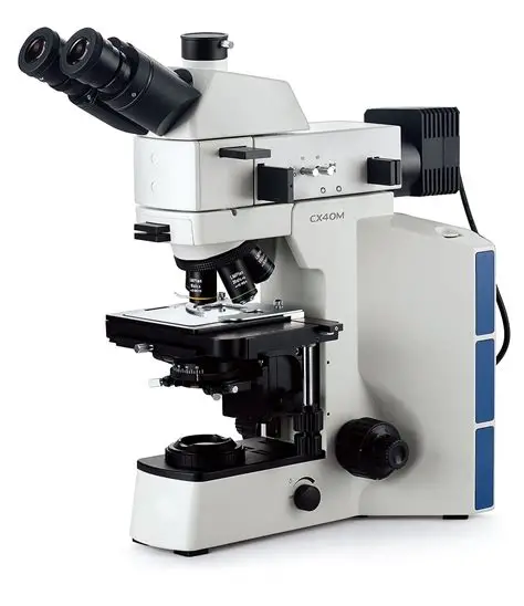
optical microscopy
use visible light to illuminate a sample and lenses to magnify the image. the light passes through or reflects off the sample and is then captured by the lenses to create a magnified image. 1000-2000x magnification up to 20 nm resolution
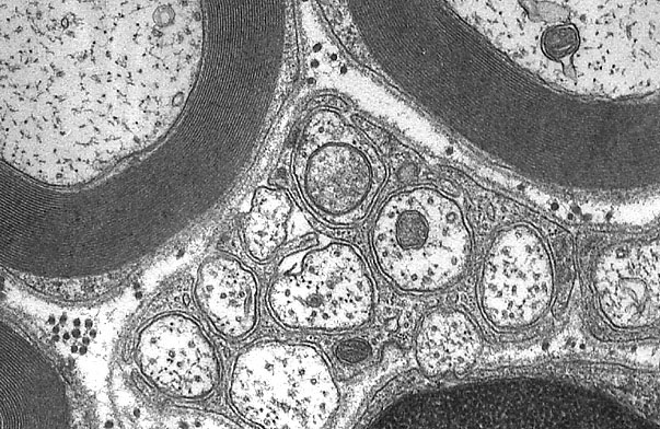
electron microscopy
uses a beam of electrons instead of light to examine the sample. electrons have much shorter wavelengths than visible light, allowing for much higher resolution. requires fixed tissue and is used to image small structures (synaptic vesicles, ion channels, etc.), with 1,000,000x magnification up to 0.1nm resolution.
magnification
how much larger a sample appears compared to its actual size
resolution
the ability to distinguish details of a sample
oil immersion
often required at higher magnification (60x), placing oil between the objective and the specimen can be used to increase the resolution
light microscopy
uses white light, good for any stains visible to the naked eye that need to be magnified (Golgi, Nissl, PLAP, etc.) in living or fixed tissue 10mm to 1uM size
fluorescence microscopy
good for any fluorescent tissues alive (calcium imaging) or fixed (immunostaining, FISH). Typical magnification is 10-80x. main limitation: photobleaching
upright microscope
fixed slides/tissues
inverted microscope
living tissue, cell culture, calcium imaging
dissection microscope or stereomicroscope
Used to provide minimal magnification for dissections (a type of white light microscope). Would be used for:
- After perfusion when you’re dissecting tissue so that it can be post-fixed in more PFA directly (what you would do before immunostaining/FISH, PLAP, or other tissue staining)
- During cryosectioning
- When collecting DRG/brain for primary neuron cell culture before calcium imaging
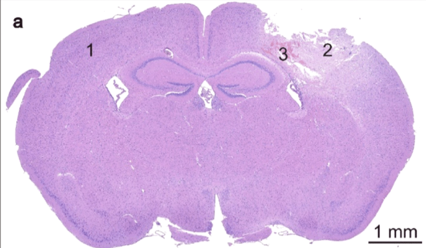
bright field microscope
a type of light microscope that is good for stained tissue - histology, PLAP, LacZ, etc.
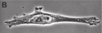
phase-contrast microscope
a type of light microscope, good for cultured cells since it doesn’t require staining
DIC/Differential interference Contrast microscope
a type of light microscope, used for unstained tissue/samples in which you want to observe structures
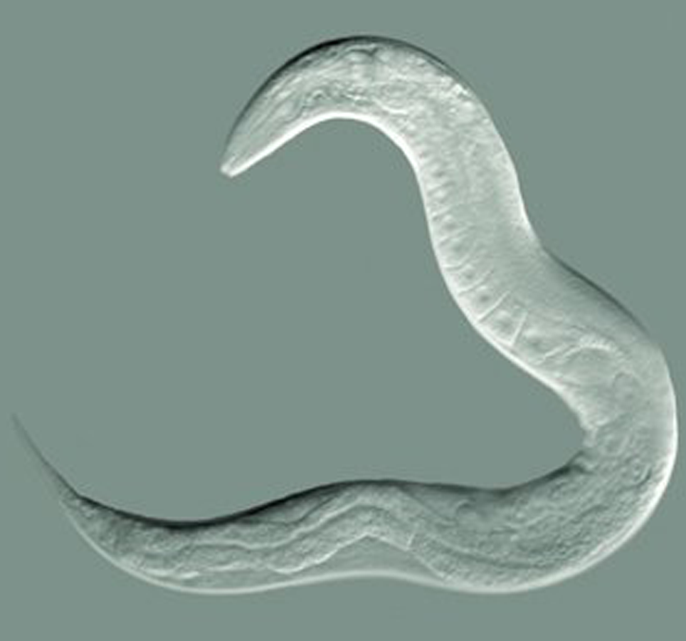
darkfield microscope
a type of light microscope, makes the cytoplasm dark, useful for small living organisms invisible in brightfield or any object that’s refractive value is similar to the background
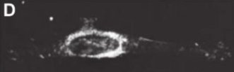
confocal microscope
a type of fluorescent microscope, living or dead tissue stained with fluorescent probes
- You can take 1uM thick ‘stacks’ or ‘zstacks’ of tissue
- You can ‘tile’ multiple tissue photos and stitch them together
- Produces more signal-to-noise ratio than the two-photon microscope
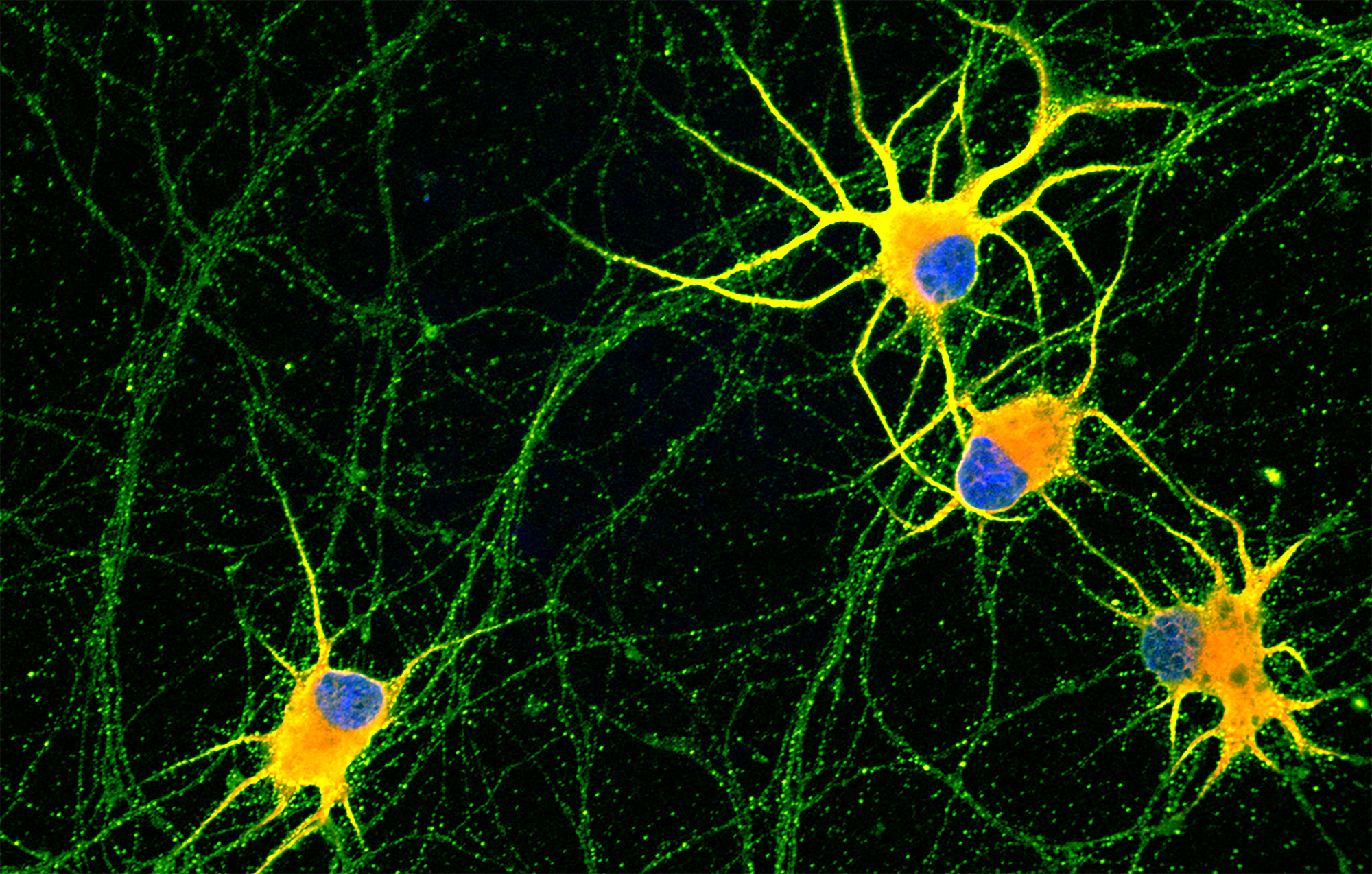
two-photon microscope
a type of fluorescent microscope, much more expensive than confocal, produces clearer images up to 500 μm, can penetrate deeper tissue – useful for CLARITY, can use living cells
light sheet microscope
a type of fluorescent microscope, living or dead tissue stained with fluorescent probes
- Unlike confocal and two-photon that use point-scanning, uses a light sheet to scan the entire specimen
- Less photobleaching and quicker
- Great for thicker samples
- Can be more difficult to prepare, not as commonly used
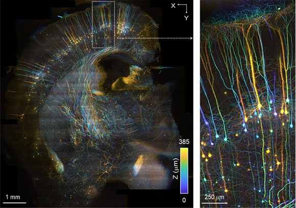
tiles
capturing multiple overlapping images of different regions of a large sample and then stitching them together to create a composite image of the entire area.
- Allows you to use high magnification and ‘stitch’ a large image together
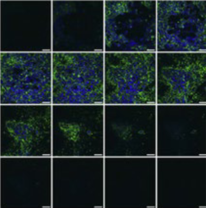
z-stacks
capturing a series of images at different focal depths (z-positions) across the vertical plane of the sample.
- Allows you to capture a more 3D image
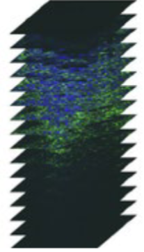
Image-J and the Fiji
most commonly used software along with Zen which is used to capture the images on the confocal
fixation
the process of preserving biological tissues by chemically stabilizing their structure to prevent decay and maintain their morphology for analysis
cross-linked fixatives
covalent bonds between proteins (ex: formaldehyde and paraformaldehyde)
- Preserve structures for light microscopy & electron microscopy
dehydrating fixatives
disrupt lipids and reduce protein solubility (menthol and acetone)
perfusion
the process of delivering a fixative such as (PFA) through an animal’s cardiovascular system (needed for immunostaining/in situ Hybridization)
post-fixation
after perfusion and dissection, you might place tissue in fixative for additional time
embedding
the process of surrounding a tissue with a substance that infiltrates and forms a protective shell around the tissue
• 24-hour soak in 30% sucrose will protect the tissue during freezing
• After sucrose, put the tissue in optimal cutting temperature (OCT) compound before cold sectioning with a cryostat
• Gelatin, paraffin wax, and plastic are also common embedding materials
microtome
used to cut frozen (PFA or ethanol > OCT/sucrose) or unfrozen tissue (PFA or ethanol > parafilm) to a medium thickness 25-100 um; best preservation of structures (electron microscope)
cryostat
uses frozen tissue (PFA > OCT/sucrose), sections easily directly mounted on slides (fast), thin to thick tissue slices 10-500um (immunostaining/FISH; thick- micro punches tissue)
vibratome
on-frozen, tissue can be live (no fixative); thick 100-400um; avoids artifacts or changes with morphology (electrophysiology)
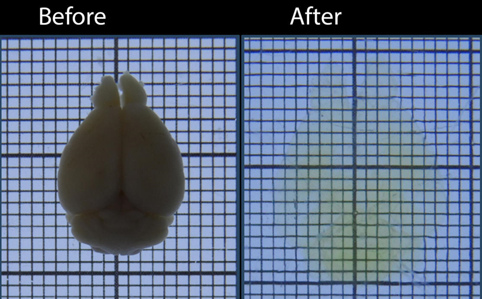
tissue clearing
the process of making intact tissue transparent (as opposed to sectioning)
CLARITY
fixes tissue to preserve the physical structures of proteins and nucleic acid by allowing light-scattering lipids to be removed. once the tissue is transparent it allows in-tact structures to be visualized (two photon microscope)
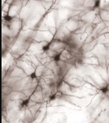
golgi stain
sparsely stains entire individual neurons (dendrites, cell body, axons)
weil stain
stains myelin black or blue in white light
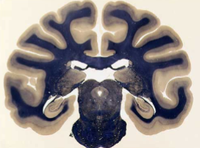
blasophilic stain
a stain that selectively binds to and highlights acidic components of cells (RNA or DNA or rough ER/ribosomes) – thus, predominantly isolating cell bodies
example: Nissl stains, Cresyl violet, DAPI, Propidium Iodine (PI), & Methylene Blue
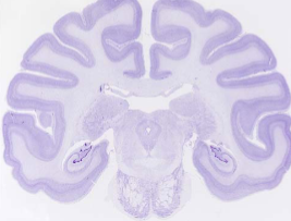
propidium iodine
red colored (green light) stain for dead cells only
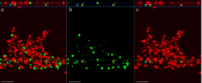
NF-200
stains axons fluorescently to a color of your choice. Double-stain with specific myelin markers
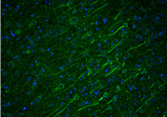
Immunohistochemistry
stains particular proteins
• Chromogenic/colorimetric probes (for light microscopy)
• Fluorescent probes
• Radioactive label (less common)
• Gold (for electron microscopy)
RNA in situ Hybridization
stains particular mRNA
• Chromogenic/colorimetric probes (for light microscopy)
• FISH = fluorescent in situ Hybridization
primary antibody
binds to the protein of interest
secondary antibody
chooses your color for fluorescence, used for visualization of the target
retrograde tracing
a substance that will be taken up by synaptic terminals and transported backwards (toward the cell body).
• Allows you to trace the ‘origin’ of connections
anterograde tracer
a substance that is taken up by the cell body and transported forward (down the axon) toward the axon terminals. This allows researchers to trace the destination of the neuronal connections (i.e., where the signals are going to)
Western blot or immunoblot
antibody binds to a protein that has been run through a gel, tells us the amount of protein in the sample (quantity), requires SDS-PAGE gel to separate proteins by size, cheap, ug
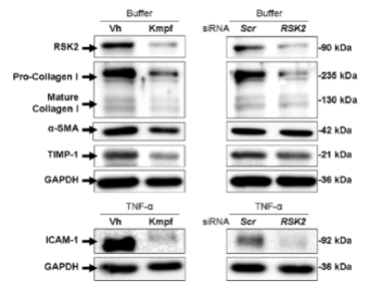
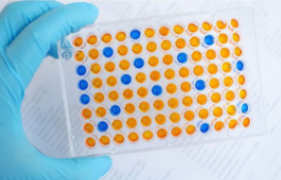
ELISA
same as a Western blot BUT is more precise/sensitive, ng of protein is all that’s required. Liquid-based samples.
radioimmunoassay
antibody binds to radioactive protein with known concentrations; these compete with nonradioactive proteins of unknown quantity to determine concentration (quantity), most precise, good for low conc. (NTs), most expensive. biological fluids.
immunohistochemistry
an antibody binds to a protein to show its spatial/physical expression in tissue/cells (doesn’t tell us about quantity)
monoclonal antibody
recognizes a single epitope of the antigen-high specificity, batches behave the same, renewable source (can make new identical batches), expensive
polyclonal antibody
antibody mixture that recognizes many different epitopes, high sensitivity, but not renewable, each batch may behave differently
aptamer
nucleic acid or peptide-based molecules engineered to bind to a particular target molecule, useful for labeling amyloid plaques for instance
cell fractionation
centrifuge sample tissue at certain speeds to collect specific organelles; separation of cellular components based on size, density, or solubility
chromatography
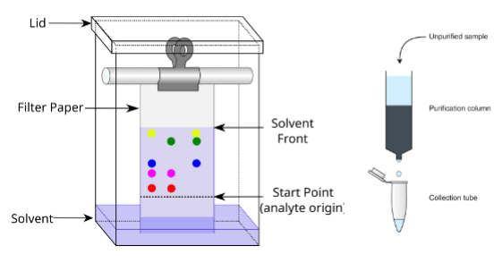
a way of separating out or isolating proteins or protein complexes based on size/charge using a filtering column. Large volumes.
immunoprecipitation
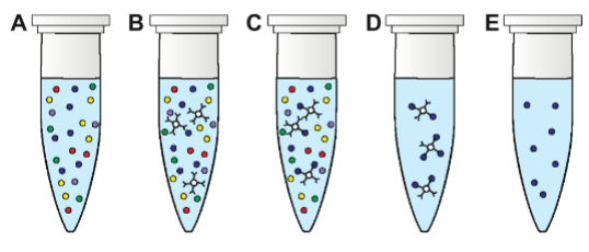
a way of purifying proteins using an antibody bound to small beads. Small volumes.
chromatin immunoprecipitation (ChIP)
an antibody is bound to beads in a column and a sample is run through the column to purify a protein which can then be checked to see if it’s bound to a specific region of DNA, uses cultured living cells but impossible to see if the protein interacts directly; Identifies DNA associated with a protein in cells
EMSA
seeing if a protein can directly interact with a short specific sequence of DNA that is radioactively labeled, shows direct interaction; not living tissue (protein - DNA)
luciferase assay
used to determine if a protein can activate/repress the expression of a target gene (directly or indirectly), establishes a functional connection / change in gene expression levels
Yeast two-hybrid assay screening
using the Gal4 protein. Divide the protein into two domains AD and BD and fuse each domain with a protein you think will interact together, if the two proteins don’t directly interact then AD and BD will stay apart and no transcription will occur - no fluorescence (protein - protein interaction)
co-immunoprecipitation (Co-IP)
regular immunoprecipitation (to isolate a particular protein) combined with a Western blot or ELISA to detect the presence of a secondary protein interacting with the bead-bound protein in some way (protein-protein interaction doesn’t have to be direct – an entire protein complex could be detected); Detects proteins found in a complex with a target protein
proximity labeling
identify which proteins come in close proximity to the protein of interest (BioID and APEX)
Fluorescence or Forster resonance energy transfer (FRET)
visualize interactions between two proteins in real-time, sensitive to concentration
Fluorescence-lifetime imaging microscopy (FILM)
similar to FRET but insensitive to concentration, tissue thickness, photobleaching, uses time for light to decay
Biomolecular fluorescence complementation (BiFC)
two halves of a single reporter protein are each fused with one of the proteins of interest
GRASP
a BiFC system to identify synapse formation between two cells. Split GFP is expressed on pre- and post-synaptic proteins. If a synapse forms, GFP reconstitutes and is visible
Post-translational modification (PTM) assays
looking for protein modifications such as the addition/removal of a phosphate group, acetylation, methylation, etc. (example: using a PTM-specific antibody)
kinase assay
determining if one protein can phosphorylate another (most common PTM assays) using radioactive ATP whose labeled phosphate will only be present if the protein of interest was phosphorylated (it was cleaved from the ATP), follow up with Western blot to show concentrations