Klein Chapter 11 - Contrast Enhancing Ultrasound Imaging
1/15
There's no tags or description
Looks like no tags are added yet.
Name | Mastery | Learn | Test | Matching | Spaced |
|---|
No study sessions yet.
16 Terms
The current FDA indication for ultrasound contrast is truly for…
improvement in LV opacification
Change in the gas molecular weight from room air gas to high molecular weight gases of UEA lead to improved…
transpulmonary passage and LV opacification because of persistence of microbubbles due to reduced diffusivity and solubility
Low mechanical index with UEA helps with… (3)
Myocardial perfusion imaging
Endocardial border resolution
Detection and evaluation of intracardiac masses
power modulation versus power pulse inversion
Contraindication to using Optison
allergy to blood products
Contraindication to all agents except Optison
Right to left intracardiac shunting
Can in hospital cardiac sonographers start an IV line?
Yes!
Why is UEA not used for PDA or intracardiac shunts?
Because you use agitated saline for those, not necessarily trans pulmonary contrast agents
The very low MI imaging techniques (<0.2 MI) are designed to detect only _____ activity
non-linear (ie distortion in the sound wave as it passes through a given medium) from micro bubbles
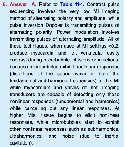
Patient has h/o CAD and MI. Given bolus injection of IV contrast with very low MI imaging. What is shown at the blue arrows?
Apical pseudoaneurysm
FYI not apical aneurysm or apical thrombus
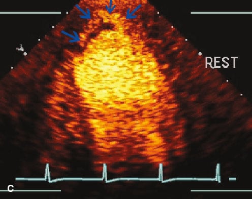
Diffuse T-wave inversions on ECG and the following end systolic images. What does this show?
Apical hypertrophic cardiomyopathy
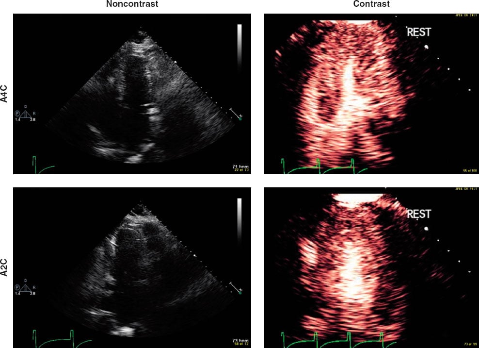
What does this LAA show
Spontaneous echo contrast but no thrombus
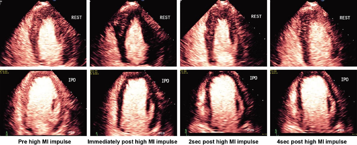
high MI impulse images with contrast — what coronary artery explains the perfusion defect?
left main
The variables that determine what the MI is includes… (2)
peak negative pressure and transmit frequency
FYI MI = peak negative pressure divided by the square root of the transmit frequency (in MHz)
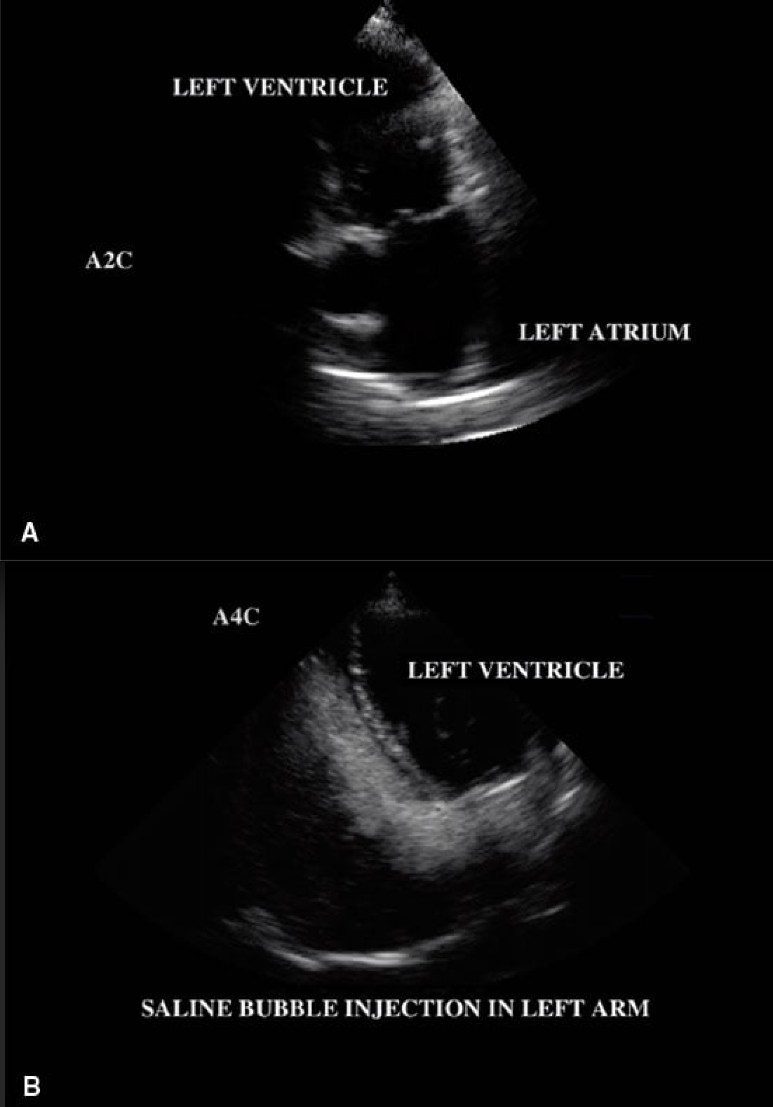
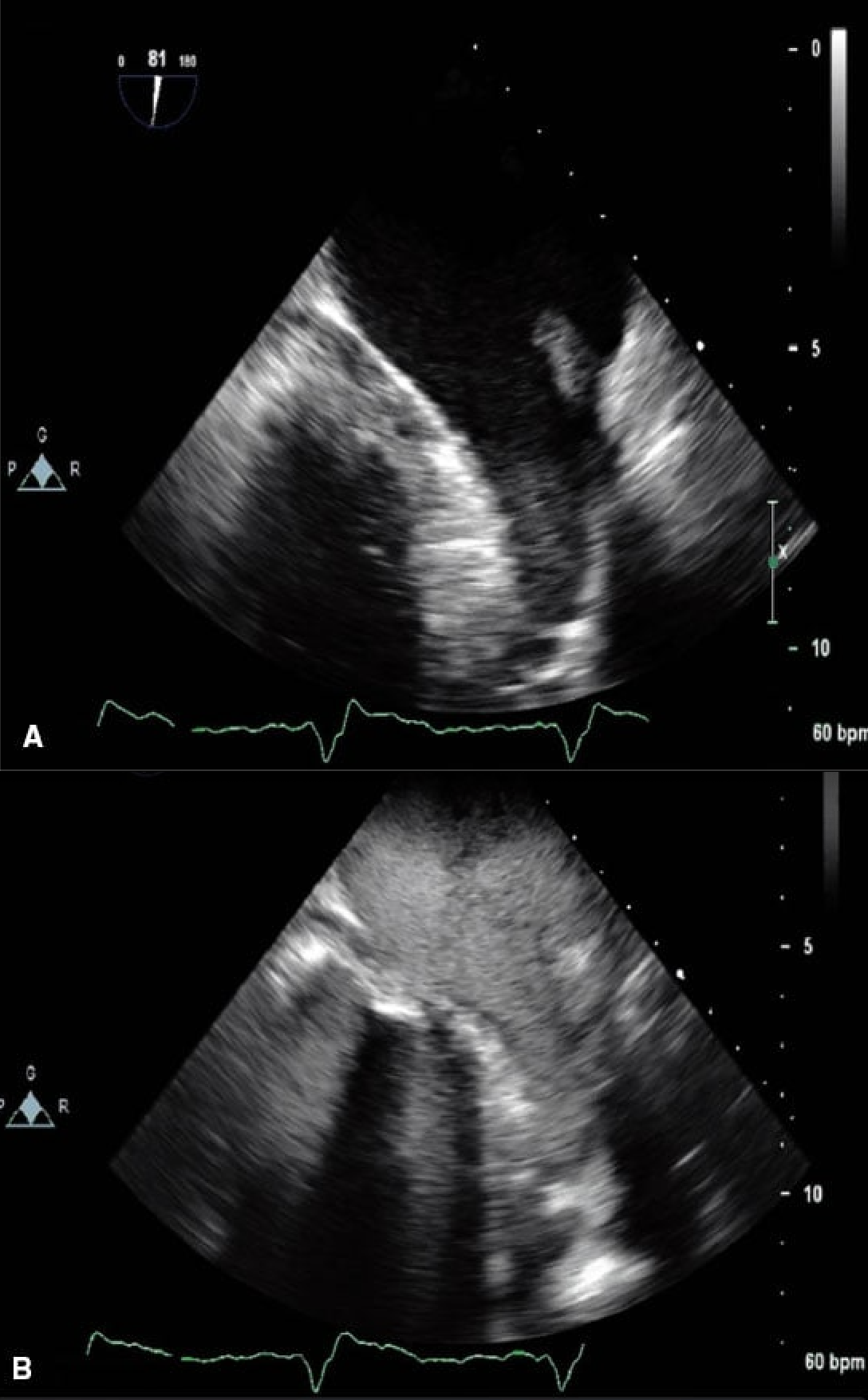
A patient with exertion SOB had a saline contrast injection performed to assess for intracardiac shunting. A right arm saline injection detected no evidence of intracardiac shunting. However, an apical 4 chamber image before and after a left arm saline contrast injection.
An unroofed coronary sinus with a left sided superior vena cava