Looks like no one added any tags here yet for you.
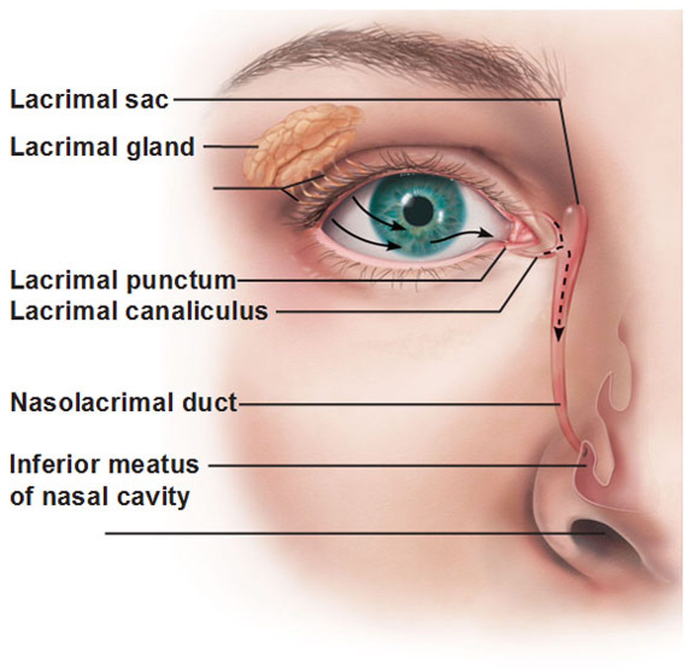
What produces and drains tears ate keep the eye moist and shield it from dust and irritants?
lacrimal apparatus
Where are palpebrae/eyelids connected?
medial and lateral canthus
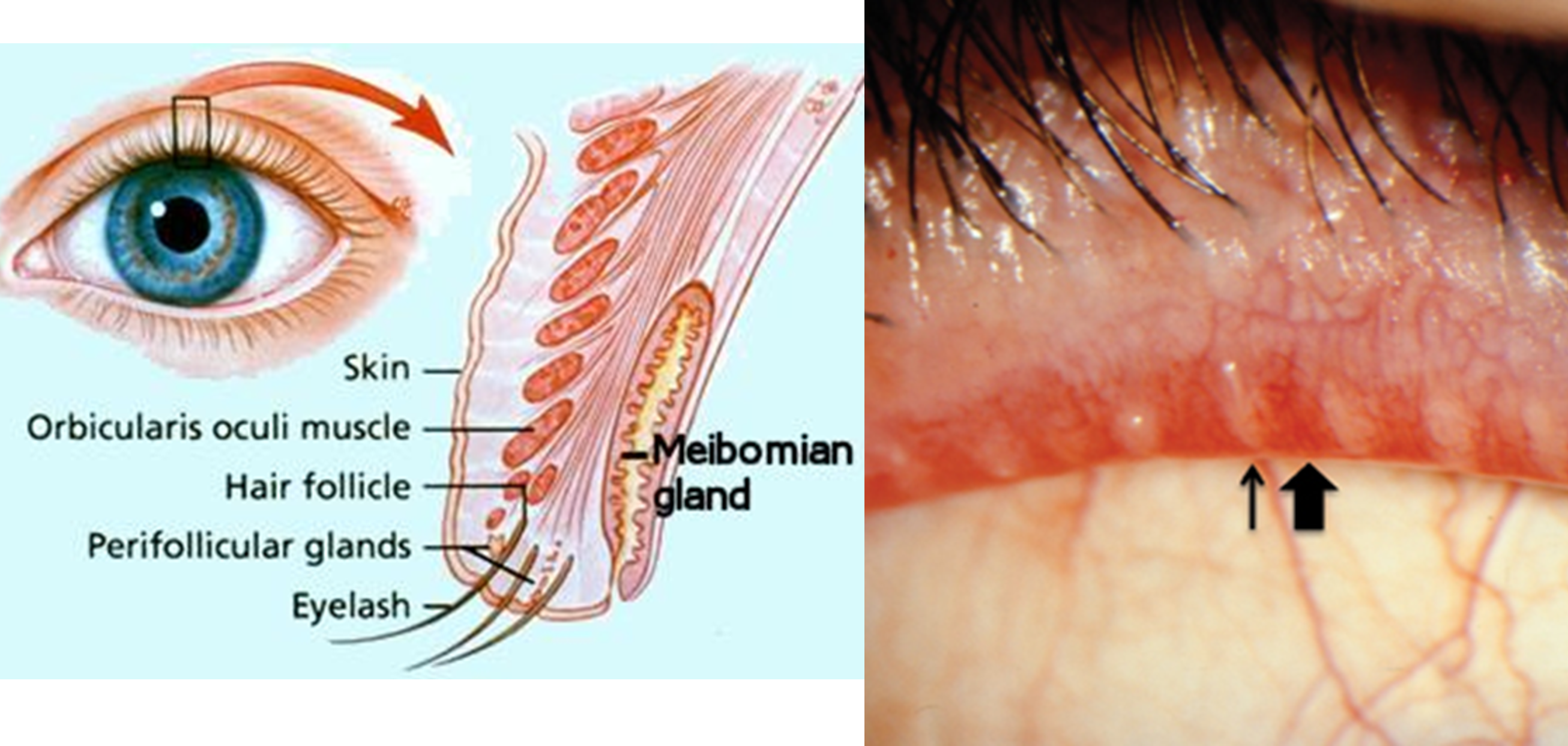
What do the meibomian / tarsal glands do?
modified sebaceous glands that secrete lipid rich product that keeps eyelid from sticking together
What produces aqueous portion of tear film?
lacrimal gland
what produces outer lipid layer to tear film?
meibomian glands
What is the small hole on the medial edge of eyelid that drains tears?
lacrimal punctum
What is the small channel tat drains tears from eye surface into nasal cavity?
lacrimal canaliculus
What is the reservoir for tear overflow and drains eye of debris and microbes?
lacrimal sac
What connects lacrimal canaliculus to lacrimal sac which drain tears into nasal cavity?
nasolacrimal duct
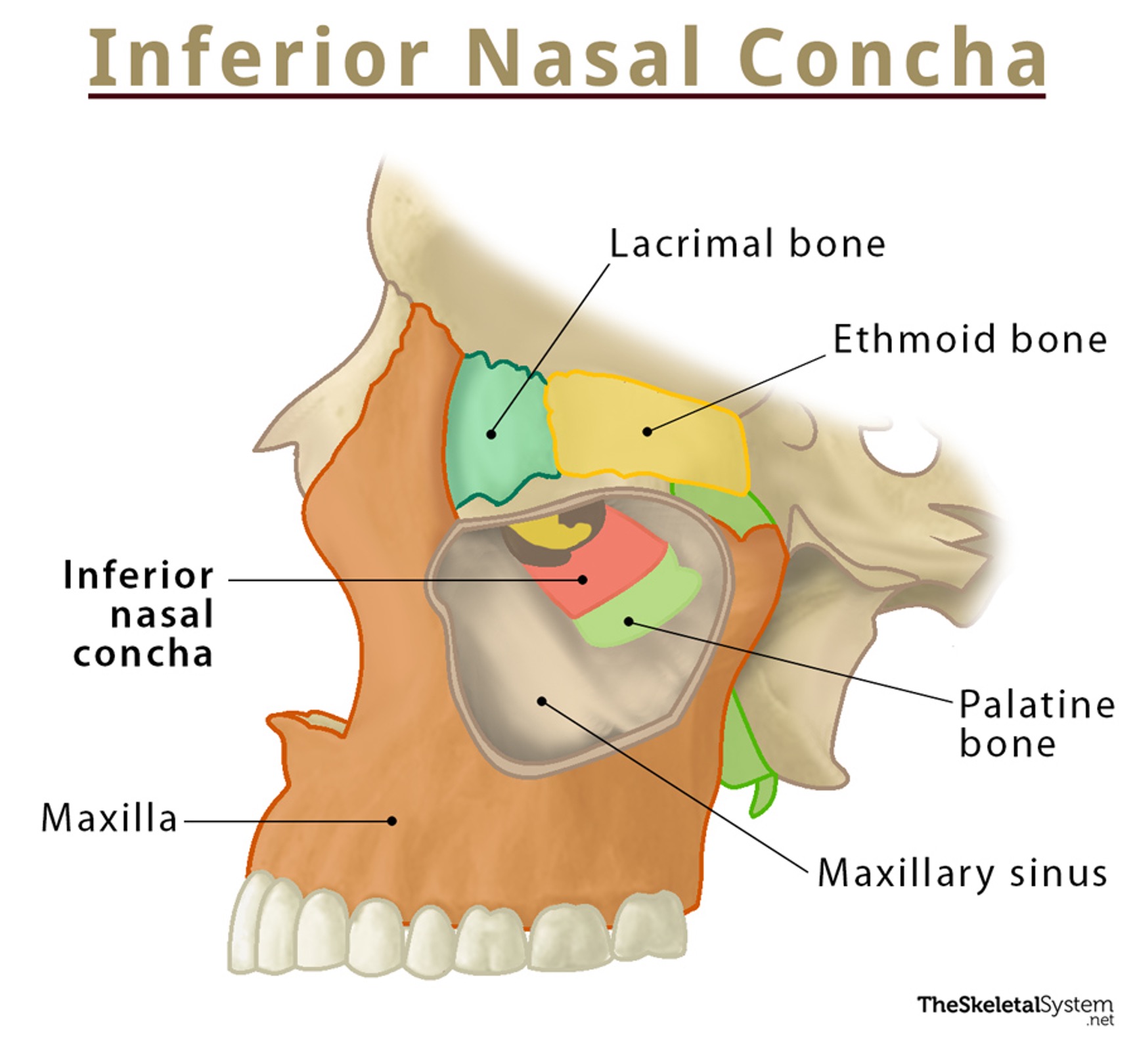
What do the inferior, middle, and superior nasal conchae do?
filter, humidify, and warm are that we breathe to prevent cold air from entering lungs
Where is lacrimal gland?
superolateral corner of orbit
What innervates the lacrimal gland?
greater petrosal nerve (CN VII)
What is the Z axis of eye movement?
horizontal axis in saggital plane with intorsion and extorsion
What is the Y axis of eye movement?
vertical axis in the sagittal place with elevation and depression
What is the X axis of eye movement?
horizontal axis in the axial plane with abduction and adduction
What eye muscles does CN III innervate?
superior rectus, inferior rectus, medial rectus, inferior oblique
What eye muscle does CN VI innervate?
lateral rectus
What eye muscle does CN IV innervate?
superior oblique
Contraction of what muscle dilates the pupil? SNS or PNS? What is it innervated by?
dilator pupillae; sympathetic; CN III
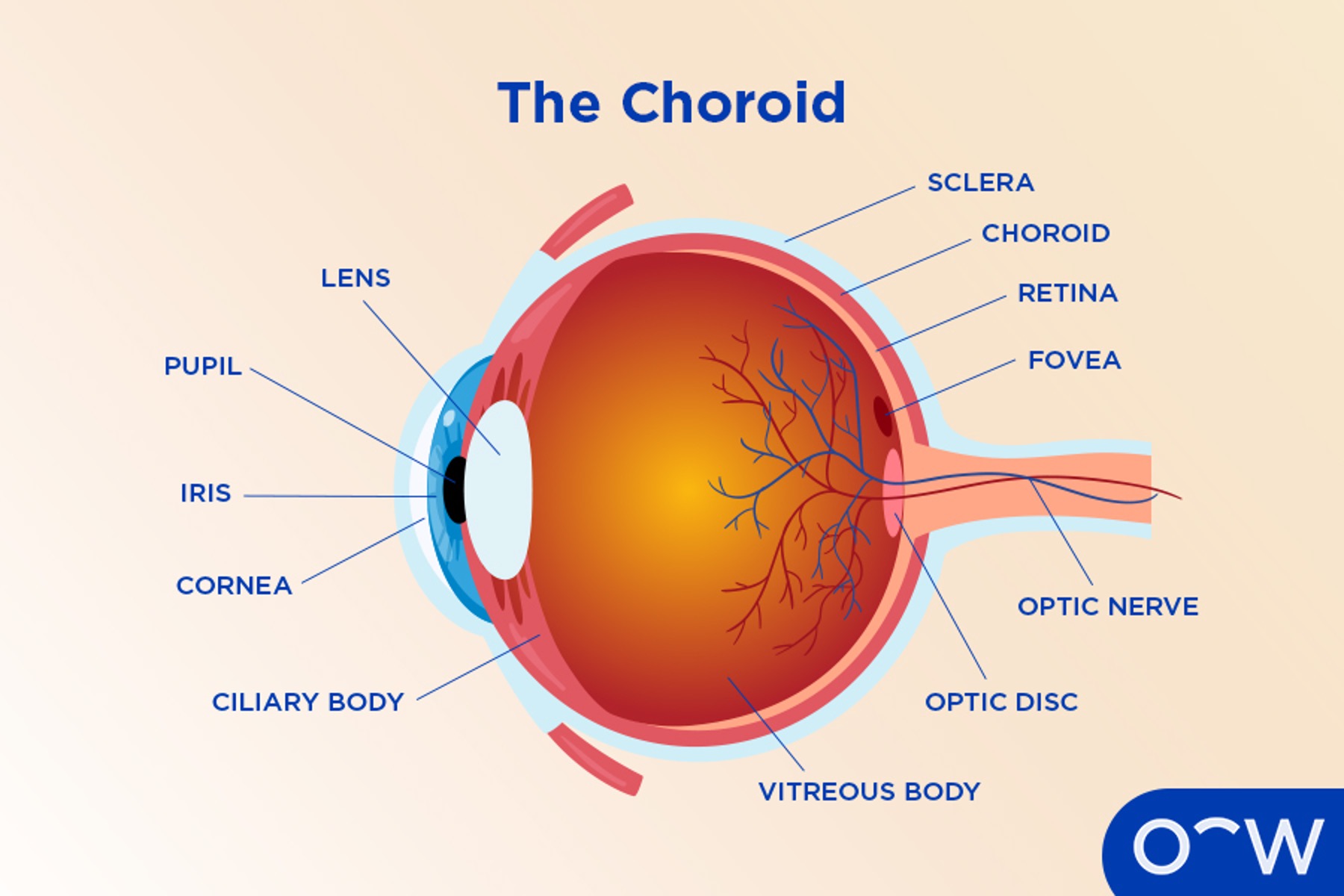
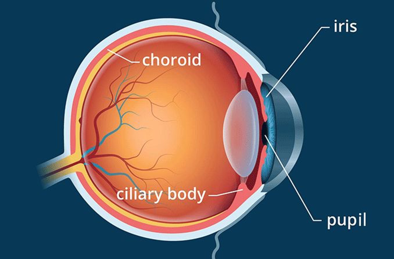
What is the visible colored part of the eye?
iris
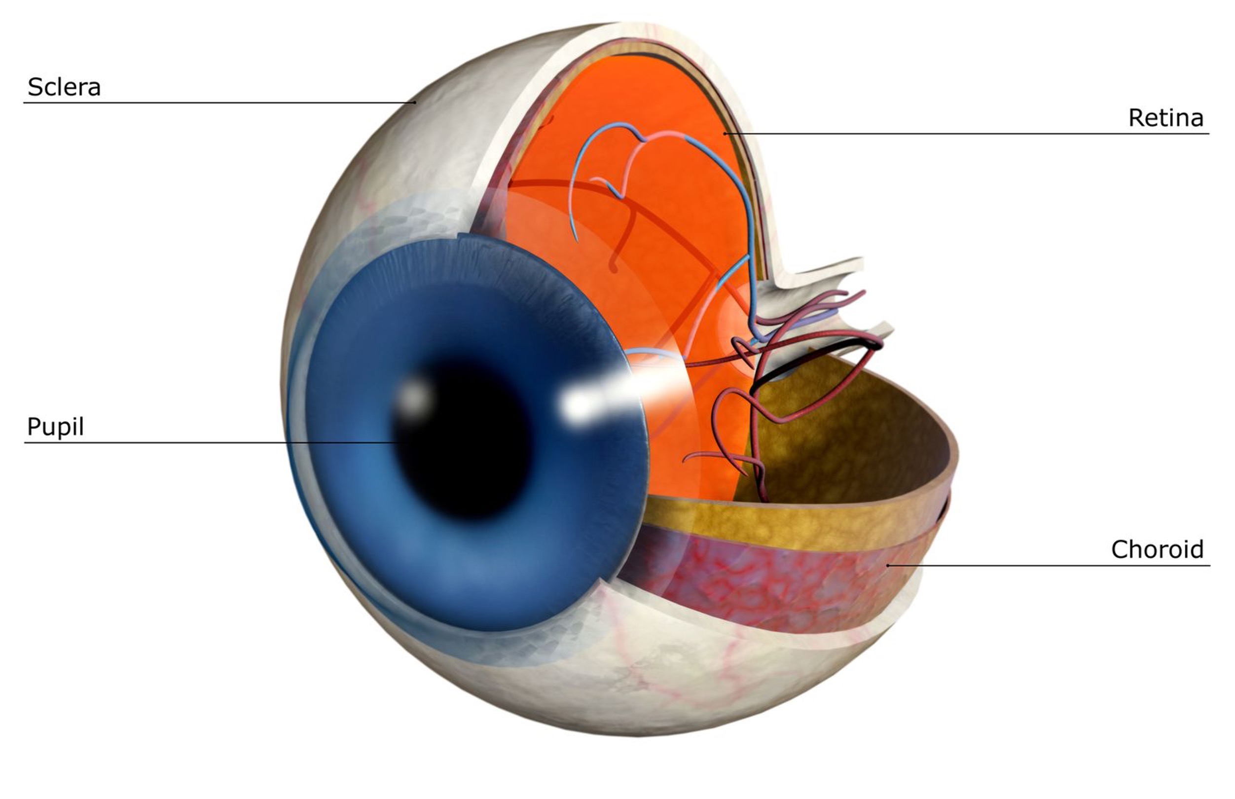
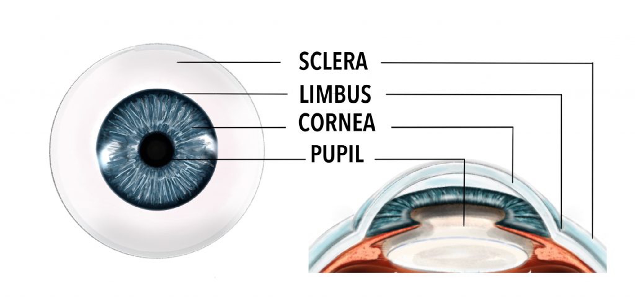
What allows light to enter the eye?
pupil
Contraction of what muscle constricts the pupil? PNS or SNS? What innervates it?
sphincter pupillae; PNS; CN III
When does the pupil constrict?
bright light and close vision
When does the pupil dilate?
dim light and distant vision
What muscle closes eye tightly? What innervates it?
orbicularis oculi; CN VII
What muscle elevates superior eyelid? What innervates it?
levator palpebrae superioris; CN III
What CN opens eyes?
CN III
What CN closes eyes?
CN VII
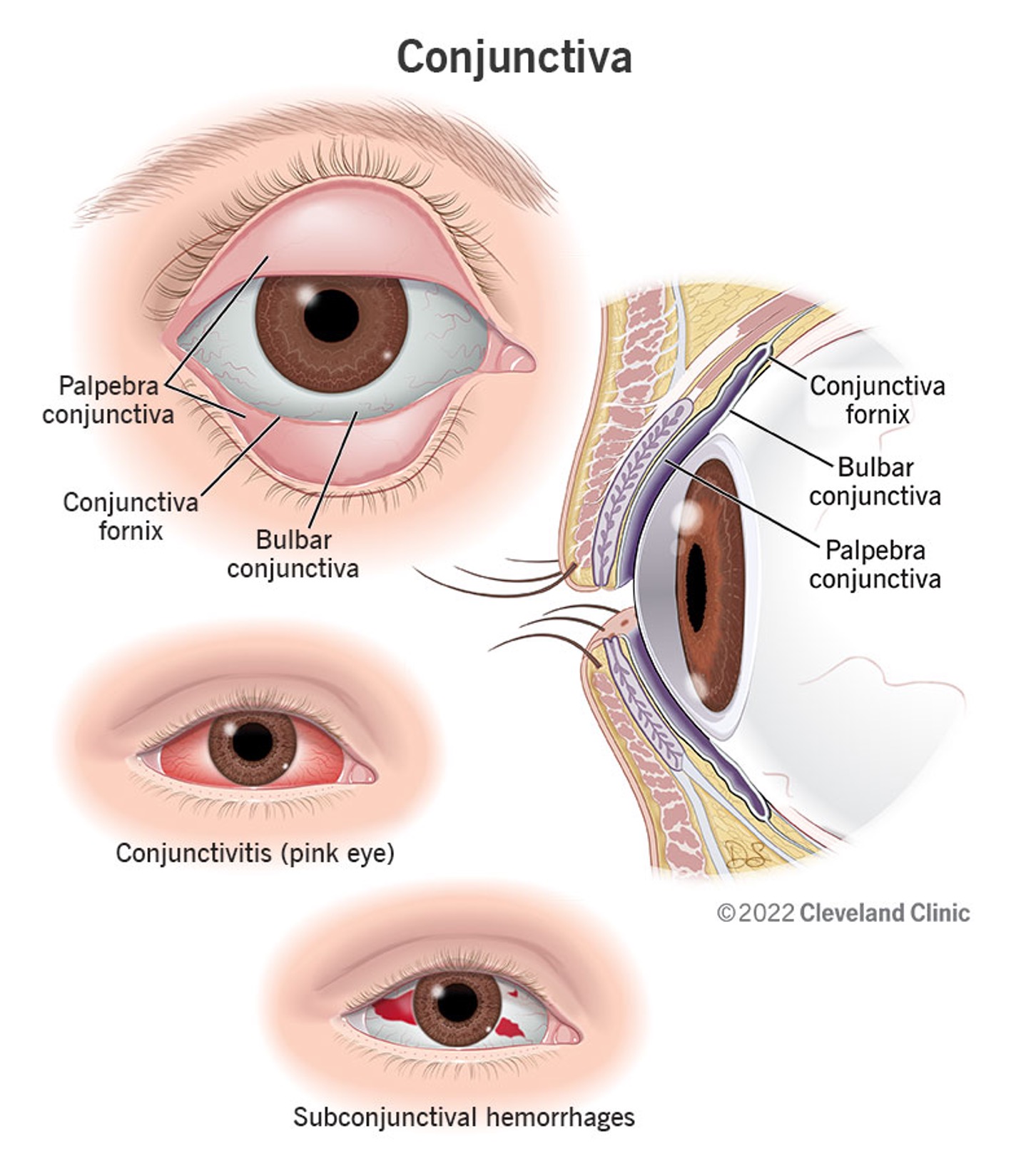
What is bulbar conjunctiva?
covers anterior aspect / sclera of eye, firm attachment to limbus
What is palpebral conjunctiva?
starts at the mucocutaneous junction and lines the eyelids
What is responsible for maintaining shape of globe?
sclera
What is sclera?
white fibrous eye covering that provides strength and flexibility to eye and provides attachment for muscles
What is the cornea?
outermost clear layer of eye; transparent bulging area connected to the sclera that refracts light upon entry
What is the choroid?
vascular, middle layer of the eye
What holds the lens for focusing at different distances?
suspensory ligaments
What do the anterior choroid and ciliary muscle do?
adjust lens diameter for focusing
What divides eye’s anterior cavity?
iris (located between cornea and lens)
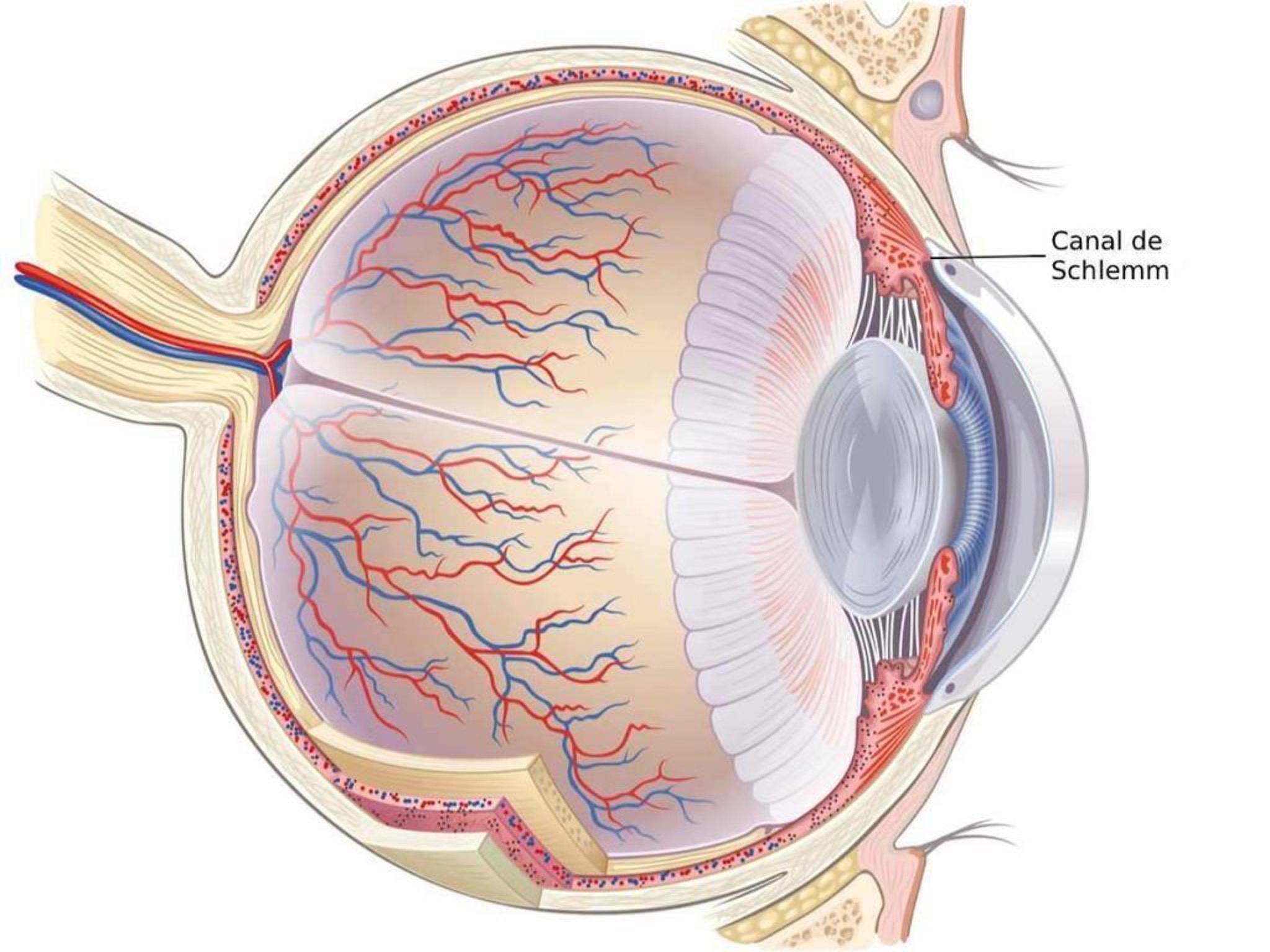
What is aqueous humor?
circulates from posterior to anterior chamber of eye and provides nutrients, taste transport, and a fluid cushion
What does aqueous humor pass through before passing to the scleral veins?
canal of schlemm
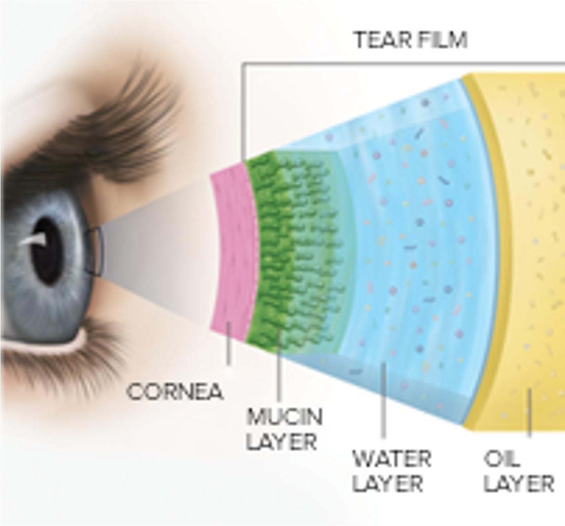
What are the 3 layers of tear film?
lipid / oily layer, aqueous / watery layer, mucin / mucous layer
What secretes the lipid layer of tear film?
meibomian glands and glands of zeis
What secretes the aqueous layer of tear film?
lacrimal gland and acessories
What secretes the mucin layer of tear film?
goblet cells in conjunctiva
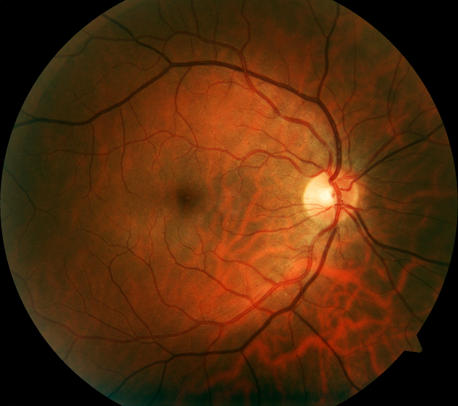
Where is the retina located?
innermost layer, back of eye
What does the retina contain?
rods, cones, macula, fovea
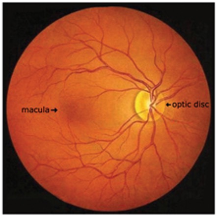
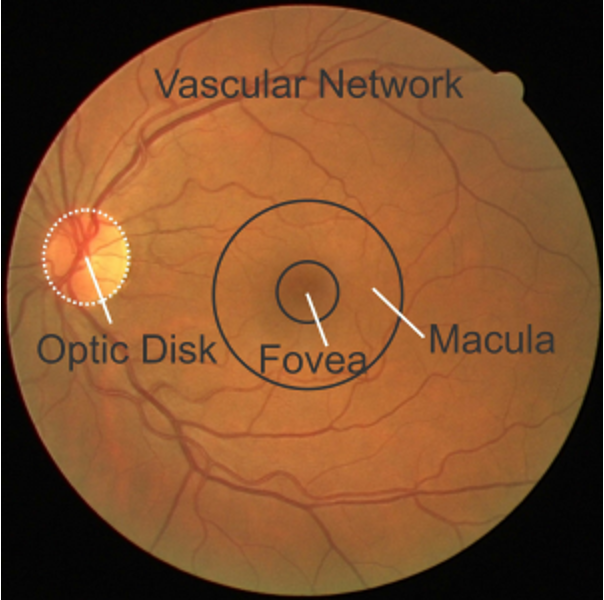
What is the optic disc?
point of entry for CN II that lacks photoreceptors and is insensitive to light; “blind spot”
What is macula lutea?
area behind optic disc that is specialized for high-acuity vision
What is the fovea centralis?
lies within the macula and contains high density photoreceptors where light rays from center of vision land
What is responsible for vision in low light?
rods
What is responsible for visual acuity and color vision?
cones
Where is clear margin and cup, central retinal artery, and veins?
optic nerve
What is the most common cause of reduced visual acuity?
refractive errors
What is presbyopia?
natural loss of accommodation due to age (can’t focus on nearby objects; starts around 40)
what is myopia?
nearsightedness
what is hyperopia?
farsightedness
Does distant vision or near vision need more refraction for focus?
near vision
What is amblyopia?
reduction of vision in one eye due to eye and brain’s inability to work together
What is color blindness?
hereditary x linked disorder characterized by defective/absense of color vision
What is diplopia?
double vision
What is emmetropia?
“normal” refractive condition / clear vision
what is asinometropia?
refractive power of one eye differs from the other
what is strabismus?
misalignment of the eye
What is the most common color blindness?
red-green
Who is color blindness more common in?
men
What do you use to test for color blindness?
ishihara plates
How does color blindness work?
based on perception of red, green, blue;
defect perception of one color results in color being perceives as a combination of the other two
what should be done before instilling any medications? (except for chemical burns)
visual acuity
what must you do to rule out foreign bodies?
flip / evert the lid to examine underneath
what is hypotropia?
downward
what is hypertropia?
upward
what is exotropia?
outward
what is esotropia?
inward
How does cornea and anterior chamber usually appear?
clear w/o crescent shadows
How does optic disc appear normally?
creamy, orange, yellow color with sharp margins
How does optic cup appear normally?
whiter, les than 0.5 cup to disc ratio
How do arteries and veins appear normally?
2:3 ratio without nicking, spasms, copper wire, silver wire, or box car segments
What is helpful in identifying ulcers vs abrasions?
fluorescein stain and woods lamp
What is a slit lamp?
high intensity light source to view front and back of eye
How do you measure interocular pressure? What is it normally?
tonopen; 10-21 mmHg