Oral histology LO 7 part 4- cementum
1/18
There's no tags or description
Looks like no tags are added yet.
Name | Mastery | Learn | Test | Matching | Spaced |
|---|
No study sessions yet.
19 Terms
50%
The cementum is ——% inorganic material, calcium hydroxyapatite.
40%
The cementum is ——% organic material, collagen.
10%
The cementum is ——% water.
Cementum
——————— 1. Seals the surface of the root dentin and covers the ends of the open tubules. 2. Embeds the ends of the periodontal ligament fibres to the tooth.
Cementoblasts
—————————— are formed from the undifferentiated mesechymal cells in the dental follicle (developing periodontal ligament space).
Matrix
Cementoblasts produce the cementum —————.
Acellular cementum
This is the first layer of cementum deposited at the DCJ. Covers the entire root surface, thicker in the cervical third near the CDJ. Produced slowly: thin layer. Width never changes.
Cellular cementum
This covers the acellular cementum. Cementoblasts become entrapped in the forming cementum. Cementocytes lie within lacunae. Cementoblasts with lacunae near the suface of the ————— cementum: active, capable of forming new cementum when necessary, reactive.
Intrinsic fibres
——————— fibres in cementum: collagen fibres within the cementum, fibres formed by the cementoblasts.
Sharpey's fibres
Extrinsic fibres in cementum: collagen fibres, endings of the PDL fibres which are embedded within the cementum are:
15%
There are 3 transitional interfaces that may be present at points along the CEJ. One situation is the cementum may overlap enamel. This happens ——% of the time.
52%
There are 3 transitional interfaces that may be present at points along the CEJ. One situation is that the enamel and cementum meet. This happens ——% of the time.
33%
There are 3 transitional interfaces that may be present at points along the CEJ. One situation is that there may be a gap between the cementum and the enamel. This happens ——% of the time.
Reversal lines
Scalloped lines where there has been resorption of the cementum. Removal of cementum by Odontoclasts.
Arrest lines
Repair of traumatic resorption involves apposition of cementum by cementoblasts in the adjacent periodontal ligament. Apposition of cementum is noted by its layer of growth called:
Cementocytes
Calcified bodies in cementum found either attached to the cementum root surface or lying free in the PDL. They form from apposition of cementum around cellular debris in the PDL.
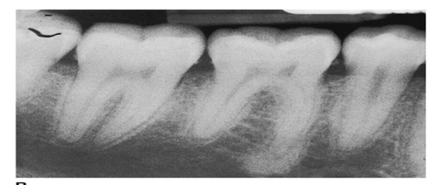
Hypercementosis
This happens at the root apex sometimes. This is due to traumatic occlusal forces of a tooth on a microscopic view with dentin, cementum, and radicular pulp tissue noted in the root.

Cementum spikes
To compensate for fibre bundles decreasing with age, there is excessive deposition of cementum around the depleted fibre bundles.
L mad;f;df'ldnfopqwbef'pojefkjbqfn'q[
Aspen;alsdnf[
Qwkefl'nqwepofj'qeorng
Qelrmg'kqernmgf'kperng;king
Vdhfbvoahbrvoibrvoqjbf