IB Biology Topic 1 past questions
1/49
There's no tags or description
Looks like no tags are added yet.
Name | Mastery | Learn | Test | Matching | Spaced |
|---|
No study sessions yet.
50 Terms
Discuss possible exceptions to cell theory. 4 marks
- Skeletal muscle fibres are larger/have many nuclei
-fungal hyphae are (sometimes) not divided up into individual cells
-unicellular organisms can be considered acellular
because they are larger than a typical cell/carry out all functions of life
-some tissues/organs contain large amounts of extracellular material
-e.g. vitreous humor of eye/ mineral deposits in bone/ xylem in trees/other example
-statement of cell theory/all living things/most tissues are composed entirely of true cells
Eukaryotic cells have intracellular and extracellular components. State the functions of one named extracellular component. 4 marks
name of component: 1 max
e.g. plant cell wall/cellulose/interstitial
matrix/basement membrane/glycoprotein/bone matrix;
functions: 3 max
EITHER
e.g. (plant cell wall) strengthens/supports the cell/plant (against gravity);
prevents the entry of pathogens;
maintains the shape of plant cells;
allows turgor pressure/high pressure to develop inside the cell;
prevents excessive entry of water to the cell;
OR
helps cells to stick together/adhere;
needed to hold cells/tissues together / example of cells/tissues holding together;
forms interstitial matrix / forms basement membrane to support single layers of cells;
e.g. around a blood capillary;
forms (part of the) filtration membrane in the glomerulus;
Explain how the surface are to volume ratio influences cell sizes. 3 marks
small cells have larger ratio (than larger cells)/ratio decreases as size increases
surface area/membrane must be large enough to absorb nutrients/oxygen/substances needed
surface area/membrane must be large enough to excrete/pass out waste products
need for materials is determined by (cell) volume
cell size is limited (by SA/Volume ratio)/cells divide when they reach a certain size
reference to diffusion across/through membrane/surface area
Outline differentiation of cells in a multicellular organism. 4 marks
differentiation is development in different/specific ways
cells carry out specialized functions/become specialized
example of a differentiated cell in a multicelluar organism
cells have all genes/could develop in any way
some genes are switched on/expressed but not others
position/hormones/cell-to-cell signals/chemicals determine how a cell develops
a group of differentiated cells is a tissue
Describe the importance of stem cells in differentiation. 3 marks
stem cells are undifferentiated cells;
embryo cells are stem cells;
stem cells can differentiate in many/all ways / are pluripotent/totipotent;
differentiation involves expressing some genes but not others;
stem cells can be used to repair/replace tissues/heal wounds;
Draw a labelled diagram to show the ultrastructure of Escherichia coli. 6 marks
1 mark for each of the following structures accurately drawn and labelled
rough endoplasmic reticulum
free ribosomes
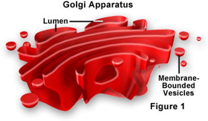
Golgi apparatus
mitochondrion
chloroplast
vacuole
nucleus
lysosome
smooth endoplasmic reticulum
https://www.youtube.com/watch?v=9Ucs0aAcPdI
State one function of each of the following organelles: lysosome, Golgi apparatus, rough endoplasmic reticulum, nucleus, mitochondrion. 5 marks
lysosome: hydrolysis/digestion/break down of materials (macromolecules)
Golgi apparatus: synthesis/sorting/transporting/secretion of cell products
rough endoplasmic reticulum: site of synthesis of proteins (to be secreted)/ intracellular transport of polypeptides to Golgi apparatus
nucleus: controls cells activities/mitosis/replication of DNA/transcription of DNA (to RNA)/directs protein synthesis
mitochondrion: (aerobic) respiration/generates ATP
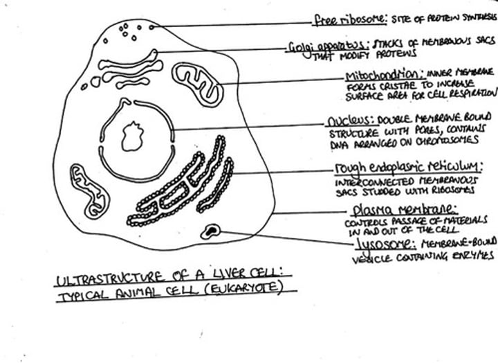
Draw a labelled diagram showing the ultra-structure of a liver cell. 4 marks
1 for each structure clearly drawn and correctly labelled. Whole cells not necessary.
(plasma) membrane - single line surrounding cytoplasm;
nucleus - with a double membrane and pore(s) shown;
mitochondria(ion) - with a double membrane, the inner one folded into internal
projections, shown no larger than half the nucleus;
rough endoplasmic reticulum - multi-folded membrane with dots/small circles on surface;
Golgi apparatus - shown as a series of enclosed sacs with evidence of vesicle formation;
ribosomes - dots/small circles in cytoplasm/ribosomes on rER;
lysosome;
https://www.youtube.com/watch?v=QS7g7RMeCvU
https://image.slidesharecdn.com/biok1-140817011307-phpapp01/95/bioknowledgy-12-ultrastructure-of-cells-31-1024.jpg?cb=1409870160
Distinguish between the structure of plant and animal cells. 6 marks
plant cells: have cell walls, animals do not
have plastids/ chloroplasts, animals do not
have a large central vacuole, animals do not
store starch, animal cells store glycogen
have plasmodesmata, animal cells do not
animal cells:
have centrioles, plant cells do not
have cholesterol in the cell membrane, plant cells do not
plant cells are generally have a fixed shape/ more regular whereas animal cells are more rounded
Using a table, compare the structures of prokaryotic and eukaryotic cells. 5 marks
DNA: P: naked/loop of DNA; E: associated with protein/histones/nucleosomes/DNA in chromosomes
location of DNA: P: in cytoplasm/nuceloid/no nucleus; E: within a nucleus/nuclear membrane
membrane bound organelles: P: none; E: present
ribosomes: P: 70S ; E: 80S
plasma membrane: P & E: same structure within both groups
cell wall: P: peptidoglycan/not cellulose/not chitin; E: cellusose/chitin/not peptidoglycan
respiratory structures: P: no mitochondria; E: mitochondria
pili: P: pili present E: pili absent;
plasmids: P: plasmids (sometimes) present E:plasmids absent;
flagella: P: flagella solid E: flagella flexible/membrane-bound;
What are the 3 principles of cell theory?
Living organisms are composed of cells. All cells come from pre-existing cells by mitosis. Cells are the smallest unit of life.
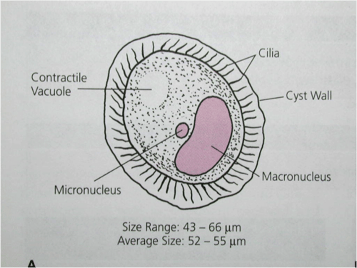
Outline how the Paramecium carries out each of the functions needed for life. 6 marks
Nutrition:
Ingest small organisms and digest these
through endocytosis (vesicles)
Growth:
Nutrients from digestion are used to provide energy and materials required for growth
Is able to grow through the absorption
of minerals and photosynthesis
Excretion :
Waste products from metabolism (e.g. CO2)
are expelled from the cell by diffusing out
the membrane
Waste products such as oxygen deriving
from photosynthesis are expelled
outside the cell through diffusion
Response :
The cilia help the cell to move around,
the paramecium moves toward or away
from external stimuli and therefore
responds to environmental changes
Has an eyespot that is able to detect the
brightest light, the chlamydomonas
moves towards the light stimuli and is
therefore capable of responding to
environmental changes
Metabolism:
Contains enzymes in the cytoplasm
which catalyse the metabolic reactions
Contains enzymes in the cytoplasm
which catalyse the metabolic reactions
Reproduction:
Asexual (mitosis) as well as sexual
reproduction (meiosis & gametes)
Asexual (mitosis) as well as sexual
reproduction (meiosis & gametes)
Homeostasis:
Contractile vacuoles in the cell fill up
with water and remove this water from
the cell by expelling it through the
plasma membrane, this maintains the
water levels inside the cell relatively constant
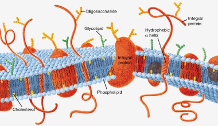
Draw a diagram to show the structure of a cell membrane
5 marks
phospholipids labelled with hydrophillic (heads) and hydrophobic (tails)
phospholipid bilayer clearly shown and labelled
proteins shown in the bilayer and labelled
transmembrane and peripheral/extrinsic proteins shown and labelled
glycoproteins shown and labelled
cholesterol shown and labelled
glycolipids shown and labelled
thickness shown as 10 nm/ + or - 2 nm
Explain how the structure and properties of phospholipids help to maintain the structure of cell membranes. 9 marks
phospholipid structure
hydrophobic tail/hydrophilic head
head made from glycerol and phosphate
tail made from two fatty acids
saturated/ unsaturated fatty acid (in tail)
arrangement in membrane
phospholipids form a bilayer
heads face outside the membrane/ tails face inside the membrane/ hydrophic interior/ hydrophilic exterior of membrane
phospholipids held together by hydrophobic interactions
phospholipid layers are stabilized by interaction of hydrophilic heads and surrounding water
phospholipids allow for membrane fluidity/ flexibility
fluidity/ flexibility helps membranes to be (functionally) stable
phospholipids with short fatty acids/ unsaturated fatty acids are more fluid
fluidity is important in breaking and remaking membranes (e.g. endocytosis/ exocytosis)
phospholipids can move about/ move horizontally/ "flip flop" to increase fluidity
hydrophilic/ hydrophobic layers restrict entry/ exit of substances
Explain the role of vesicles in transportation of materials within cells. 8 marks
vesicles are membrane bound packages/droplets
formed by pinching off/budding off a piece from a membrane
can carry proteins
rough ER synthesizes proteins
proteins enter/accumulate inside the ER
transported to Golgi apparatus for processing
targeted to/transported to specific cellular organelles
fuse with membrane of organelle so contents of vesicle join the organelle
transported to the plasma membrane
fuses with plsma membrane releases/secretes contents
exocytosis
MS
vesicles formed from rER transport proteins to Golgi apparatus; these vesicles fuse with membranes of Golgi apparatus; proteins are processed as they move through Golgi apparatus; (transport) vesicles bud off/leave Golgi apparatus; vesicles move through cytoplasm; (vesicles) fuse with plasma membrane; contents released to outside of cell / exocytosis; cells use vesicles to secrete substances such as hormones/digestive enzymes/other appropriate example; vesicles may contain cell products other than proteins;
Describe the process of active transport. 4 marks
uses/ requires energy/ ATP
goes against concentration gradient/ lower to higher concentration
requires a protein in the cell membrane/ pump/ carrier protein (reject channel)
hydrolysis of ATP/ ATP --> ADP + phosphate
involves a conformational change in the pump/ protein/ diagram to show this
Outline the ways in which substances move passively across membranes. 5 marks
diffusion (is a method of passive transport across the membrane)
pore/ channel proteins for facilitated diffusion/ to allow hydrophilic particles across
movement from high to low concentration/ down the concentration gradient
membrane must be permeable to the substance diffusing
oxygen/ other named example of a substance than can diffuse through membranes
osmosis is movement of/ diffusion of water through a membrane
from a region of lower to a region of higher solut concentration/ higher to lower water potential
membranes are (nearly) always freely permeable to water
Distinguish between active and passive movements of materials across plasma membranes, using named examples. 4 marks
passive: diffusion / osmosis / facilitated diffusion, whereas, active transport: ion pumps / exocytosis / pinocytosis / phagocytosis
a second passive method (from above), whereas, active transport: a second active method; (from above)
passive: does not require energy, whereas, active transport: requires energy/ATP;
passive: down concentration gradient, whereas, active transport: against concentration gradient;
passive: no pumps needed, whereas, active transport: requires protein pumps;
passive: oxygen across alveoli / other example, whereas, active transport: glucose absorption in ileum / other example;
Outline, with an example, the process of exocytosis. 5 marks
vesicles carry material to plasma membrane;
vesicle fuses with membrane;
(by joining of) phospholipid bilayers;
aided by the fluidity of the membrane;
material released/expelled from the cell;
membrane flattens;
name of example e.g. exocytosis of neurotransmitter / exocrine secretion/endocrine secretion / hormone secretion / release of cortical granules;
outline of example: (in the presence of calcium), neurotransmitter vesicles release their contents into the synapse / hormones released from one cell have an effect on another cell etc.;
Explain the reasons for cell division in living organims. 8 marks
to increase the number of cells in an organism
to allow differentiation/ cell specialization
for greater efficiency
to replace damaged/ lost cells
example
binary fission
asexual reproduction of unicellular organisms
gamete/ spore formation
cells only arise from pre-existing cells
refer to Virchow
cells cannot grow beyond a certain size
surface area to volume ratio becomes too small
transport across the membrane too slow
example
nucleus cannot control the cell
control of cell division sometimes lost
tumor formation
Outline the processes that occur in a cell during interphase, including those needed to prepare for mitosis.
4 marks
DNA replication
DNA transcription
enzyme/ protein synthesis
biochemical reactions/ example of a biochemical reaction
cell respiration
growth
organelles replicated
What cell parts are visible and not visible using a light microscope?
Visible: cell walls, vacuoles, cytoplasm, mitochondria,chloroplasts, nucleus and cell membrane.
not visible:ribosomes, endoplasmic reticulum, lysosomes, centrioles, golgi bodies
By what process do most bacteria divide?
binary fission, 2 identical daughter cells
Drawing of the ultrastructure of prokaryotic cells based on electron micrographs.
show the cell wall, pili and flagella, and plasma membrane enclosing cytoplasm that contains 70S ribosomes and a nucleoid with naked DNA.
https://image.slidesharecdn.com/biok1-140817011307-phpapp01/95/bioknowledgy-12-ultrastructure-of-cells-10-1024.jpg?cb=1409870160
Drawing of the ultrastructure of eukaryotic cells based on electron micrographs.
show a plasma membrane enclosing cytoplasm that contains 80S ribosomes and a nucleus, mitochondria and other membrane-bound organelles are present in the cytoplasm. Some eukaryotic cells have a cell wall
https://image.slidesharecdn.com/biok1-140817011307-phpapp01/95/bioknowledgy-12-ultrastructure-of-cells-14-638.jpg?cb=1409870160
Organelles function (based on pancreatic exocrine cell)
Nucleus
Mitochondrion
Ribosome
Rough ER
Golgi
Vesicles
Lysosomes
Vacuoles
Flagellum
Cilia
Microtubules
Centrioles
Nucleus - double membrane, pores present in membrane, contains genetic information in the form of chromosomes, mRNA is transcribed in the nucleus prior to translation in the cytoplasm. it leaves through the pores
Mitochondrion - has a double membrane, a smooth outer membrane and a folded inner membrane. the folds are cristae. site of ATP production. fat is digested here if used as an energy source
Free ribosomes - 80S, larger than in prokaryotes. no membrane, synthesise ribosomes
Rough ER - consist of flattened membrane sacs, called cisternae. often near nucleus. 80S ribosomes are attached to the outside of the cisternae. rEr synthesise protein which are transported by vesicles to the golgi apparatus for modification before secretion outside the cell.
Golgi apparatus - also consists of flattened membrane sacs called cisternae. no attached ribosomes. often close to plasma membrane. cisternae are shorter and more curved than those of the rER. Golgi modifies/processes proteins from the rEr and repackaged in vesicles f secretion outside the cell.
Vesicles - a single membrane containing a fluid, very small, used to transport materials inside of a cell
Lysosomes - formed from Golgi, contain digestive enzymes f breakdown of ingested food in vesicles, unwanted/damaged organelles, cell itself
Vacuoles - single membrane with fluid inside, in plant cells often large and permanent, in animals vacuoles are smaller and temporary
Flagellum - in animal cells only. Thin projection, usually singular, from cell surface. Contain microtubules. Move cell.
Cilia- in animal cells only.
Thin projections from surface. Contain microtubules. Eitehr move cell or move fluids adjacent ot the cell
Microtubules
- small cylindrical fibres, have a variety of functions e.g. involved in structure of flagella and cell divsion
Centrioles
- 2 groups of 9 triple microtubules
- mainly found in animal cells, not present in vascular plants or fungi
Why are eukaryotes compartmentalized?
Efficiency of metabolism - enzymes and substrates can be localized and much more concentrated.
Localised conditions - pH and other such factors can be kept at optimal levels. The optimal pH level for one process in one part of the cell. Toxic substances can be isolated, e.g. digestive enzymes that could digest the cell itself are stored in lysosomes. Numbers and locations of organelles can be changed dependent on the cell's requirements.
Structure and function of
chloroplast
cell wall
double membrane, inside are stacks of thylakoids, site of photosynthesis and glucose production, starch grains may be present
Cell wall- extracellular component, consists mainly of cellulose which is permeable, strong, prevents the plasma membrane from bursting when under pressure, hard to digest so it is resistant to be broken down
Identify a cell from an electron micrograph
https://image.slidesharecdn.com/biok1-140817011307-phpapp01/95/bioknowledgy-12-ultrastructure-of-cells-32-1024.jpg?cb=1409870160
https://image.slidesharecdn.com/biok1-140817011307-phpapp01/95/bioknowledgy-12-ultrastructure-of-cells-36-1024.jpg?cb=1409870160
https://image.slidesharecdn.com/biok1-140817011307-phpapp01/85/bioknowledgy-12-ultrastructure-of-cells-21-320.jpg?cb=1409870160
https://image.slidesharecdn.com/biok1-140817011307-phpapp01/95/bioknowledgy-12-ultrastructure-of-cells-35-1024.jpg?cb=1409870160
https://image.slidesharecdn.com/biok1-140817011307-phpapp01/95/bioknowledgy-12-ultrastructure-of-cells-37-1024.jpg?cb=1409870160
function of cholesterol in cell membranes?
support, effective barrier to ions, fluidity, provide mechanical stability to prevent cells from bursting
Can you label/identify structures from the lipid bilayer? Draw a diagram to show the fluid mosaic structure of a plasma membrane, indicating the hydrophilic and hydrophobic regions
https://s14-eu5.ixquick.com/cgi-bin/serveimage?url=http%3A%2F%2Ffrontalcortex.com%2Fgallery%2Fpics%2F1_3351_1409629078.jpg&sp=536862e97df78d31050930a3af12fcfd
https://www.youtube.com/watch?v=n8ZvK0r9HIY
Compare and contrast the Davson-Danielli and Singer-Nicholson models of cell membranes and
describe the evidence that disproves the Davson-Danielli model. 4 marks
Danielli and Davson proposed a model whereby two layers of protein flanked a central phospholipid bilayer
The model was described as a 'lipo-protein sandwich', as the lipid layer was sandwiched between two protein layers
The dark segments seen under electron microscope were identified (wrongly) as representing the two protein layers
It did not account for the permeability of certain substances (did not recognise the need for hydrophilic pores)
new model was proposed by Seymour Singer and Garth Nicolson in 1972
According to this model, proteins were embedded within the lipid bilayer rather than existing as separate layers
This model, known as the fluid-mosaic model, remains the model preferred by scientists today (with refinements)
What route is used to export proteins from the cell?
Rough endoplasmic reticulum → Golgi apparatus → plasma membrane
Define osmosis
Osmosis is the passive movement of water from a high water concentration to a low water concentration through a partially permeable membrane.
Compare exocytosis and endocytosis
Exocytosis releases materials out of the cell, endocytosis takes material into cell
Similarities: both need energy, both use a vesicle, both transport molecules across the membrane
Exocytosis: vesicle forms in the Golgi, then fuses with the membrane
Endocytosis: Vesicle forms from the cell membrane which then gets ino the cytoplasm
Exocytosis increases size of cell membrane, endocytosis does the opposite
Compare and contrast simple diffusion with facilitated diffusion as mechanisms to transport
solutes across membranes 5 marks
Both do not require ATP
Both mean that molecules will go from a region of high concentration to a region of low concentration
Both need substances permeable to the membrane
Simple diffusion involves diffusion of molecules through the phospholipid bilayer
Facilitated diffusion uses channel proteins embedded in the membrane
Hydrophobic molecules will pass through the cell membrane by simple diffusion, hydrophilic molecules and charged particles will use facilitated diffusion because the cell membrane is hydrophobic inside
Describe the process of endocytosis. 5 marks
Endocytosis allows substances to enter the cell
It requires energy
Plasma membrane is pulled inwards
Decreases size of plasma membrane
Vesicle forms from the cell membrane and is pinched off
Vesicle can carry its content into the cytoplasm
Definition of mitosis
Division of the nucleus into 2 genetically identical daughter nuclei
List 4 processes needed for the spontaneous origin of life. 2 marks
1. The non-living synthesis of simple organic molecules (from primordial inorganic compounds)
2. The assembly of these organic molecules into polymers
3. The formation of polymers that can self-replicate (enabling inheritance)
4. Packaging of these molecules into membranes with an internal chemistry different from their surroundings
Outline the evidence that spontaneous generation of cells does not now occur. 3 marks
Pasteur designed an experiment to test whether sterile nutrient broth could spontaneously generate microbial life. He set up two experiments. In both he added nutrient broth into flasks, each cell was heated to boil the broth so that all existing microbial life was killed. One flask was exposed to air and the other one was not. Microbe growth occured only in the flask exposed to air
1. Cells are highly complex structures and no mechanism has been found f producing them from simpler subunits
2. All known examples of growth, e.g. tissue, organism, population are result of cell division
3.Viruses are produced from simpler subunits, not of cells and can only be produced inside of host cells they have infected
4. Genetic code is universal so there must be a single common ancestor
State two properties of RNA that would allow it to play a role in the origin of life.
Self replicating, acts as a catalyst
Outline the experiment of Miller and Urey into the origin of organic compounds. 3 marks
Miller and Urey recreated the conditions of pre-biotic Earth in a closed system consisting of a series of flasks and tubes
These conditions included a reducing atmosphere (low oxygen), high radiation levels, high temperatures and electrical storms
Water was boiled to form vapour and then was mixed with methane, ammonia and hydrogen
The mixture of gases was exposed to an electrical discharge (sparks) to simulate lightning
The mixture was then allowed to cool and after one week was found to contain some simple amino acids and complex oily hydrocarbons
Based on these findings, it was concluded that under the hypothesised conditions of pre-biotic Earth, organic molecules could be formed
Discuss the endosymbiotic theory for the origin of eukaryotes. 6 marks
The theory states that organelles, e.g. mitochondria and chlorplasts, originated as symbioses between unicellular organisms. A prokaryote grows in size and develops folds in its membrane to maintain an efficient SA to vol ratio. Infoldings are pinched off forming an internal membrane. Nucleoid region is enclosed in internal membrane and hence becomes nucleus
What is the sequence of stages during the cell cycle?
G1, S, G2, mitosis, cytokinesis
G1 - growth
S- DNA replication
G2 - protein synthesis, growth
What processes need mitosis?
GATE - Growth, asexual reproduction, tissue repair, embryonic development
Distinguish between cytokinesis in animals and plants?
Cytokinesis is the last stage during which the cytoplasm divides to create two daughter cells. In animal cells the cell is pinched in two while plant cells form a plate between the dividing cells.
Explain how cyclins affect control the progression of a cell through the cell cycle. 4 marks
Cells cannot progress to the next stage of the cell cycle unless a specific cyclin reaches its treshold.
Cyclins bind to enzymes called cyclin-dependent kinases.
These kinases then become active and attach phosphate groups to other proteins in the cell.
The attachmemt of phosphate triggers the other proteins to become active and carry out tasks (specific to one of the phases of the cell cycle)
3 types of mutagens
High energy radiation such as X-rays, UV light, some viruses
Distinguish between primary and secondary tumours, 2marks
Primary tumor grows at the site where the abnormal growth first occured. Secondary tumours develop at different locations due to the circulating cancerous cells
Explain enzyme substrate specificity 3 marks
enzymes fit together with substrates similar to a lock and key; active site has shape that gives specificity; enzymes catalyze a reaction with a specific substrate; example of named enzyme and its substrate; substrate held precisely in (optimum) position to make/break bonds/carry out reaction / chemical interaction occurs between enzyme and substrate;