COMD 4700 - Basic Audiology Exam 4 Definitions
1/71
There's no tags or description
Looks like no tags are added yet.
Name | Mastery | Learn | Test | Matching | Spaced |
|---|
No study sessions yet.
72 Terms
Acoustic Immittance
A term coined to refer to either acoustic impedance or acoustic admittance.
Acoustic ohms and mhos
A measure of electrical current resistance named for a German physicist Georg Simon Ohm. The ohm is labeled sometimes with the symbol Ω. There is one ohm of resistance when one ampere of current is produced by one volt. Inter-electrode impedance in evoked response measurement is optimally 5,000 ohms or less, and balanced (difference of less than 2000 ohms between electrodes). The reciprocal of an ohm is a mho.
Admittance
Ease of energy flow through the middle ear system.
DecaPascals (daPa)
A unit of pressure used in acoustic immittance measurements, such as tympanometry.
Electroacoustic procedures
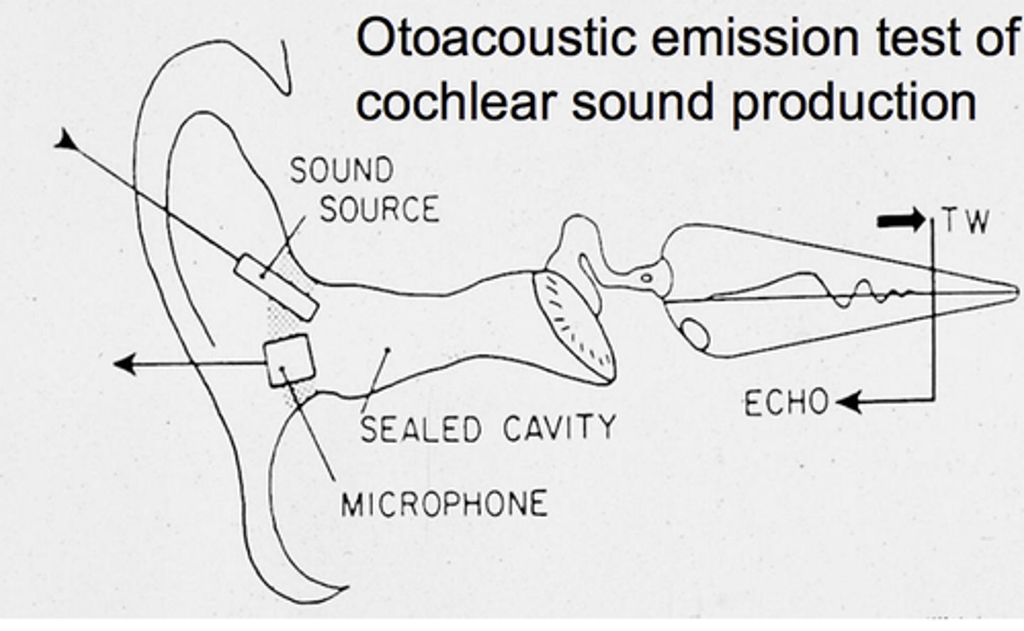
Auditory tests involving the presentation of a sound stimulus and detection of an auditory response by the measurement of sound in the ear canal. Acoustic immittance measurements and otoacoustic emissions are examples of these
Hermetic seal
In audiology, an airtight seal or connection between a rubber probe tip and the walls of the external ear canal.
Impedance
Electrical; a measure of total opposition to energy (current) flow in an an electrical circuit. There are also mechanical and acoustic forms of impedance. Inter-electrode impedance in evoked response measurement is the opposition to current flow between a pair of electrodes, reported as electrical resistance in ohms.
mm H2O
A unit of pressure expressed as millimeters of water pressure
Objective auditory procedures
Auditory tests that do not require a behavioral response from a patient. Otoacoustic emissions, acoustic immittance measures, and auditory evoked responses are examples of these
Manometer
A meter for measuring air pressure. Acoustic immittance devices include one to measure air pressure in the ear canal during tympanometry.
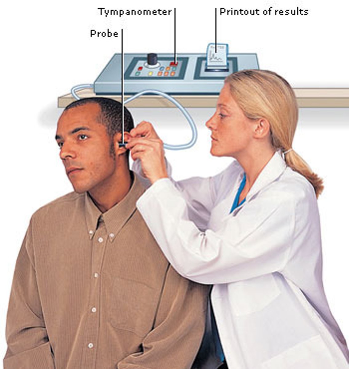
Probe tone
A tone presented to the ear via a probe in the external ear canal that is used in acoustic immittance measurements.
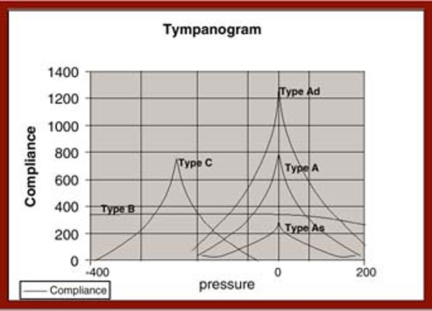
Tympanogram
A graph showing measurement of tympanic membrane mobility as a function of air pressure changes within the ear canal. Tympanograms are classified into types, e.g., type A, type B, and type C.
Tympanometry
Measurement of tympanic membrane mobility as a function of air pressure changes within the ear canal.
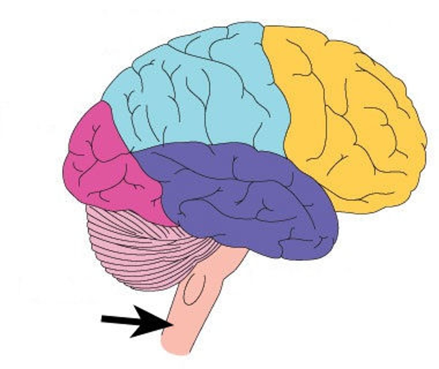
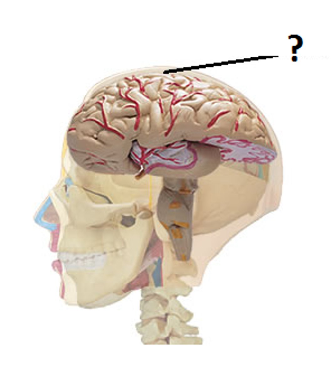
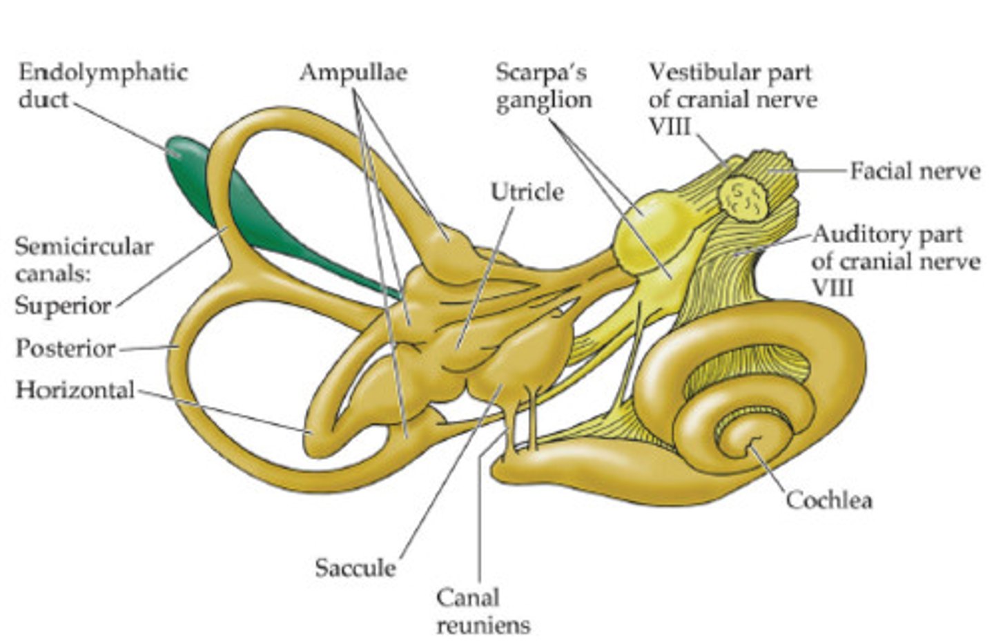
Brainstem
Part of the central nervous system above the spinal cord that contains important auditory centers as well as centers that control vital life functions like breathing and heart activity.
Central auditory nervous system
The auditory system beginning with the cochlear nucleus in the brainstem and including also pathways and auditory regions in the thalamus and the cerebrum.
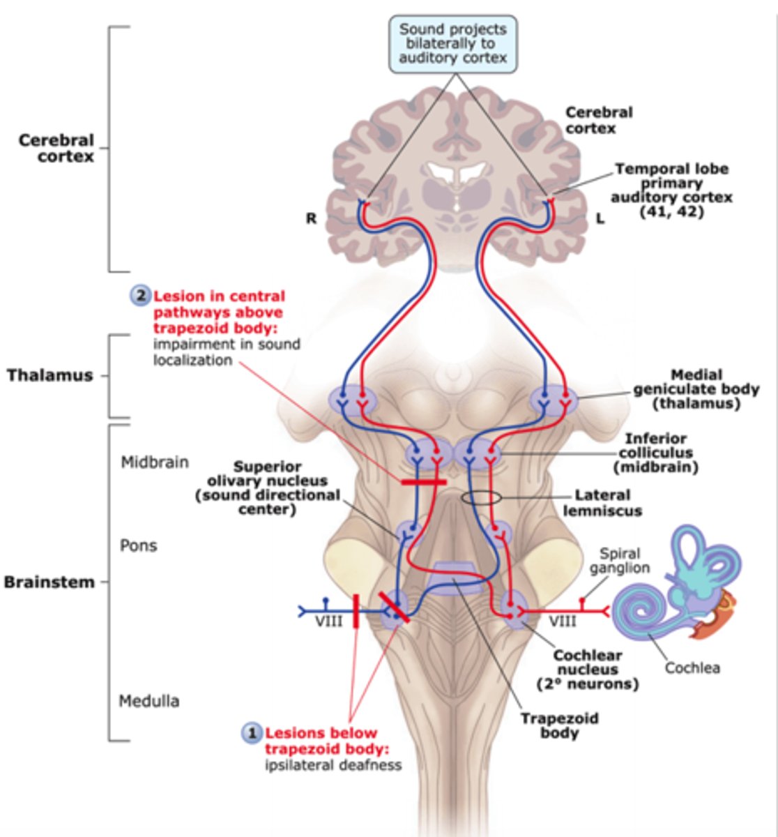
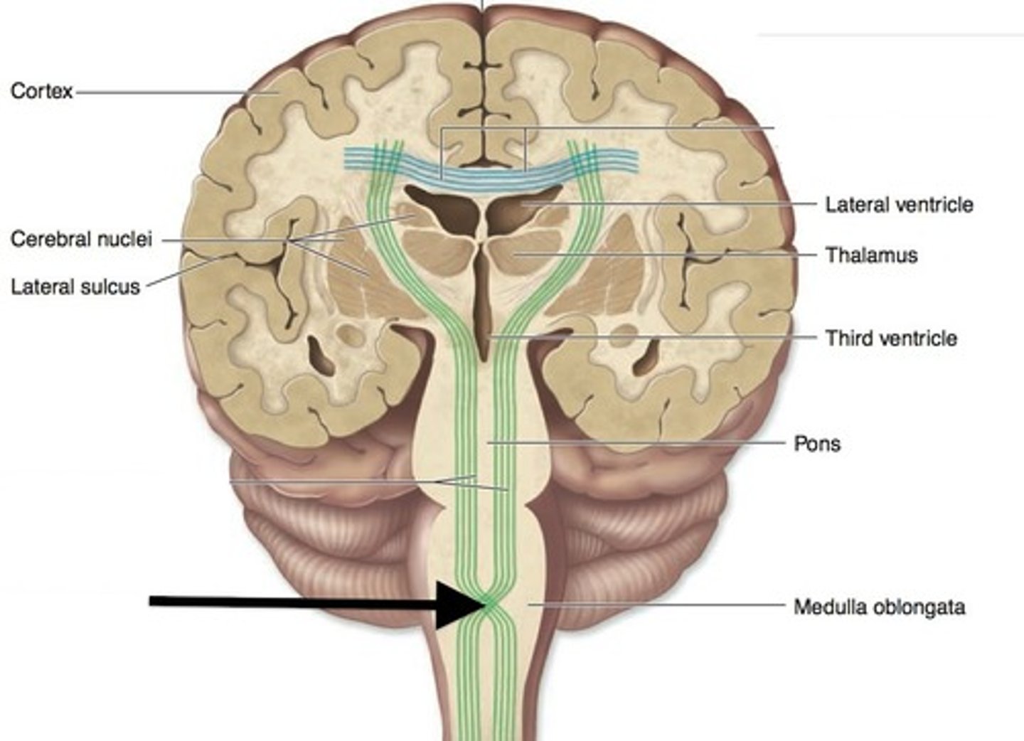
Cerebral cortex
The thin outermost layer of the cerebrum. The auditory cortex is located on the superior surface of the temporal lobe in the cerebrum.
Decussation
A crossing of fibers from one side of the central nervous system to the other. A portion of the corpus callosum is a major one of these within the auditory system.
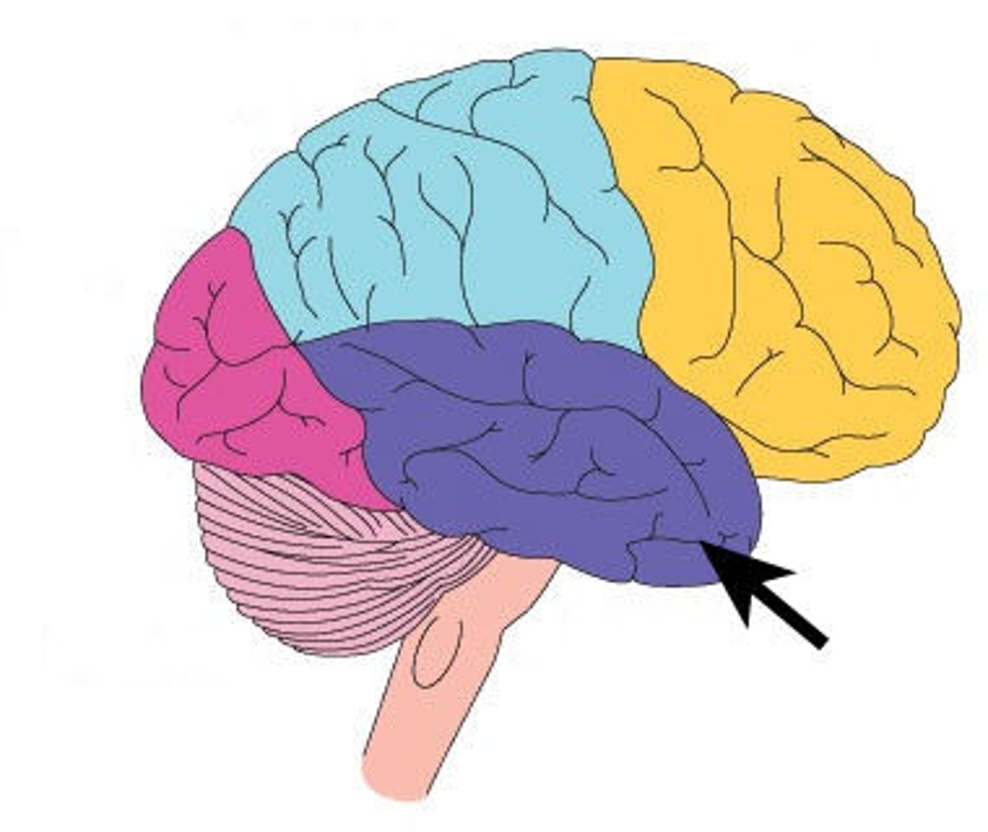
Temporal lobe
One of the four major lobes of the brain. Auditory regions are located in the here.
Thalamus
A sub-cortical oval shaped structure on each side of the central nervous system that serves as a major relay station for sensory pathways (auditory, visual, somatosensory) between the brainstem and cortex. The medial geniculate body, an important auditory structure, is located on the posterior portion of this
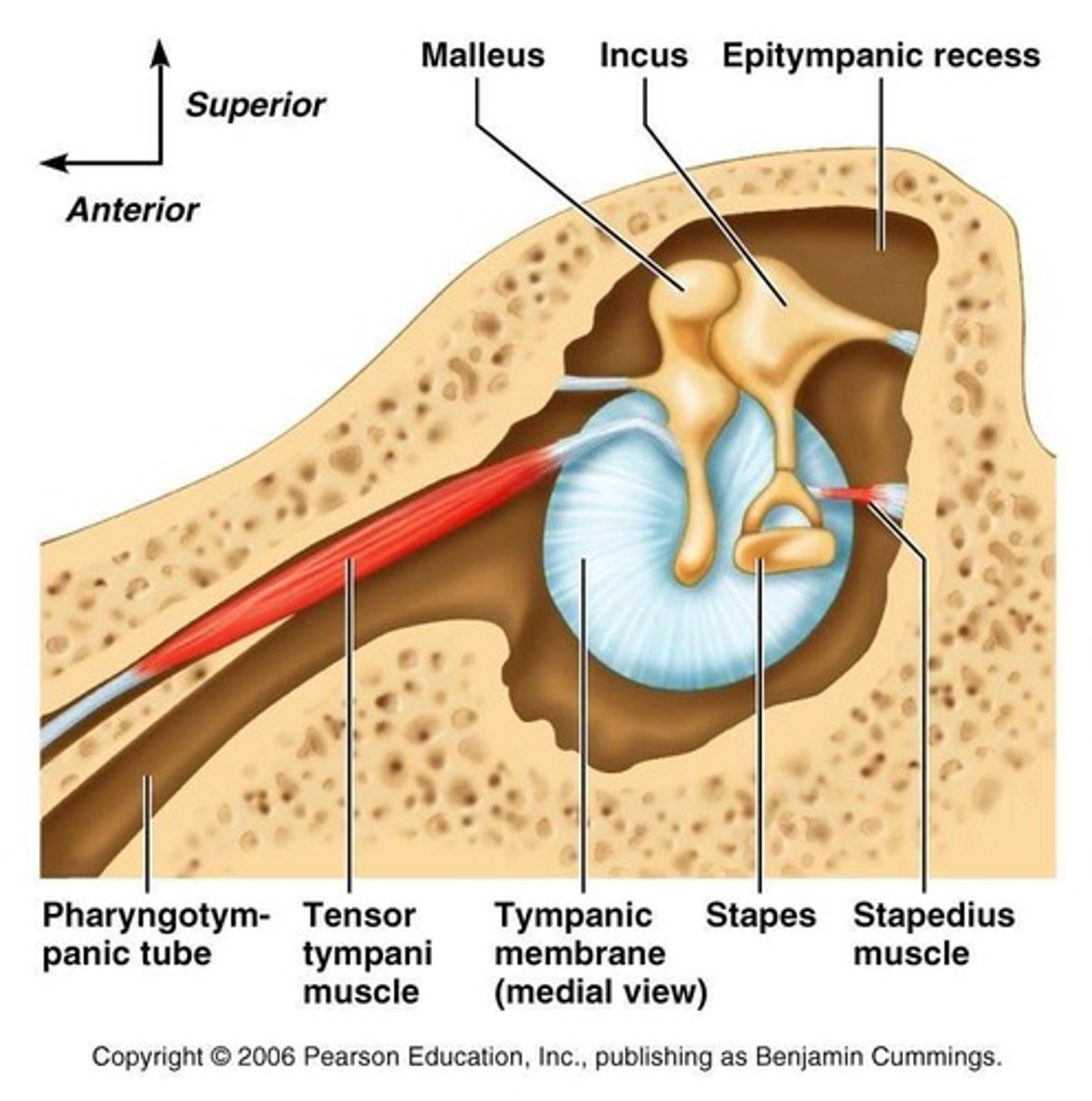
Acoustic reflex
Contraction of one or both middle-ear muscles in response to an intense sound. Contraction of the stapedius muscle is typically measured in clinical audiology.
Distortion product otoacoustic emissions (DPOAE)
Sounds recorded in the external ear canal associated with activation and movements of the outer hair cells in the cochlea in response to stimulation with two closely-spaced pure tones. Distortion products are evidence of normal outer hair cell functioning.
DPgram
A graph showing the amplitude of distortion product otoacoustic emissions plotted as a function of the stimulus frequency (usually the f2 stimulus). See distortion product otoacoustic emissions.
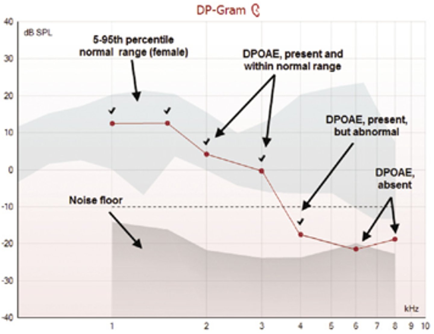
Otoacoustic emissions (OAEs)
Acoustic energy measured within the ear canal that is produced by the cochlea, specifically movement or motility of outer hair cells. Evoked OAEs, including transient (TEOAEs) and distortion product (DPOAE) otoacoustic emissions are generated by acoustic stimulation of the ear.
Spontaneous otoacoustic emissions (SOAEs)
Energy produced by outer hair cells and detected in the external ear canal in the absence of any outside acoustic stimulus.
Stapedius muscle
The smallest muscle in the human body, attached to the posterior portion of the neck of the stapes and innervated by a branch of the seventh (facial) cranial nerve. This muscle contracts in response to high intensity sounds (see acoustic stapedial reflex).

Tensor tympani muscle
Connected to the malleus in the middle ear this muscle contracts with bodily activities like chewing and also in a startle type response to very high intensity sounds. This muscle is innervated by the trigeminal (5th cranial) nerve. The other smaller muscle in the middle ear is the stapedius muscle.
Transient evoked OAEs (TEOAEs)
Energy associated with stimulation of outer hair cells in the cochlea in response to very brief sounds like clicks.
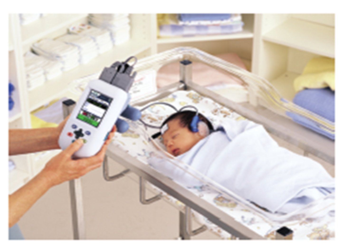
Auditory brain stem response (ABR)
Electrical activity evoked (stimulated) by very brief sounds that originates from the eighth nerve or auditory portions of the brainstem. Consists of five waves and is usually recorded from the surface of the scalp and external ear. Used in hearing testing of infants and young children and in detection of auditory abnormalities affecting the auditory nerve and brain.
Auditory evoked response (AER)
Electrical activity evoked (stimulated) by sounds arising from auditory portions of the peripheral or central nervous system and recorded with electrodes. These include but are not limited to the electrocochleography (ECochG), auditory brainstem response, auditory middle-latency response, auditory late response and P300 response.
Electrode
A device that makes contact with the body that conducts bio-electrical activity from the body via a wire lead to recording equipment in all sensory evoked responses. May consist of different materials including metal in the shape of a small cup or an adhesive material integrated with conducting gel. May also be used to deliver electrical stimulation to the body in a test of facial nerve function called electroneuronography (ENoG).
Tone bursts
A very brief (usually less than 1 second) tone stimulus with no specific duration characteristics. These stimuli are effective in eliciting an auditory brainstem response.
Aural atresia
Abnormal absence of the outer ear and/or closure of the ear canal.
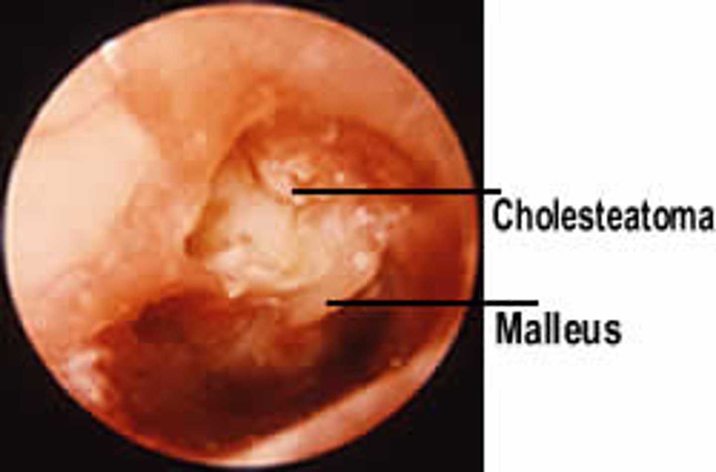
Cholesteatoma
A growth of skin-like material from the tympanic membrane and within the middle ear space. A cholesteatoma is a serious ear disease that requires medical attention.
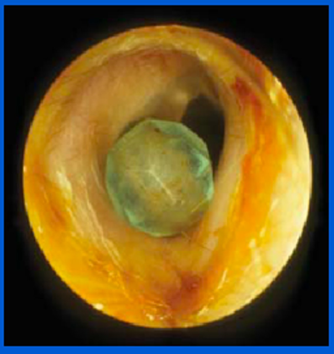
Foreign body
Term used to describe anything out of the ordinary in the external ear canal such as raisins, pebbles, insects, and other assorted small objects.
Hyperacusis
Abnormal intolerance to everyday loud sounds.
Malignant
The tendency of a pathology like a tumor to progressively worsen and potentially cause death. In describing tumors the term malignant often refers to cancer.
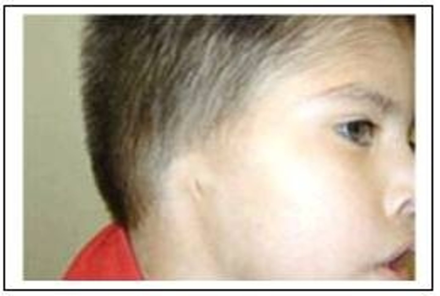
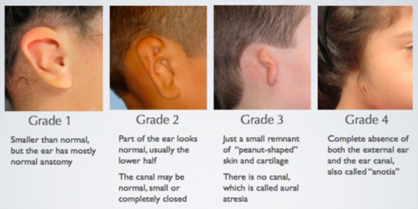
Microtia
Abnormally small and/or deformed outer ear.
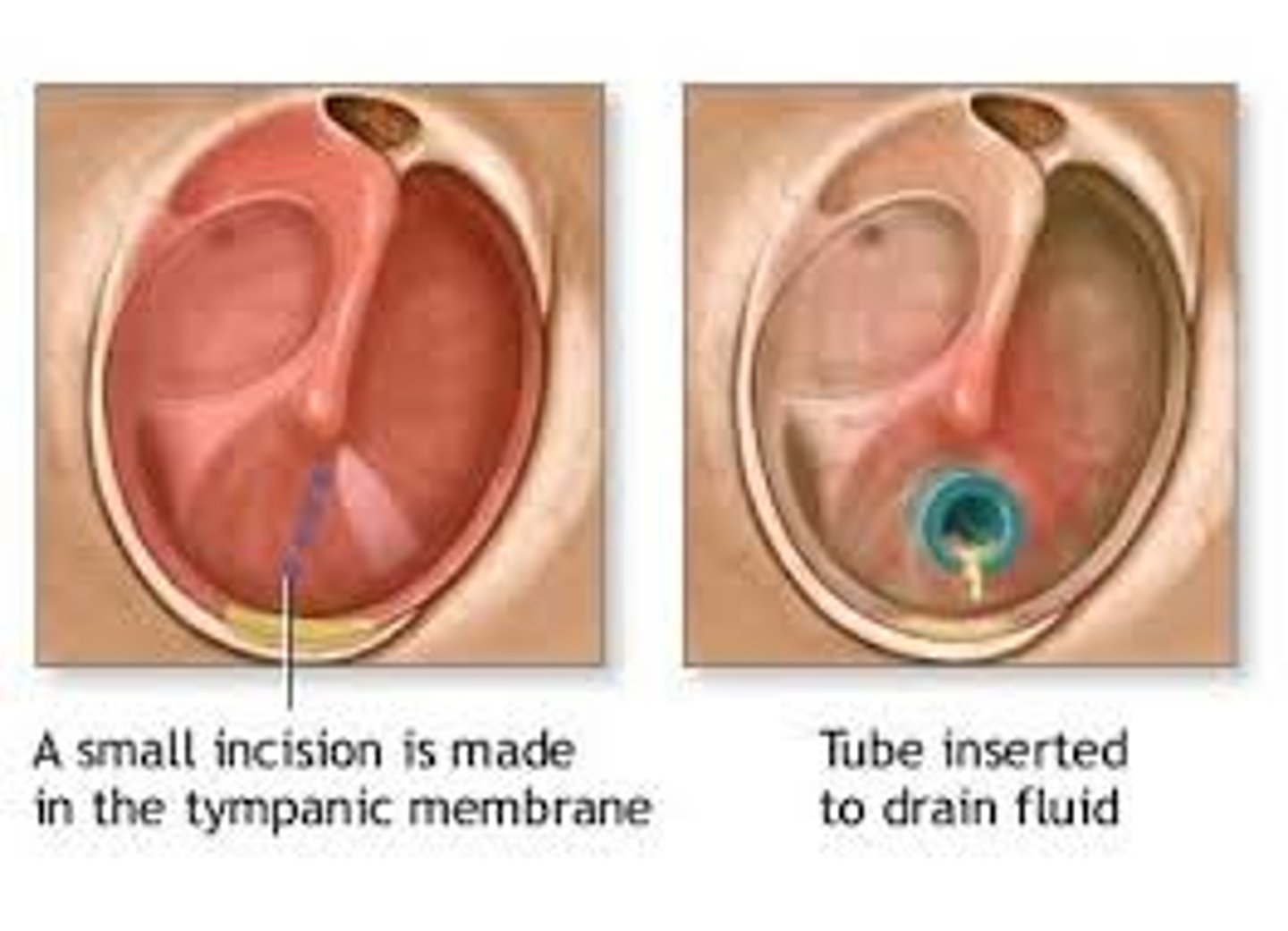
Myringotomy
A surgical procedure that involves making a small incision or cut in the tympanic membrane usually to drain fluid from the middle ear space. A myringotomy is performed before insertion of tympanostomy tubes into the tympanic membrane.
Neoplasm
A tumor within the body, literally, a new growth. An anti-neoplastic drug is given to stop, or reduce, the growth of a tumor.
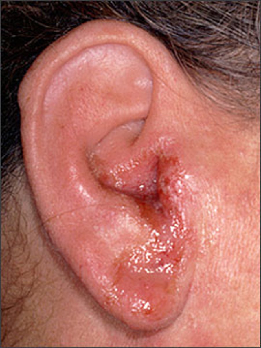
Otitis externa
Infection involving the external ear canal walls.
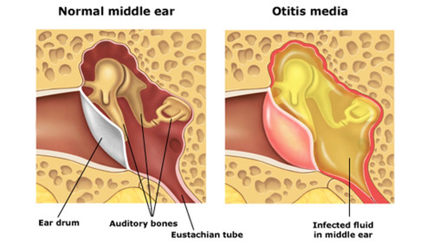
Otitis media
A general term for various forms of middle ear disease, such as serous otitis media, otitis media with effusion, purulent otitis media, and chronic otitis media. Otitis media is one of the most common childhood diseases and usually produces a conductive hearing loss.
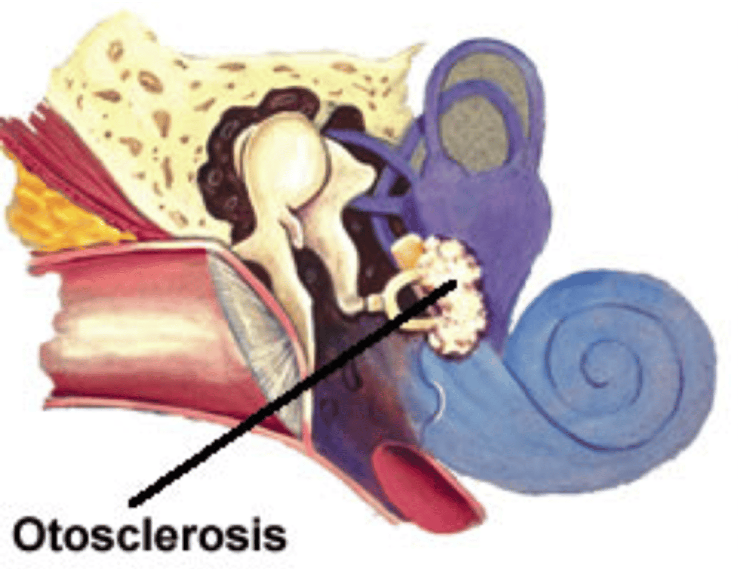
Otosclerosis
A bony degenerative disease process that can involve the stapes footplate and/or the cochlea; also called otospongiosis.
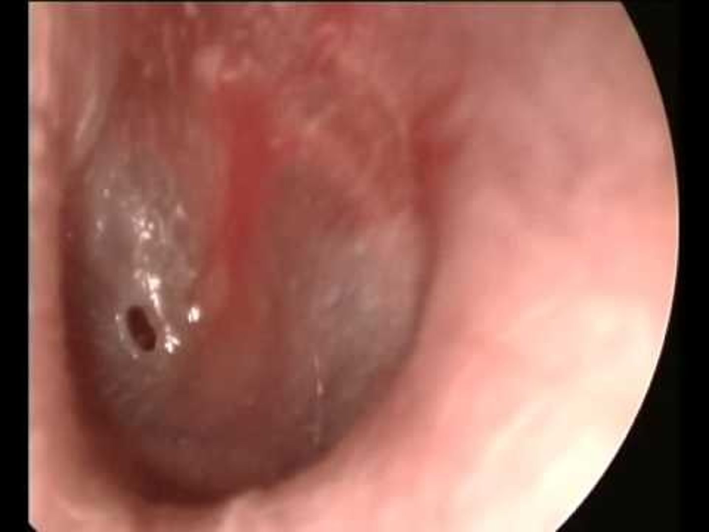
Perforation
A hole in the ear drum. may result from infection and rupture of the tympanic membrane due to build up of infected fluid or traumatic damage to the tympanic membrane.
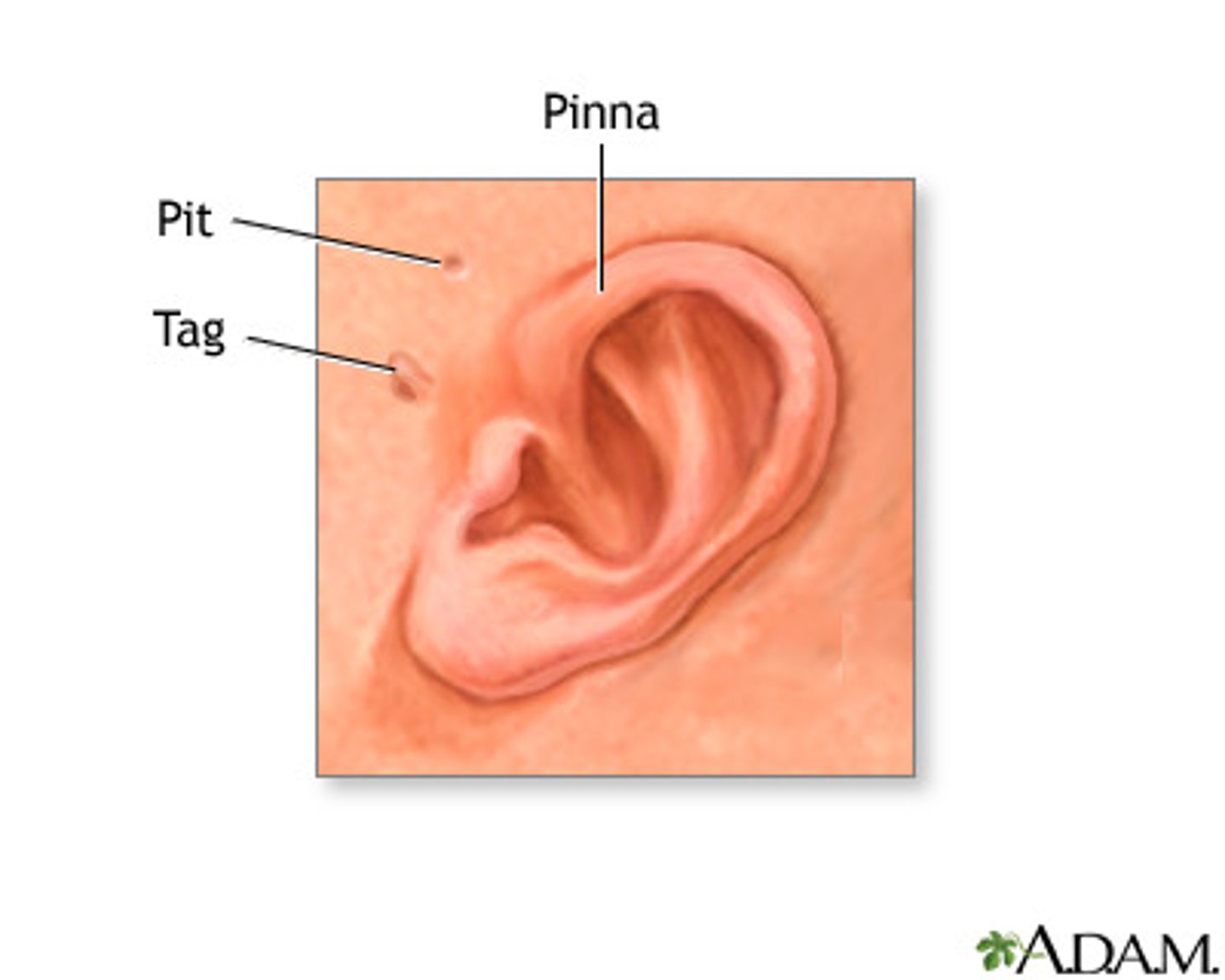
Preauricular pits and tags
A small indentation or outgrowth of skin in front of the ear.
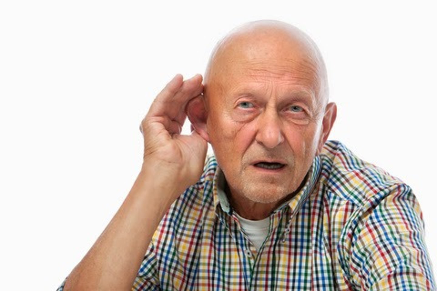
Pseudohypacusis
False or exaggerated hearing loss.
Stenosis
In audiology, a restriction or even total occlusion of the external auditory canal.
Tinnitus
The perception of a noise in the ear like ringing, cricket sound, roaring even when there is no external sound; a phantom sound. not a disease but, rather, a symptom associated with many disorders of the auditory system.
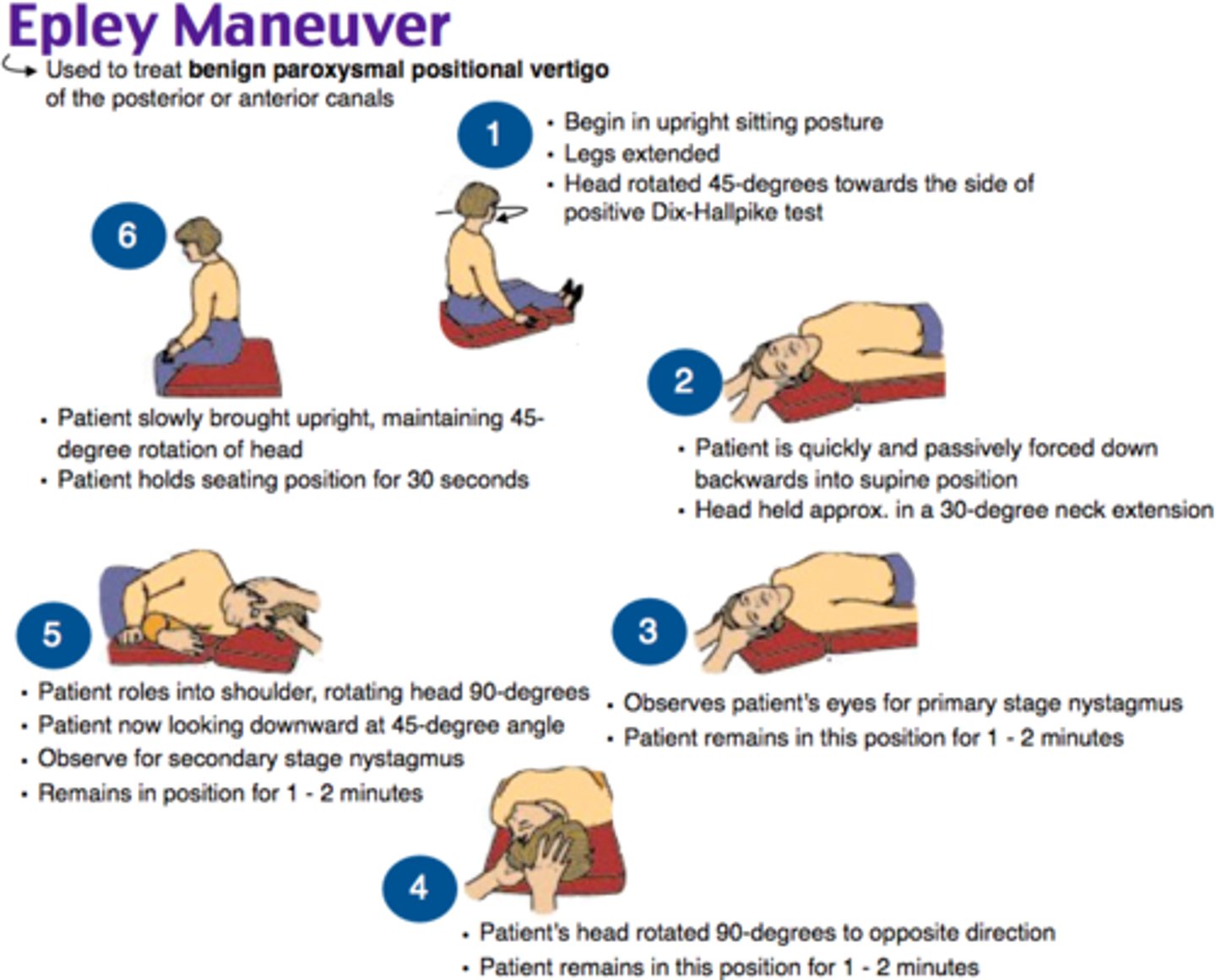
Benign paroxysmal positional vertigo (BPPV)
A vestibular disorder characterized by vertigo associated with changes in head position. can be successfully treated by audiologists and physicians in a clinic office with special maneuvers.
Canalith repositioning procedure
Maneuvers of the head that are used in treating the a disorder known as benign paroxymal positional vertigo (BPPV).
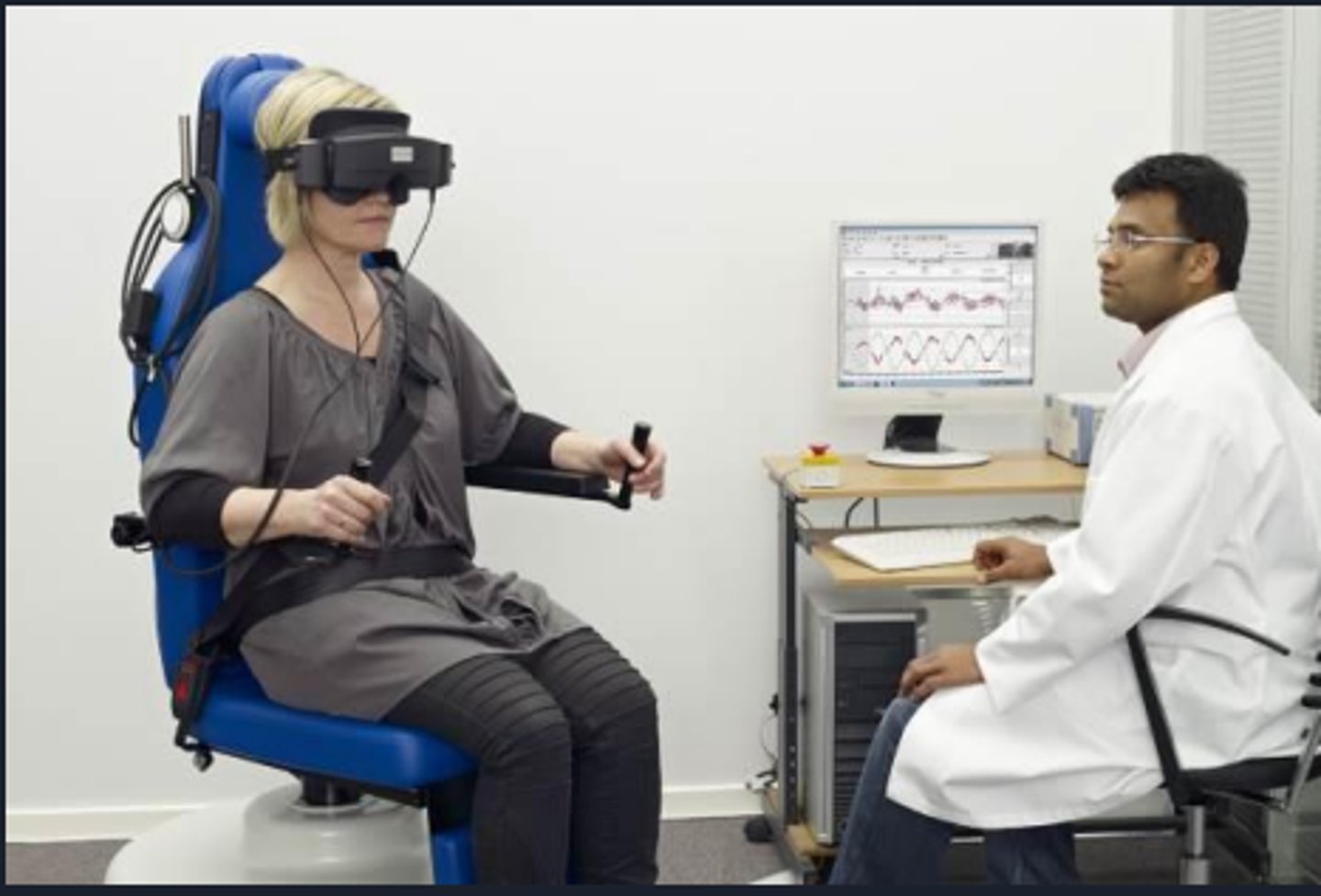
Electronystagmography (ENG)
A group of vestibular tests that involve measurement of eyeball movements (nystagmus) that are spontaneous or that follow different types of stimulation of the vestibular system.
Epley maneuver
A clinical repositioning maneuver performed in an office and used to treat a disorder known as benign paroxymal positional vertigo (BPPV) in which floating particles in a semicircular canal are relocated. Named after Dr. John Epley.
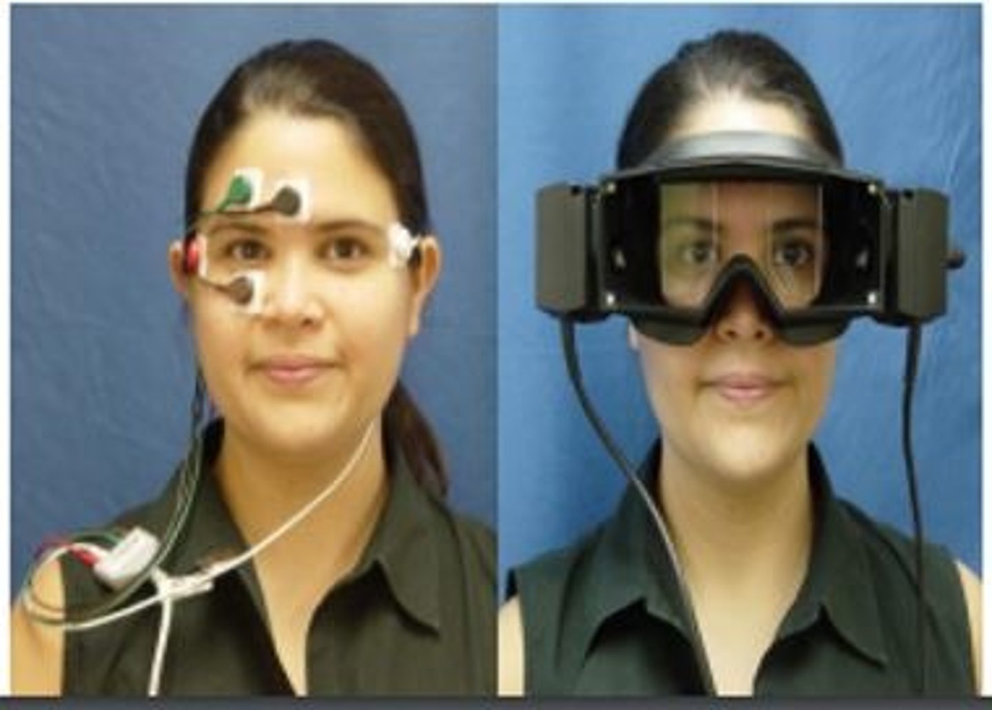
Frenzel goggles
Special googles that magnify the appearance of the eyes that are used in testing vestibular functions.
Ménière's disease
Pathology affecting the cochlea and resulting in sensory (sensorineural) hearing impairment. Characteristic signs and symptoms are tinnitus, vertigo, sensation of ear fullness, and a fluctuating, often low frequency, sensorineural hearing loss.
Meningitis
An inflammation of the meninges, the tissue that is a protective covering of the brain and the spinal cord.
Nystagmus
Horizontal or less commonly vertical movements of the eyeballs. Nystagmus may result from stimulation of the vestibular (balance) system. Recording nystagmus with electrodes placed around the eyes during stimulation of the vestibular system is technique called electronystagmography (ENG).
Ototoxic
Drugs or medications that potentially damage the cochlea and cause hearing loss. Most of these drugs interfere with metabolism of the hair cells in the cochlea.
Presbycusis
Decrease in hearing sensitivity associated with aging. Although hearing may first begin to show aging effects at 20 years, presbycusis usually does not cause speech perception difficulty and serious hearing impairment until age 60 or older.
Syndromes
A collection of signs and symptoms associated with a specific disease or condition.
Vertigo
A symptom of balance and specifically vestibular disorder. The patient experiences a spinning sensation, or senses that the environment is spinning around him/her.

Vestibular evoked myogenic potential (VEMP)
Momentary reduction in contraction of the sternocleidomastoid muscle in response to high intensity low frequency stimulation of an ear and activation of the saccule within the vestibular apparatus.
Vestibular system
Portion of the nervous system that is responsible for maintaining a person's equilibrium and position in space. The vestibular system consists of structures in the ear, vestibular nerves, and collections of vestibular nerves in the brainstem with connections also to eye muscles and large muscle groups in the trunk of the body.
Video-nystagmography (VNG)
Measurement of nystagmus (eye movements) with special magnifying goggles during various vestibular tests.
Normal ear canal volume (ECV)
adult = 1.0 to 2.0 ml or cc
child - 0.3 to 1.0 ml or cc
what does abnormal ECV indicate?
low = TM blocked with wax, craniofacial abnormalities
high = TM perforation or PE tube
Normal middle ear pressure
-150 to +50 daPa
What does abnormal middle ear pressure indicate?
eustachian tube dysfunction. referral needed
Type A tympanogram
tympanogram peaks at 0 daPa. normal

Type Ad (deep or disarticulated) tympanogram
tympanogram with unusually high peak-high compliance (off the chart). ossicular dislocation (ossicular chain becomes dislodged)/monomeric TM (healed perforation that inflates when pressurized)

Type As (shallow or stiff) tympanogram
tympanogram with reduced peak- low compliance. ossicular fixation/tympanosclerosis. gluier or bonier TM

Type B tympanogram
tympanogram that is flat, no peak. No movement usually a sign of middle ear fluid. Look at volume for diagnosis
Normal volume = otitis media
High volume = TM perf or PE tube open
Low volume = faulty probe placement
Type C tympanogram
tympanogram with negative pressure- peak is -150 to -200. caused by eustachian tube dysfunction or developing/resolving otitis media
Tympanogram x and y axis
X axis = air pressure measured in daPa or mm H2O
Y axis = compliance measured in cc, ml, or mmho