bio - unit 3 aos 3
1/71
There's no tags or description
Looks like no tags are added yet.
Name | Mastery | Learn | Test | Matching | Spaced | Call with Kai | Chat |
|---|
No analytics yet
Send a link to your students to track their progress
72 Terms
7A - pathogens
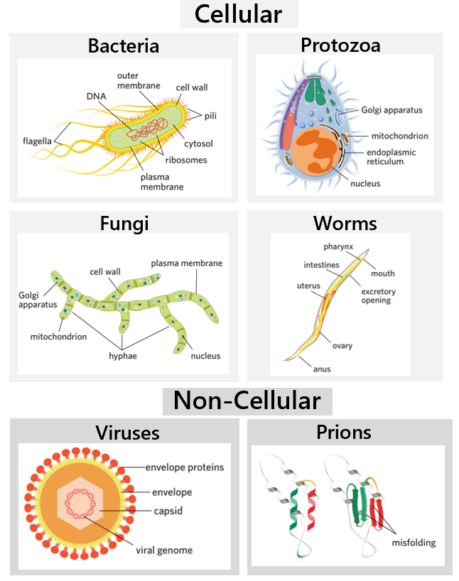
pathogens - sources of non-self antigens that can cause disease. toxins that some pathogens secrete can also act as antigens.
cellular pathogens - living organisms with a cellular structure that contain genetic material that encode proteins + does so.
bacteria
fungi
protozoa
nematodes (worms)
non-cellular pathogens - don’t have a cellular structure + are non-living. no metabolic activity + can’t reproduce without host cells.
DNA viruses
RNA viruses
prions
7A - cellular pathogen - bacteria
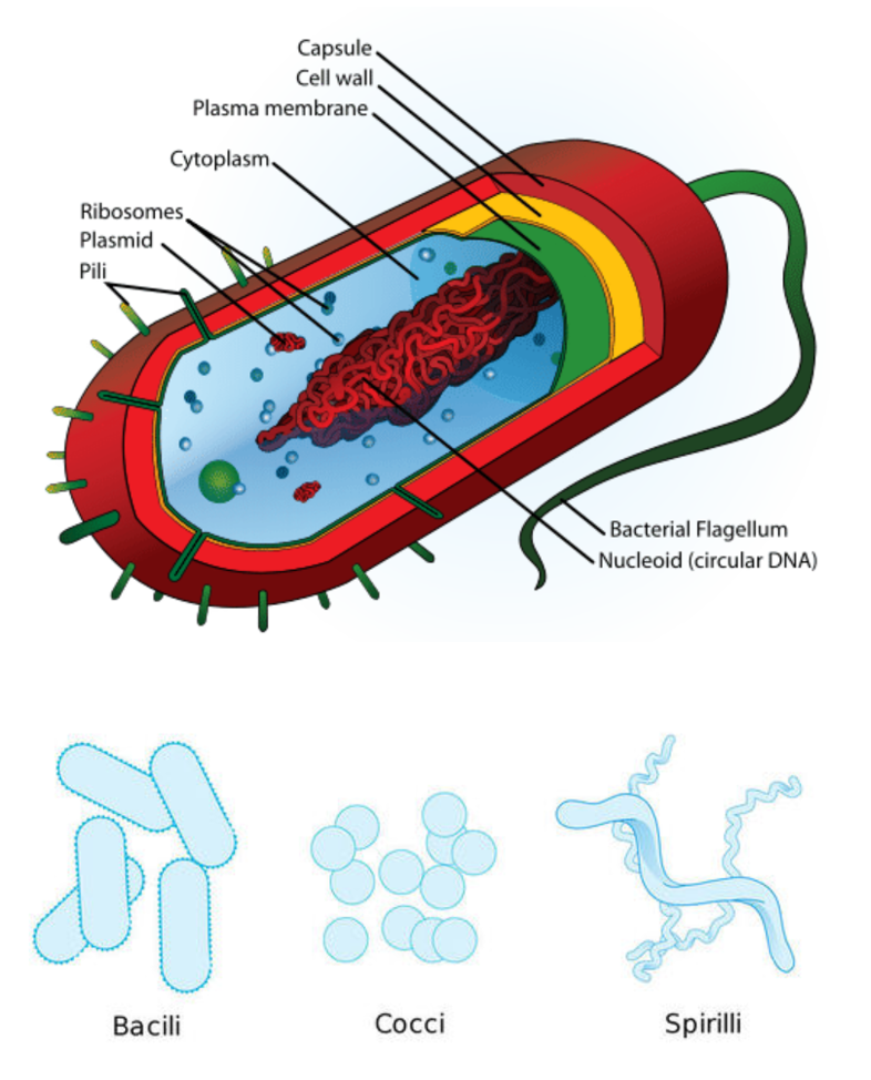
bacteria - prokaryotic, unicellular organisms that reproduce asexually through binary fission. only some are pathogenic.
bacteria shapes
bacilli - rod shaped
cocci - ball shaped
spirilli - spiral shaped
some bacteria have a protective capsule that prevents the bacteria being recognised by the immune system + engulfed by macrophages
ex. Clostridium tetani causing tetanus
7A - bacteria classification
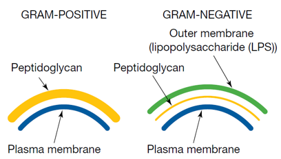
gram staining - if bacteria cells have peptidoglycan in cell wall, they are gram positive, and become stained purple. if bacteria cells have lPS, they are gram negative, and become stained pink.
7A - how bacteria cause harm
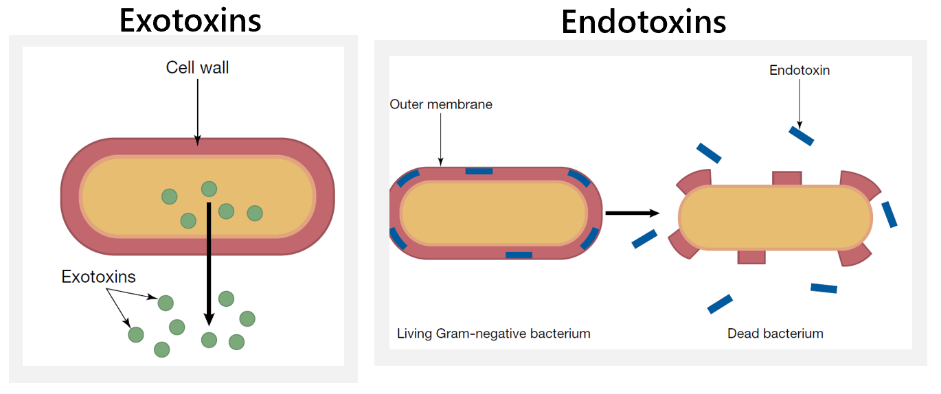
pathogenic bacteria - live outside the organism’s cells.
most pathogenic bacteria produce toxins to damage/kill cells.
exotoxins - secreted by living bacteria and spread through the body
endotoxins - part of the cell wall of gram-negative bacteria and are released when bacteria die
some bacteria reproduce so rapidly that they crowd other cells
some bacteria kill cells outright.
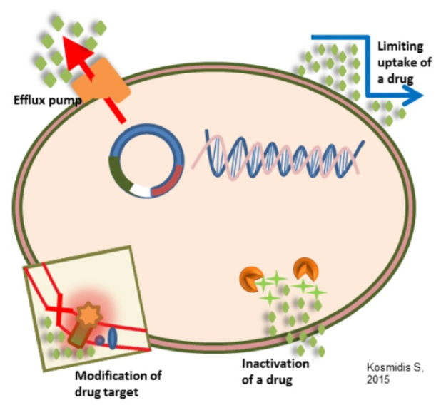
7A - antibiotics
antibiotics - medications that destroy or slow down the growth of bacteria + are specific for the bacteria. they recognise the cell wall of bacteria.
antibiotic resistance (image) - however, bacteria can evolve to develop resistance to antibiotics. this can happen quickly as bacteria divide rapidly (in 20 minutes). there are many antibiotic resistance mechanisms.
7A - cellular pathogen - fungi
fungi - eukaryotic, uni/multi-cellular organisms that reproduce both asexually and sexually, through spore formation. contain long branching filaments called hyphae.
ex. yeasts, molds. thrush, ringworm
7A - cellular pathogen - worms
worms - eukaryotic, multicellular invertebrate parasites that reproduce sexually.
ex. tapeworm
7A - cellular pathogen - protozoa
protozoa - eukaryotic, unicellular pathogens that reproduce both asexually and sexually. most protists are spread by vectors. ex. plasmodium protist which causes malaria is spread by mosquitoes
ex. Plasmodium causing malaria
7A - non-cellular pathogen - viruses
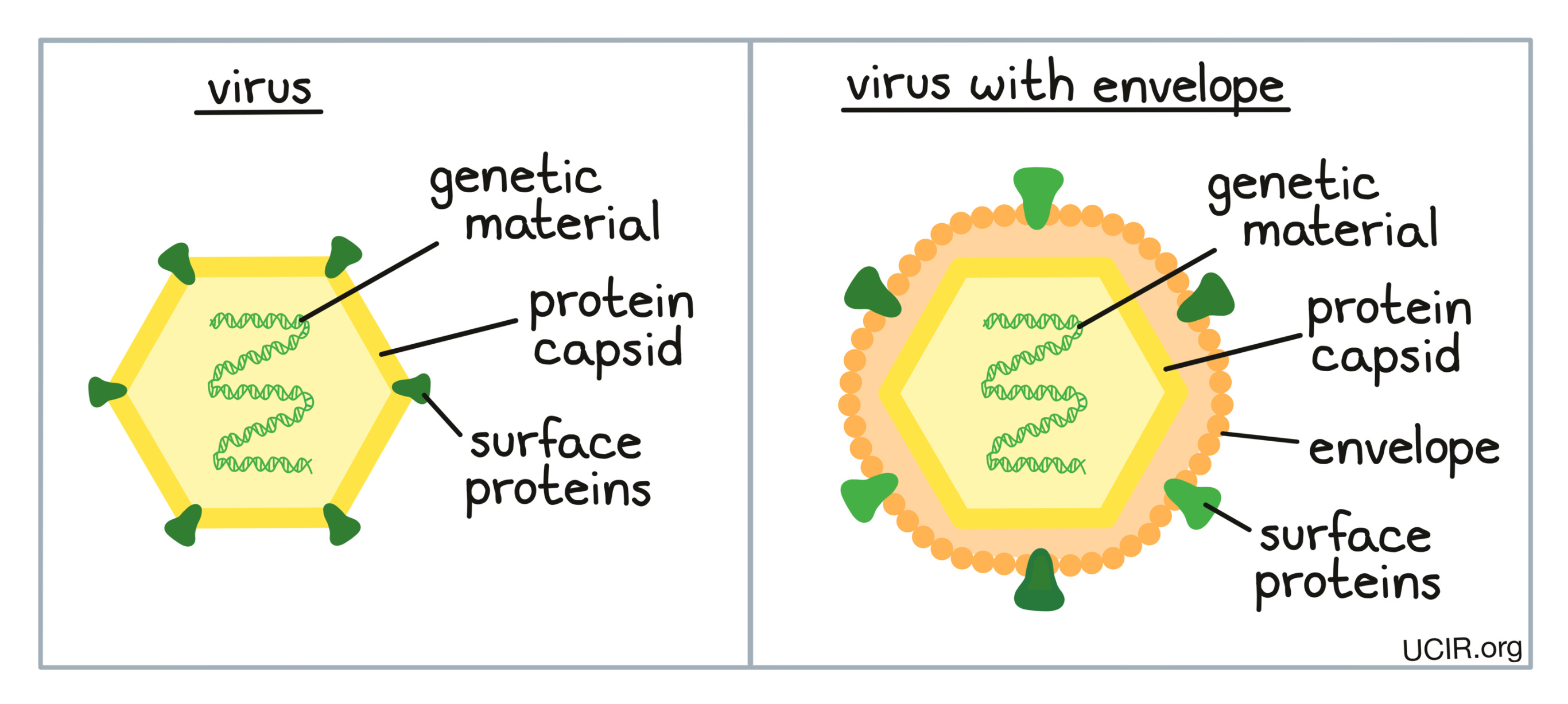
viruses - non-living, non-cellular pathogens composed of a nucleic acid (DNA or RNA) within a protein coat (capsid) + sometimes lipid + protein envelope which protect the nucleic acid. proteins on capsid or envelope. viruses have no metabolic activity + can’t reproduce independently, but contains genetic material that encodes proteins.
replication - they lack the structures required to reproduce independently → to replicate, they must take over a host cell. viruses have preferred target cells that they enter + replicate in. (more later)
viruses cause disease by
infecting target cells + disrupting their normal function. + direct damage to cells/death → lead to symptoms
some DNA viruses can alter the DNA of host cells, leading to the development of cancers
viruses are continually evolving, and crossing species barriers
ex. influenzea virus (causes flu), ebola virus (causes ebola)
7A - virus infection
attachment - virus attaches to host cell. specific viral proteins detect + attach to specific receptor proteins on the membrane of the target host cell. (virus only affects some cells in the body bc only some cells have the specific receptor).
entry - after attachment, virus breaches plasma membrane. three types:
membrane fusion - if a virus has an envelope, it can use a protein to puncture the cell membrane, allowing the envelope to fuse with the membrane → capsid enters cell → capsid breaks down by enzyme action, releasing viral genome
endocytosis - if a virus doesn’t have an envelope, it can become engulfed in a vesicle by the membrane → virus enters through endocytosis → virus exits vesicle → capsid breaks down by enzyme action, releasing viral genome
genetic injection - after attachment, some viruses simply inject the viral genome into the cytoplasm of the cell, leaving the rest of the viral structure outside the cell.
replication - the host cell’s machinery is taken over, using the inserted viral genome, begins genome replication + protein expression → lots of viral nucleic acids + proteins produced. host cell uses all it’s energy + it’s own amino acids.
assembly - viral particles produced are assembled into new viruses.
release - assembled viral particles exit the host cell, and continue to infect more cells. three ways:
cell lysis - host cell bursts, releasing all viral particles. cell membrane is left ruptured.
budding - virus pushes through host cell’s plasma membrane, which is how enveloped viruses aquire their lipid envelope.
exocytosis - viruses are packaged into a vesicle, transported to the membrane + exported through exocytosis
7A - plant viruses
plant viruses are usually transmitted using a vector (usually an insect that feeds on the plant)
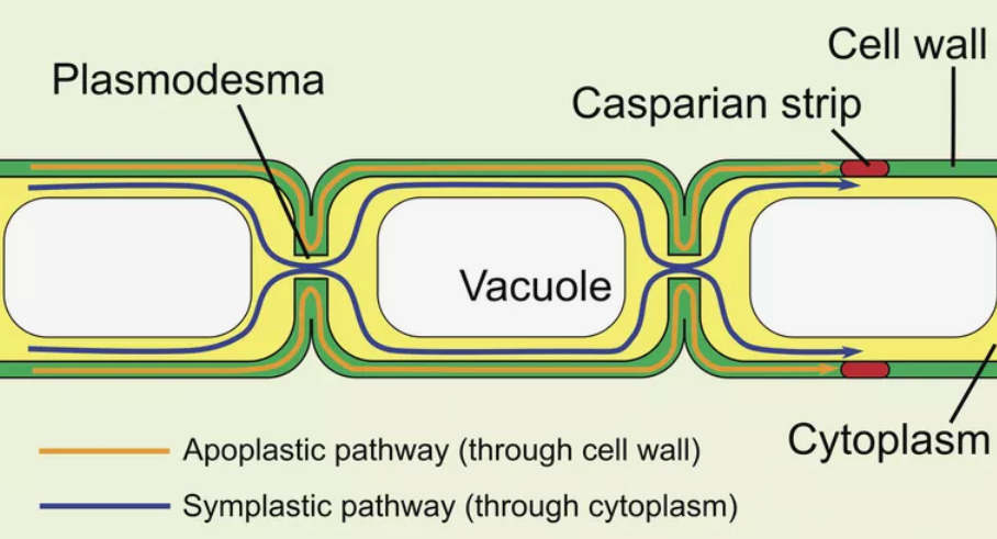
due to the cell wall, a plant virus enters cells using plasmodesmata (channel between cells)
plants only have one way of dealing with viruses + infections - preventing the virus from entering the plant. ex. waxy cuticle + chemicals. once virus enters, plant tries to prevent spread by dropping leaf
7A - non-cellular pathogen - prions
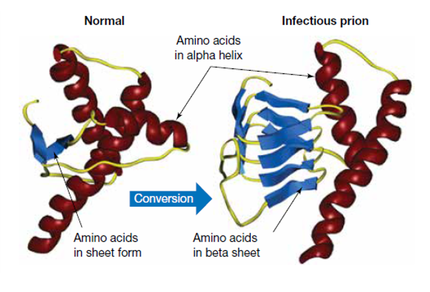
prions - non-living, non-cellular pathogens. misfolded proteins that can induce nearby normal proteins to become misfolded. they contain no genetic material.
prion disease - the protein PrPc is found in nerve cells, and it’s normal form is not harmful. however, it can spontaneously convert into a different secondary structure, with more beta sheets. this misfolding turns the protein into the prion PrPSc. when these prions interact with normal ones, they convert them into misfolded ones. if this infection spreads, you get prion disease.
prion disease - all prion diseases result in:
a long incubation time (measured in years)
a progressive deterioration of brain function resulting in fatality
changes in the brain including loss of neurons and development of lesions (holes)
there is no immune system response to prion disease, they are also non-treatable and result in death.
7A - infections + diseases
infection - when a pathogen enters the body and begins to multiply
disease - when an infection affects the normal function of an organism. only labelled a disease once there are symptoms
non-communicable/non-infectious - cannot be transmitted from one individual to another. ex. diabetes, heart disease, most cancers, multiple sclerosis, arthritis
communicable/infectious - transmitted from one individual to another
host - any organism containing the pathogen
vector - a living organism that carries and transmits a pathogen from an infected host to another host. can spread pathogen within a species or to another species. ex. mosquito, tick
immunity - resistance to an infectious disease
herd immunity - ?
epidemic - rapid spread of infectious disease within a single population
pandemic - an outbreak of infectious disease over a wide geographical area, affecting a large number of people
endemic - present in the population at a constant low level
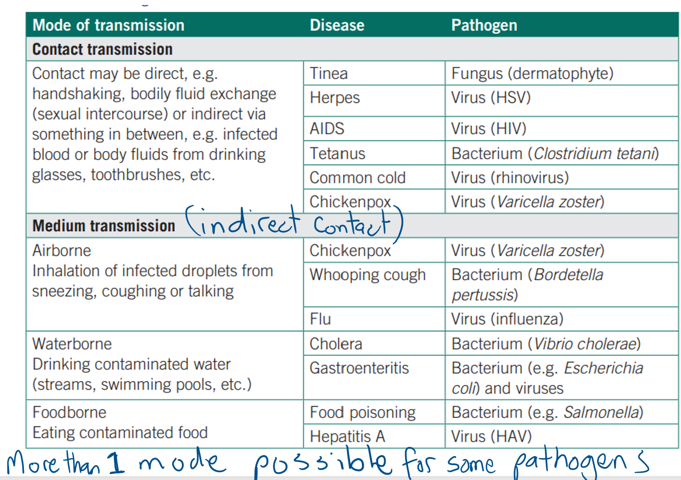
mode of transmission
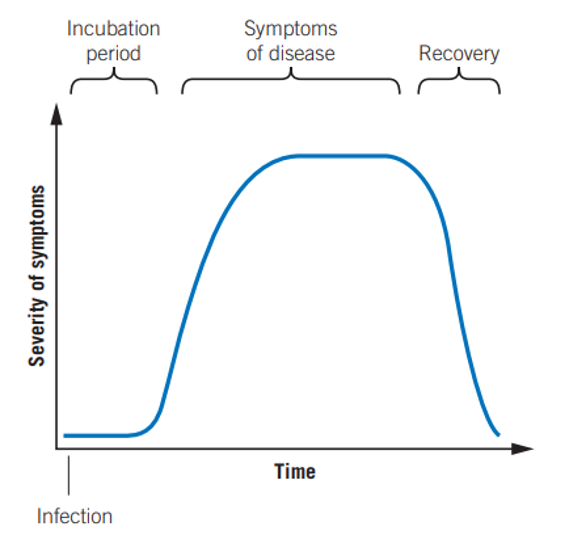
infection timeline
incubation period - pathogen multiplying and travelling to target tissue, toxins made and released.
symptoms of disease - pathogen results in symptoms in the body. when the immune system recognises the presence of non-self antigens from the pathogen, it begins producing an immune response to eliminate the infection. an innate response is quickly initiated, and if the infection continues and specific antigens from the pathogen are recognised by lymphocytes, an adaptive response is initiated.
recovery - pathogen eliminated by immune system
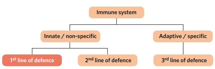
7B - immune system
immune system - the cells and tissues involved in resistance to infection. the immune system identifies the difference between its own cells (self antigens) and foreign cells + molecules of pathogens (non-self antigens)
innate immune system - composed of non-specific defences and responses + doesn’t keep a memory of pathogens. responds to injury + antigens quickly. includes first + second line of defence.
adaptive immune system - composed of specific defences and responses + keeps a memory of specific pathogens → response to re-infection is faster + larger. includes third line of defence.
7A - antigens
antigens - molecules that are recognized by the immune system, usually found on the surface of cells or pathogens.
self antigens - originate inside the body + are found on the surface of cells. they mark the cells of an organism as ‘self’, the immune system recognizes them as their own → no immune response is initiated
non-self antigens - originates outside the body. found on the surface of foreign cells or viruses. the immune system recognizes these as ‘non-self’ (foreign) → immune response is initiated.
7A - MHC markers
in humans, self antigens take the form of MHC markers. they are a group of proteins that are made in the cell (encoded by the Major Histocompatibility Complex) and embedded into the membrane of all self-cells
class I MHC markers - present on the surface of all body cells that have a nucleus (all except red blood cells).
class II MHC markers - present on the surface of antigen-presenting cells (only macrophages, dendritic cells and B lymphocytes).
MHC markers show a high degree of variability between individuals → enables the immune system to distinguish self from non-self material.
this is because there are 6 main gene loci on our chromosomes that code for MHC markers. each person inherits 2 alleles for each gene locus, and each gene has many possible alleles, (ex. HLA-A has 350) → very unlikely that two individuals have same combination of alleles + therefore very unlikely two individuals have same type of MHC markers. genes:
HLA-A - class I
HLA-B - class I
HLA-C - class I
HLA-DP - class II
HLA-DQ - class II
HLA-DR - class II
7A - malfunctions involving antigens
autoimmune disease - if immune system incorrectly recognize self antigens as non-self antigens → immune system attacks self-cells
allergies - an overreaction to the presence of an allergen. allergens are antigens that the immune system recognises as non-self, even though they are non-pathogenic and harmless. this unwarranted immune response is called an allergic reaction.
7B - the first line of defence
the first line of defence - consists of 3 types of barriers that prevent pathogens from entering the body. part of the innate immune system.
physical barriers - physically block pathogens from entering the body
chemical barriers - chemicals that destroy pathogens or inhibit their growth
microbiota barriers - competes with pathogen for resources and space, preventing pathogen growth
plants only have the first line of defence
7B - physical barriers
physical barriers - physically block pathogens from entering the body. entry = crossing a membrane
examples
animals
mucous membranes + hairs line respiratory tract that trap foreign material, + cilia move pathogen to throat to be swallowed + destroyed
intact skin - the outer layer of our skin is dead. this means when viruses land on the dead outer surface of the skin and insert their genome, the cells won’t do anything
mechanical defences - sneezing, coughing, diarhea, vomiting, flushing action of tears, flushing action of urine
plants
cellulose cell wall - thick barrier hard for pathogens to penetrate
thick bark
presence of thorns + trichomes (small hairs) to deter pathogens + insects
formation of galls (abnormal outgrowth of tissue) to prevent the spread of infectionb
waxy cuticle on leaves - prevents water accumulation (reduces water-based pathogens) and protects epidermal cells from exposure to pathogens
closing of stomata to prevent pathogen invasion
7B - chemical barriers
chemical barriers - inhibit the growth of or destroy pathogens through the production of chemicals
examples
animals
presence of enzyme lysozome in tears + saliva which destroys bacterial cell walls, therefore killing bacteria
stomach lining cells secrete strong acids to kill pathogenic bacteria that have been swallowed
plants
production of chemicals (often toxins) ex. defensins - small peptides that are toxic to microbes + fungi
7B - microbiota barriers
microbiota barriers - the presence of non-pathogenic bacteria ‘normal flora’ that competes with pathogens for resources and space/adhesion sites, limiting pathogen growth
these bacteria live in a mutualistic relationship with the person, ‘normally’ living on/in an individual. the immune system has adapted to not respond to them despite being non-self.
examples
animals
bacteria in the digestive tract which prevent pathogenic bacteria from growing by outcompeting them for nutrients and space + also secrete antimicrobial chemicals that prevent growth.
bacteria on skin
plants
non-pathogenic bacteria living in and around roots (ex. citronella oil, peppermint oil) form a barrier against pathogens
7C - the second line of defence
second line of defence - part of the innate immune response, which consists of non-specific responses to pathogens which have breached the first line of defence and entered the body, + injury. the responses are via a variety of cells + molecules. the innate immune response is non-adaptable, does not change during an individual’s lifetime and doesn’t keep a memory.
cellular responses
mast cells
eosinophils
natural killer cells - degranulation
phagocytes - phagocytosis
non-cellular responses
interferons
complement proteins
both
inflammatory response → interactions with third line of defence
7C - leukocytes
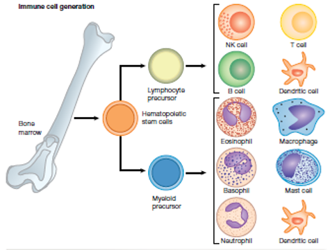
leukocytes (white blood cells) - all immune cells are a type of leukocyte. leukocytes are responsible for protecting the body against pathogens and foreign material. they are found in the blood, tissues, lymph and lymphatic organs, and derived from multipotent stems cells in the bone marrow. types to know:
mast cell
eosinophil
natural killer cell
phagocytes
dendritic cell
macrophage
neutrophil
7C - cellular response - mast cells
mast cells - mast cells are found in connective tissues, and contain granules rich in histamine. mast cells are involved in early recognition of pathogens, as part of the inflammatory response - when a mast cell detects damage to surrounding cells (through cytokines), it degranulates, releasing all it’s histamine into the extracellular environment.
histamine does three main things as part of the inflammatory response:
causes vasodilation (dilation of blood vessels) - increases blood flow so more immune cells can go to the area
increases permeability of blood vessels, causing fluid to leak out of blood vessels → releases leukocytes needed for response
attract phagocytes - the more phagocytes involved, the better
7C - cellular response - eosinophils
eosinophils - eosinophils are found in the blood, and contain granules with toxic chemicals.
when a large parasite (ex. worms) is too large to be attacked by phagocytosis, eosinophils release their granules onto the parasite as the major defence.
also have a role in allergic reactions and inflammation response
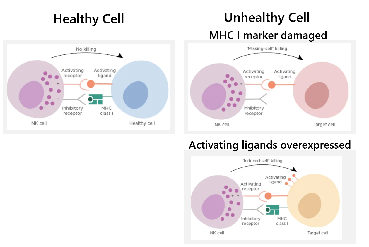
7C - cellular response - natural killer (NK) cells + detection
natural killer cells - NK cells patrol tissues, looking for body cells with abnormal or missing MHC I markers (don’t rely on specific antigens), thus identifying cells infected with a pathogen (intracellular virus) or cells that are cancerous. NK cells detect unhealthy cells, then apoptose them.
NK cells check body cells all the time. the NK cell has two receptors which detect whether the cell is healthy or unhealthy
healthy - the activating receptor on the NK cell binds to its receptor on the healthy cell, and the MHC I marker on the healthy cell binds to the inhibitory receptor on the NK cell. the cell is recognised as ‘self’ and no activation of the NK cell occurs
unhealthy (damaged MHC I marker) - the activating receptor on the NK cell binds to its receptor on the infected cell, but the MHC I marker on the infected cell is damaged/missing and cannot bind to the inhibitory receptor on the NK cell. the cell is recognised as ‘non-self’ → NK cell is activated + starts apoptosis pathway
unhealthy (overexpressed activating ligands) - NK cells can also detect when activating ligands are overexpressed, which can occur in both cancerous cells and virally infected cells.
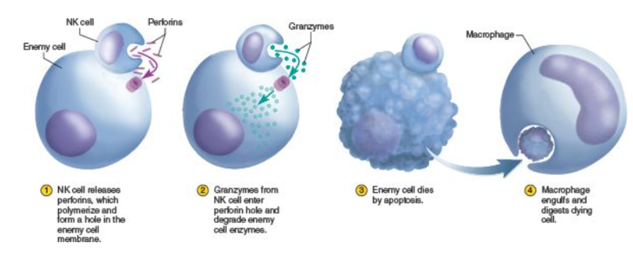
7C - NK cells - apoptosis
apoptosis - when the NK cell identifies an unhealthy cell, the NK cell releases cytokines (to attract phagocytes), perforin and granzymes which enter the cell and cause apoptosis (programmed cell death).
the NK cell releases perforins, which bind to the unhealthy cell’s membrane, where they polymerise, forming a ring that makes a hole in the membrane.
granzymes released by the NK cell enter through the hole. they induce apoptosis of the cell.
the unhalthy cell dies by apoptosis.
a macrophage engulfs and digests dying material
7C - cellular response - phagocytes
phagocytes - leukocytes that engage in phagocytosis, a strategy used to attack extracellular pathogens in tissue fluid or blood. phagocytes include:
neutrophils - most abundant leukocyte circulating in the blood
macrophages - in tissue
dendritic cells - in tissue, also act as APC
cytokines - phagocytes also release signalling molecules to communicate with the immune system. ex. cytokine, helps protect against pathogens + guide immune cells to site of infection or injury
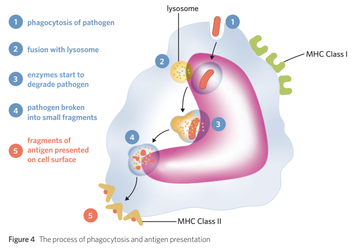
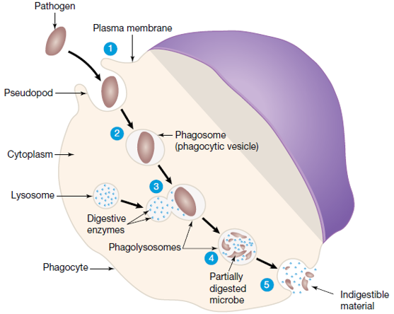
7C - cellular response - phagocytosis
phagocytosis - a strategy used to attack extracellular (in space outside cells) pathogens in tissue fluid or blood.
for phagocytosis to occur, the pathogen must be recognised as non-self. phagocytes have pattern recognition receptors that are complimentary in shape to things they recognise such as: LPS of cell wall in gram negative bacteria, acid on cell wall of gram positive bacteria, glycoproteins on virus envelope, flagella (tail) in bacteria
process
pattern recognition receptors on membrane of phagocyte recognise an extracellular pathogen + engulf it through out-foldings of membrane
pathogen is completely enclosed in vesicle (phagosome)
lysosomes fuse with the vesicle, releasing their digestive enzymes
pathogen undergoes digestion through enzyme action
indigestible material is released from the phagocyte by exocytosis
if dendritic cell, takes sample, and presents it’s antigens on their MHC II markers→ activates third line of defence
cytokines
cytokines - cytokines are signalling molecules of the immune system (non-cellular).
there are many types of cytokines:
when a cell is damaged, it releases cytokines, which mast cells require to recognise damaged cells + undergo degranulation.
cytokines are released by immune cells to act on other specific immune cells to activate further adaptive immune responses
interferons
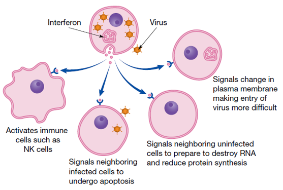
7C - non-cellular response - interferons
interferons - interfere with virus replication. if NK cell can’t make it in time, cells infected with virus particles secrete interferons which are recieved by neighbouring cells. interferons are a cytokine. interferons act as a signal to nearby cells to prepare in advance for possible viral infection.
if a cell is going to die from a virus infection and NK cells can’t make it in time, the infected cell will send interferons (type of cytokine) to help neighbouring cells, and interfere with how they function.
signals fir nearby cells
shut down protein synthesis
attract NK cells
change palsma membrane
tell cells to undergo apotposis
7C - non-cellular response - compliment proteins + complement system
complement system - part of the humoral innate immune system. consists of about 30 proteins made in the liver that circulate in the blood in an inactive form. they are activated when they contact bacteria.
3 different functions of complement system
opsonisation of bacteria - label bacteria for phagocytes
chemotaxis - complement proteins are activated by contact with bacteria. this involved cleavage of the complement protein. these then diffuse from the bacteria, creating a trail of signals (chemoattractants) which immune cells detect + follow to find the bacteria
lysis of bacteria - membrane attack complex (MAC) is formed from different complement proteins combining together. this puts a hole in the bacteria membrane causing the bacteria to undergo lysis.
7C - cellular + non-cellular response - the inflammation response
when cells and molecules work together, get an inflammation response
an early and rapid response to infection (ex. abcteria entering through an open cut)
usually a short-term response localised to the site of pathogen entry.
4 symptoms of inflammation - redness, heat, swelling, pain
also a response to cell damage by thermal burns, sunburn, frostbite, acid spills onto skin
purpose
to localise and prevent spread of pathogen
recruit all cells + molecules needed to eliminate pathogen + remove damaged tissue..
+ repair damaged tissue
stages
vascular stage
cellular stage
resolution stage
histamine
functions
results in vasodilation, so blood flow increases → redness + heat
results in increased capillary permeability, so fluids enter site of inflammation → swelling + pain
vascular stage
dilation of blood cells
increased permeability of local capillaries
cellular stage
escape of immune cells from capillaries. dilation of capillaries allows neutrophils to get through capillaries into tissue
migration of neutrophils to infection site. attracted by cytokines released by damaged cells
macrophages from nearby tissues arrive next. macrophages release cytokiunes and histaminee to attract more phagocytosise
resolution stage
7C - identifying extracellular pathogens
7C - mast cell
mast cell -
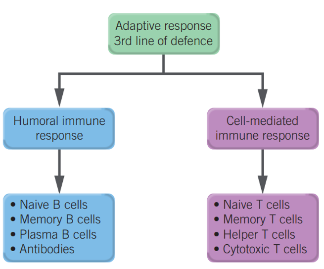
7D - the third line of defence
adaptive immunity - initiated when an antigen (on a MHC II marker of an antigen presenting cell) is presented to a T helper cell. initiated when innate immunity fails to stop an infection
slower than innate immunity
specific to pathogen
a long-lasting memory is made so if re-infection occurs, the response is quicker.
involves a humoral immune response + cell mediated immune response
7D - cells of adaptive immune system
T helper cells
humeral
Naive B cells
B memory cells
Plasma cells (Antibodies)
cell medicated /cellular response
Naïve T cells
helper T cells
Cytotoxic T cells
memory T cells
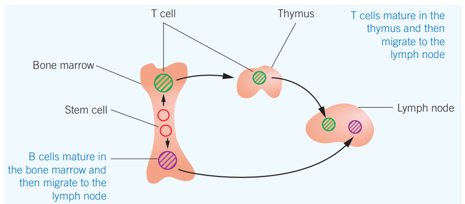
how B cells vs T cells made
B cells - mature in bone marrow → matures in bone marrow → goes to lymph nodes
T cells - T cell precursors are made in bone marrow → migrate to thymus to mature into naiive T cells → migrate to lymph nodes
lymph nodes - nodules of the lymphatic system where immune cells accumulate
cell mediated response
T cells are made in bone marrow and mature into naive T cells in the thymus, which then travel to the lymph nodes. every naïve T cell has a different shaped receptor, (randomly made).
antigen presenting cells - cells which display non-self antigens on their surface MHC II markers, they then bring them to the lymph node, to present the antigens to naiive T cells. dendritic cells and macrophages display antigens after phagocytosis. B cells are also APC.
adaptive immunity is initiated when the antigens on the APC are perfectly complementary to the receptors of a T helper cell in the lymph nodes. the T helper cell releases cytokines, and then starts dividing (no longer naive), to expand that particular cell type.
7D - stages of cell mediated immune response
APCs - when pathogens at the site of infection are recognised as non-self, they can be phagocytosed by dendritic cells, macrophages (or B cells?); which are antigen presenting cells. after phagocytosis, the phagocyte will present antigens from the consumed pathogen on their MHC II markers. this APC then travels to a lymph node to interact with the adaptive immune system.
T cells - T cells are made in bone marrow and mature into naϊve T cells in the thymus, which then travel to the lymph nodes. naïve T cells are always being made, and each has a different shaped receptor, (randomly made).
APCs present the antigen to the naϊve T cell population at the lymph nodes.
when a naϊve T helper (Th) cell with a receptor that is complementary to the antigen is found, it’s receptor binds to the antigen on the APC → the naϊve Th cell releases cytokines, which activates itself + Tc cells, and produces lots more identical Th cells (clonal expansion), to increase chances…
when the naϊve cytotoxic T (Tc) cell with a receptor complementary to the antigen is found (which can take several days), it’s receptor binds to the antigen on the APC. this first naϊve Tc cell does nothing once binded. it must be activated, which is done when a Th cell binds to the same APC, and secretes cytokines (explains why Th cells undergo clonal expansion, to increase chances of binding to the APC with the Tc cell).
When the Tc cell + a Th cell binds to the APC, the Tc cell is activated, and undergoes clonal expansion, producing more Tc cells (which no longer require activation by Th cells) and also memory T cells.
Tc cells travel to the site of infection. there they recognise infected host cells (most often virally infected), due to Tc’s receptor complementary to the antigen presented on pathogen’s MHC class I marker. Tc cell induces apoptosis of infected cells through the release of perforin and granzyme, or by releasing death ligand.
memory T cells remain in the lymph nodes, and are activated if they bind to the APC in the future, quickly activating adaptive immune system and producing lots of Tc cells.
7D - humeral response
2 ways
APC (which has phagocytosed pathogen + displaying it’s antigens on the MHC II markers) goes to the lymph nodes, presenting the antigens to naive B cells. it binds to the B cell with complementary receptors. T helper cells activate the B cell (using cytokines) → B cells then undergo clonal expansion + differentiation
when there is enough antigen buildup in the body, pathogens themselves (presenting antigens on their class I markers) can find their way to naive B cells in the lymph nodes, and binds. the B cell acts as an APC. it phagocytoses the pathogen, and displays it’s antigens on their MHC II markers. the B cells get activated by Th cells → B cells then undergo clonal expansion + differentiation
activation of a B cell results in:
producing plasma cells - plasma cells stay in the lymph nodes, but they produce specific antibodies that are released from the lymph node and travel around the body, and bind to the antigen.
producing Memory B cells - stay in the lymph node. activated if they bind to the antigen in the future.
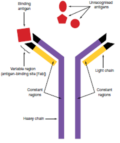
antibodies
antibodies are proteins. they are made from more than one amino acid chain, they contain primary, secondary, tertiary, quaternary and quintenary structures
each type of antibody is made from a selected plasma cell
each antibody has a unique antigen-binding site that can recognize and bind to just one specific antigen.
functions of antibodies
neutralisation - bind to antigens on pathogens and block pathogen receptors from attaching to and infecting body cells
agglutination - antibodies bind together with antigens forming antibody-antigen complexes, to hold them in place, so phagocytes can easily come and phagocytose them all
immobilisation - the antibodiy-antigen complexes formed have restricted movement through the body
opsonisation
activation of compliment proteins
antibody structure
variable region - site where specific antigen binds. on an antibody, the variable region on each arm is the same.
constant regions -
7D - classes of antibodies
IgG - breastmilk, placenta
IgE - allergies
IgD
IgM
IgA
7D - stages of humeral response
allergies
allergy - a substance in the environment that is harmless to most people but causes some people’s immune systems to react abnormally
allergic responses involve both specific and non-specific immune systems
allergen - a substance that causes an allergic reaction. many allergens are small, highly soluble proteins present on the surface of dry particles, food substances, pharmaceutical drugs and plant products
the allergic reaction
sensitisation
the body is exposed to a potential allergen. ex, you inhale pollen and it lodges in the mucous membrane of the airway
cells of the immune system identify antigens on this pollen as non-self and an immune response is activated
specific antibodies against the antigens are produced by plasma cells
the IgE antibodies on allergen attach to surface receptors of mast cells. these mast cells are primed with antibody. the next exposure to the allergen will cause an allergic reaction.
activation of mast cells
next exposure to the allergen results in primed mast cells (have IgG antibodies on surface)
binding to the allergen and
causing degranulation of mast cells (releasing of chemical mediators, including histamine)
7E - the lymphatic system
the lymohatic system is a transport network that:
transprots immune cells throughout the body
is where antigen recognition by lymphocytes occurs
returns fluid that seeps out of the circulatory system back into blood
provides a place for lymphocytes to mature
primary lymphoid oragns - bone marrow and thymus. sites where lymphocytes (B cells and T cells) mature
secondary lymphoid oragns - lymph nodes and spleen. site where mature B cells and T cells are activated by meeting complementary antigens.
8A - ways of aquiring immunity
specific immunity - using antibodies to resist specific diseases. antibodies do neutralisation, aggutiration, opsinisation.
active immunity - the person’s own adaptive immune system has developed antibodies + memory cells to a particular antigen which provide lifelong protection against the antigen.
natural active - pathogens enter the body naturally
artificial active - the pathogen is introduced into the body as a vaccine
passive immunity - antibodies enter the person’s body from an external source. no memory cells created → not lifelong protection
natural passive - antibodies enter a person naturally.
breastfeeding - human milk contains antigens which enter the baby’s bloodstream + protect against pathogens. important bc babies have poorly developed adaptive immune systems + can’t protect themselves against pathogens for a few months.
placenta - during pregnancy, some antibodies produced by the mother cross the placenta + enter the baby’s bloodstream through the umbilical cord to give the baby protection against pathogens while in the womb.
artificial passive - antibodies are injected into a person
8A - aquiring immunity
active immunity - Creates memory cells for long-lasting protection.
passive immunity - Immediate protection, but no memory.
vaccines - Contain inactivated/weakened pathogens or antigenic fragments to stimulate adaptive immunity.
vaccines
vaccines - medical treatments that contain components that resemble a certain pathogen’s antigens,
memory cells specific to antigens from the pathogen are present in vaccinated people. this results in the repid production of antibodies to neutralise the virus if it infects the body. the virus is destroyed vefore severe virus symptoms develop
8A - herd immunity
herd immunity - the indirdct protection of a population against an infectious disease by the presence of a high proportion of individuals who are vaccinated against the disease
not every person in a population can be vaccinated against a pathogen - ex. newborns,people with autoimmune diseases or acquired conditions (ex AIDS), elderly, chemotherapy patients, immunosuppressed people (from organ transplant)
emerging diseases vs re-emerging diseases
emerging disease
caused by a newly identified or previously known agent
has existed in other species but whos incidence in humans has increased in the past 20 years, either locally or intenrationally
re-emerging disease
reappears after a significant decline in its incidence. once controlled but has increased to a level that causes significant health issues.
disease outbreak - what people need to know
identity of the pathogen that causes the disease
how the pathogen spreads (transmission)
defence strategies against pathogen spread
ways to identify pathogen
physical methods (using microscopy - electron microscopy)
biocgemical tests
immunological methods (uses antibodies)
molecular techniques (RNA or DNA sequencing)
factors that influence diease spread, emergence and re-emergence
human demographic changes, such as increasing population and urbanisation → overcrowding/high population density → speeds up the chances of spreading of diseases
increase in international travel, especially without taking appropriate vaccines and other protective measures, leads to increased infection in travellers who then bring the infection home
sanitation and/or contaminated water supply may cause higher rates of transmission, both dirrect (ex. unable to wash hands) and indirect (ex. pathogens in water)
ecological damage - more people live closer to wild animals, making it more likely that pathogens will be passed from animals to humans
transport of food - moving food long distances makes it harder to trace the origins of pathogens
intensive farming
climate change
changhes in vaccination programs
reasons for re-emergence
drop in proportion of population vaccinated
evolution of pathogen (including resistance to treatments such as antibiotics)
public health measures
ongoing efforts
safe water supply for drinking and cleaning
sewage treatment and disposal
food safety regulations
food processing and farming standards
border control of exotic pest species
pest and animald controls
vaccinations programs
quarantine procedures (for people, animals and plants)
Aboriginakl
when Australia was colonised, there was a wave of epidemic diseases, as Aboriginal people had no previous exposure. the European colonists had resistance to many of these diseases as they had already been exposed to the pathogens.
epidemic - the rapid spread of an infectious disease to a large number of people within a population
reservoir - the site an infection originates or is usually found in - can be people, water, soil, animals
zoonosis/zoonotic disease - an infectious disease transmitted from animals to humans. (doesn’t include malaria, mosquitoes are vectors that get disease from humans)
emerging disease - a disease that has not occurred in humans before, or has only previously affected a small number of people
re-emerging infectious diseases - diseases that were once present and have had a dramatic decline in cases, but have returned and are affecting a significant proportion of the population
reasons for emerginfginfectious diseases
most relate to changes in human behaviour
population growth - more people living in closer proximity to each other, providing more opportunities for pathogens to be transmitted between hosts
international and domestic travel - spreading disease is easier and tracking the origins of disease outbreaks is harder
poverty - sanitation and water conditions may be inaccessable or lower in quality, causing higher rates of transmission, both indirect (ex. lack of water for washing after contact with an infected person) and direct transmission (ex, water-borne pathogens in untreated water). food sources - some people can only afford to hunt for bush meat
ecological damage - the spread of human populations means that more humans live closer to wild animals, making it more likely that pathogens will be passed from animals to humans
food supply chains - transporting food long distances makes tracing the origins of pathogens associated with food more difficult.
intensive farming - more animals are kept in smaller spaces, meaning a higher chance of transmission. when close to humans, this also increases the chance of transmission to humans.
human contribution to climate change - warmer climates are causing animal habitats to change, causing disease to be introduced to new areas
reasons for re-emerging diseases
resistance to treatments caused by evolution of a pathogen. ex, antibiotic resistance
drop in the number of people vaccinated against a pathogen
melting of permafrost releasing preserved pathogens
preventing spread of pathogens
things people do
good hygiene (washing hands)
social distancing
reducing movement of people to reduce chance of exposure
face mask
testing if feeling unwell
isolation if infected or possibly infected
public health measures
ensuring a safe supply of water for drinking and cleaning
sewage treatment and disposal
food safety standards and regulations
food processing and farming
border control of entry of exotic species
pest and animal ontrols
vaccination programs
quarantine procedures
8B - emergence of pathogens
8C - controlling pathogen spread
8D - immunotherapy