Lange Mock Exam 1
1/14
There's no tags or description
Looks like no tags are added yet.
Name | Mastery | Learn | Test | Matching | Spaced | Call with Kai |
|---|
No study sessions yet.
15 Terms
The portion of the remnant x-ray beam representing anatomical details having desirable quality is called
A) photoelectric
B) Compton
C) signal
D) noise
C) signal
Feedback:
The portion of the x-ray beam striking the IR and representing image anatomy is called the signal. Some of the initial x-ray beam is absorbed via photoelectric interaction; some is scattered via Compton scatter (creating noise).
Signal-to-noise ratio (SNR) is an important factor in all of medical imaging. Noise impairs image resolution; a high SNR is desirable (more signal, less noise).
Generally speaking, SNR increases as milliampere-seconds (mAs) value increases; however, this is at the expense of patient dose.
It is the responsibility of the radiographer to select technical factors and techniques that will provide a quality diagnostic image while keeping the ALARA concept in mind and minimizing patient dose.
Which type of error results in grid cutoff at the periphery of the radiographic image?
A) Off-focus
B) Off-center
C) Off-level
D) Off-angle
A) Off-focus
Feedback:
The lead strips in a focused grid are angled so as to parallel the x-ray beam. Therefore, scattered radiation, which radiates in directions other than that of the primary beam, will be absorbed by the grid.
When the x-ray beam does not parallel the lead strips, some type of grid cutoff occurs. If the x-ray beam is not centered to the grid (off-center error), or if the x-ray tube and grid surface are not parallel (off-level error), there will be a fairly uniform decrease in receptor exposure across the entire image.
However, if the grid is not used within its recommended SID (focus) range (i.e., if the SID is too great or too little), there will be a decrease in density at the periphery of the image.
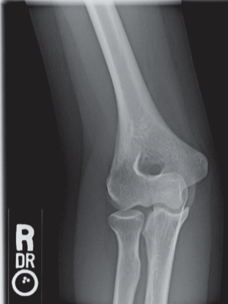
In which of the following projections was the image in Figure 6-1 made?
A) AP
B) Medial/internal oblique
C) Lateral/external oblique
D) Acute flexion
C) Lateral/external oblique
Feedback:
Figure 6-1 illustrates an oblique view of the proximal radius and ulna and distal humerus with epicondyles 45° to the IR—the external oblique (lateral rotation) projection is shown demonstrating the radial head free of superimposition as well as the radial neck, tuberosity, and the humeral capitulum.
The medial oblique (internal rotation) projection of the elbow is particularly useful to demonstrate the coronoid process in profile, the trochlea, and the medial epicondyle.
The acute flexion projection (Jones method) of the elbow is a two-projection method demonstrating the elbow anatomy when the part cannot be extended for an AP projection.
During measurement of blood pressure, which of the following occurs as the radiographer controls arterial tension with the sphygmomanometer?
A) The brachial vein is collapsed
B) The brachial artery is temporarily collapsed
C) The antecubital vein is monitored
D) Oxygen saturation of arterial blood is monitored
B) The brachial artery is temporarily collapsed
Feedback:
A stethoscope and a sphygmomanometer are used together to measure blood pressure. The sphygmomanometer’s cuff is placed around the midportion of the upper arm.
The cuff is inflated to a value higher than the patient’s systolic pressure to temporarily collapse the brachial artery. As the inflation is gradually released, the first sound heard is the systolic pressure; the normal range is 110–140 mm Hg.
When no more sound is heard, the diastolic pressure is recorded. The normal diastolic range is 60–90 mm Hg.
Elevated blood pressure is called hypertension. Hypotension, low blood pressure, is not of concern unless it is caused by injury or disease; in that case, it can result in shock.
As the CR laser scanner/reader recognizes the phosphostimulated luminescence (PSL) released by the PSP storage plate, it constructs a graphic representation of pixel value distribution, called a/an
A) processing algorithm
B) histogram
C) lookup table
D) exposure index
B) histogram
Feedback:
As the CR laser scanner/reader recognizes the phosphostimulated luminescence (PSL) released by the PSP storage plate, it constructs a graphic representation of pixel value distribution called a histogram.
The photostimulable storage phosphor (PSP) is the image receptor (IR) and is a europium-doped barium fluorohalide-coated storage plate. When exposed by x-ray photons, the PSP crystals emit a small amount of visible light, but most of the x-ray energy is stored (hence, the term storage plate). This stored energy represents the latent image.
In the CR scanner/reader, a helium–neon, or solid-state, laser beam scans the PSP and its stored energy is released as blue-violet light (phosphostimulated luminescence [PSL]). This light signal represents varying tissue densities and the latent image that is then transferred to an analog-to-digital converter (ADC).
The PSL values result in numerous image brightness values representing various tissue densities (i.e., x-ray attenuation properties). The CR scanner/reader recognizes these values and constructs a representative grayscale histogram of them corresponding to the anatomical characteristics of the imaged part.
Thus, all PA chest histograms will be similar, all lateral chest histograms will be similar, all pelvis histograms will be similar, and so on. A histogram of the actual imaged part is compared with the programmed representative histogram for that part. Over time, if required diagnostic image characteristics change, a histogram can be updated to reflect the latest required characteristics.
An accurately positioned oblique projection of the first through fourth lumbar vertebrae will demonstrate the classic “Scotty dog.” What bony structure does the Scotty dog’s “eye” represent?
A) Superior articular process
B) Pedicle
C) Transverse process
D) Pars interarticularis
B) Pedicle
Feedback:
The 45° oblique position of the lumbar spine generally is performed for demonstration of the zygapophyseal joints. In a correctly positioned oblique lumbar spine, “Scotty dog” images are demonstrated.
The Scotty’s ear corresponds to the superior articular process, its nose to the transverse process, its eye to the pedicle, its neck to the pars interarticularis, its body to the lamina, and its front foot to the inferior articular process.
The term used to describe the gradual decrease in exposure rate as an x-ray beam passes through the matter is
A) attenuation
B) absorption
C) scattered radiation
D) secondary radiation
A) attenuation
Feedback:
The gradual decrease in exposure rate as radiation passes through matter is called attenuation.
Attenuation is attributed to the two major types of interactions that occur in tissue between x-ray photons and matter in the diagnostic x-ray range. In the photoelectric effect, absorption and secondary radiation occur. In the Compton effect, scattered radiation is produced.
With each of these occurrences, there is a decrease in the exposure rate that is called attenuation.
Which three of the following elements must be included on an x-ray image for it to be considered as legitimate legal evidence?
Name of facility where examination performed
Examination date
Date of birth
Referring physician
Patient identification
Radiographer initials
A) 1, 4, and 5
B) 1, 2, and 5
C) 4, 5, and 6
D) 2, 3, and 4
B) 1, 2, and 5
Feedback:
X-ray images are often subpoenaed as court evidence in cases of medical litigation. To be considered legitimate legal evidence, each x-ray image must contain certain essential and specific patient information.
Essential information that must be included on each image is patient identification, the identity of the facility where the x-ray study was performed, the date when the study was performed, and a right- or left-side marker.
Other useful information may be included, but is not considered essential, such as additional patient demographics like date of birth, the identity of the referring physician, the time of day that the study was performed, and the identity/initials of the radiographer performing the examination.

Referring to Figure 6-2, the pulmonary veins empty blood into which chamber of the heart?
A) 2
B) 4
C) 5
D) 6
E) 8
D) 6
Feedback:
Figure 6-2 illustrates the component parts of cardiopulmonary circulation. Deoxygenated blood enters the RA (2) from the superior and inferior venae cavae and coronary sinus.
Blood flows from RA through the right atrioventricular/tricuspid valve into the RV (4). RV contraction opens the pulmonary semilunar valve (1) and blood flows into the pulmonary artery (8) to the lungs and undergoes oxygenation.
Newly oxygenated blood enters LA (6) via four pulmonary veins (7). Blood flows from the LA through the left atrioventricular/mitral valve into the LV (5). LV contraction opens the aortic semilunar valve and blood flows into the ascending aorta.
The aorta and its branches distribute oxygenated blood to all body tissues. CO₂ is collected by the venous system, and deoxygenated blood is returned via the superior and inferior venae cavae to the RA (2).
Late effects of radiation include late tissue reactions. Examples of late tissue reactions include
organ atrophy
genetic effects
malignant disease
cataract formation
reduced fertility
A) 1, 3, and 4 only
B) 2, 3, and 5 only
C) 1, 4, and 5 only
D) 1, 2, and 3
C) 1, 4, and 5 only
Feedback:
Late somatic effects of radiation can occur in tissues as a result of chronic exposure or in tissues that have survived a previous irradiation months or years earlier.
Late somatic effects can be either stochastic effects or tissue reactions. Stochastic effects, such as cancer, are usually determined at the time of radiation exposure and generally require years to manifest themselves.
Stochastic effects do not have a threshold dose, meaning any dose, however small, can cause an effect. Increasing the dose will increase the likelihood of occurrence but not its severity.
Examples of tissue reactions include organ atrophy, reduced fertility, stability, fibrosis, and cataract formation.
Which of the following procedures requires that contrast medium is injected into the ureters?
A) Cystogram
B) Urethrogram
C) Retrograde pyelogram
D) Intravenous urogram
C) Retrograde pyelogram
Feedback:
Contrast-medium injection into the ureters can be achieved only by first catheterizing the bladder, locating the ureteral orifices, and then injecting the contrast agent into the ureters.
This procedure is called a retrograde pyelogram because the contrast is introduced against the normal direction of flow.
A cystogram examines the bladder. A cystourethrogram examines the bladder and urethra. An intravenous urogram requires that the contrast agent be injected intravenously, demonstrating function, whereas retrograde urograms demonstrate structure/anatomy.
The unit for kerma is
A) R
B) rad
C) gray
D) coulomb
C) gray
Feedback:
The term kerma is used to express kinetic energy released in matter. For radiography purposes, that matter can be air or tissue.
X-rays expend kinetic energy as they ionize the air or matter. The metric unit used to express the release of kinetic energy in air or tissue is joule/kilogram.
Gray is the unit for kerma, in both air and tissue. Gya describes gray in air and Gyt describes gray in tissue.
Which of the following combinations would pose the greatest hazard to a particular anode?
A) 0.6-mm focal spot, 75 kVp, 30 mAs
B) 0.6-mm focal spot, 85 kVp, 15 mAs
C) 1.2-mm focal spot, 75 kVp, 30 mAs
D) 1.2-mm focal spot, 85 kVp, 15 mAs
A) 0.6-mm focal spot, 75 kVp, 30 mAs
Feedback:
Radiographic rating charts enable the radiographer to determine the maximum safe milliamperage, exposure time, and peak kilovoltage for a particular exposure using a particular x-ray tube.
An exposure that can be made safely with the large focal spot may not be safe for use with the small focal spot of the same x-ray tube. The total number of heat units (HU) that an exposure generates influences the amount of stress (in the form of heat) imparted to the anode.
The product of milliampere-seconds and peak kilovolts determines HU. Groups (A) and (C) produce 2250 HU; groups (B) and (D) produce 1275 HU. Groups (B) and (D) deliver less heat load, but group (D) delivers it to a larger area (actual focal spot), making this the least hazardous group of technical factors.
Groups (A) and (C) produce more heat, but group (A) delivers it to a smaller focal spot, making it the most hazardous group of technical factors for the anode.

What is the structure indicated by the letter A in Figure 6-3?
A) Greater tubercle
B) Coronoid process
C) Coracoid process
D) Acromion process
D) Acromion process
Feedback:
The radiograph in Figure 6-3 illustrates an AP projection of the scapula. Abduction of the arm moves the scapula away from the rib cage, revealing a greater portion of the scapula than would be visualized with the arm at the side.
A number of bony structures are identified: the acromion process (A), the humeral head (B), glenoid fossa (C), scapular spine (D), clavicle (E), supraspinatus fossa (F), acromioclavicular joint (G), scapular notch (H), coracoid process (I), inferior angle/apex (J), body/costal surface (K), lateral/axillary border (L), and base of the scapula (M).

Which of the following indicates the glenoid cavity seen in Figure 6-3?
A) B
B) C
C) H
D) M
B) C
Feedback:
The radiograph in Figure 6-3 illustrates an AP projection of the scapula. Abduction of the arm moves the scapula away from the rib cage, revealing a greater portion of the scapula than would be visualized with the arm at the side.
A number of bony structures are identified: the acromion process (A), the humeral head (B), glenoid fossa (C), scapular spine (D), clavicle (E), supraspinatus fossa (F), acromioclavicular joint (G), scapular notch (H), coracoid process (I), inferior angle/apex (J), body/costal surface (K), lateral/axillary border (L), and base of the scapula (M).