Overview of Organ Systems/Anatomy
1/26
There's no tags or description
Looks like no tags are added yet.
Name | Mastery | Learn | Test | Matching | Spaced | Call with Kai |
|---|
No analytics yet
Send a link to your students to track their progress
27 Terms
remember, how do you study anatomy?
common sense
learn how things connect
learn what the names of the structures are AND what they mean
we are studying God’s creation.
(ALL LEARNING OBJECTIVES AT ONCE)
what is the lymphatic system?
what does the lymphatic system transport?
lymphatic capillaires & plexuses
lymphatic vessels
lymph
lymph nodes (contains macrophages AND lymphocytes)
lymphoid organs (primary and secondary)
Part 1: The Lymphatic System
1. Components of the Lymphatic System
The lymphatic system is a network of tissues and organs that help rid the body of toxins, waste, and other unwanted materials.
Its primary function is to transport lymph, a fluid containing infection-fighting white blood cells, throughout the body.
Lymphatic Capillaries & Plexuses:
Definition: Microscopic, blind-ended tubes that weave between cells and blood capillaries in most tissues. A network of these capillaries is called a plexus.
plexus: a network of lymph capillaries.
Function: They have highly permeable walls that allow them to collect excess interstitial fluid (tissue fluid), proteins, pathogens, and fat molecules from the digestive tract.
Lymphatic Vessels:
Definition: These are formed by the convergence of lymphatic capillaries. They have a structure similar to veins, including valves to prevent the backflow of lymph.
Function: They form a one-way system that collects lymph from the capillaries and eventually returns it to the bloodstream.
Lymph:
Definition: The clear-to-white fluid carried by the lymphatic vessels.
Composition: It is essentially interstitial fluid that has entered the lymphatic capillaries. It contains water, white blood cells (especially lymphocytes), proteins, fats, and sometimes bacteria or viruses.
Lymph Nodes
Definition: Small, bean-shaped organs that cluster along the length of lymphatic vessels.
Function: They act as filters. As lymph passes through a node, macrophages destroy pathogens and debris, and lympho-cytes are activated to mount an immune response.
Lymphoid Organs:
Primary Lymphoid Organs: Where lympho-cytes are produced and mature.
Red Bone Marrow (B-cells): Produces all lymphocytes (B-cells mature here).
Thymus: Where T-cells mature.
lymphoid organs: what is the difference between B-cells and T-cells? next flashcard
Secondary Lymphoid Organs: Where lymphocytes become activated and mount immune responses.
Spleen: Filters blood; removes old RBCs and pathogens.
Tonsils & Adenoids: Trap pathogens entering through the mouth and nose.
Appendix: Acts as a reservoir for beneficial gut bacteria.
Peyer's Patches: Located in the small intestine; monitor intestinal bacteria.
lymphoid organs: what is the difference between B-cells and T-cells?
This is a fundamental concept in immunology. The difference between B-cells (B lymphocytes) and T-cells (T lymphocytes) lies in their origin, function, and method of attack.
Here’s a breakdown of the key differences:
At a Glance: B-Cells vs. T-Cells
Feature | B-Cells (B Lymphocytes) | T-Cells (T Lymphocytes) |
|---|---|---|
Maturation Site | Bone marrow | Thymus |
Primary Function | Humoral Immunity (Antibody-mediated) | Cell-Mediated Immunity |
Target | Extracellular pathogens (e.g., bacteria, viruses in bloodstream) | Intracellular pathogens (e.g., virus-infected cells, cancer cells) |
Method of Attack | Produce and secrete antibodies | Directly destroy cells or orchestrate the immune response |
Antigen Recognition | Recognize intact, free-floating antigens | Recognize antigens presented on cell surfaces (via MHC molecules) |
Key Types | Plasma Cells, Memory B-cells | Helper T-cells (CD4+), Cytotoxic T-cells (CD8+), Regulatory T-cells |
Detailed Breakdown1. B-Cells (B Lymphocytes)
Origin of Name: Originally studied in an organ in birds called the Bursa of Fabricius. In humans, they mature in the Bone marrow.
How They Work:
Activation: A naive B-cell's surface receptors bind to a specific, free-floating antigen.
Help Needed: Most B-cells require confirmation from a Helper T-cell to become fully activated (this is called T-dependent activation).
Differentiation: Once activated, a B-cell undergoes clonal expansion and differentiates into two main cell types:
Plasma Cells: These are antibody factories. They mass-produce and secrete millions of copies of a specific antibody into the blood and lymph.
Memory B-cells: These long-lived cells "remember" the antigen, allowing for a much faster and stronger antibody response if the pathogen returns.
Function - Antibody-Mediated (Humoral) Immunity: The antibodies secreted by plasma cells circulate throughout the body and:
Neutralize pathogens by binding to them.
Mark pathogens for destruction by other immune cells (a process called opsonization).
Activate the complement system to destroy pathogens.
In short: B-cells are your body's long-range artillery, producing antibodies to attack invaders in the body's fluids.
2. T-Cells (T Lymphocytes)
Origin of Name: Mature in the Thymus.
How They Work: T-cells cannot recognize free-floating antigens. They only recognize small fragments of antigens that are "presented" to them on the surface of other cells by structures called Major Histocompatibility Complex (MHC) molecules.
Key Types and Functions:
Helper T-Cells (CD4+): The "leaders" of the immune system. They don't kill pathogens directly. Instead, they:
Orchestrate the immune response by releasing cytokines (chemical signals).
Activate B-cells to produce antibodies.
Activate Cytotoxic T-cells and macrophages.
Cytotoxic T-Cells (CD8+): The "assassins" or "killers." They:
Identify and directly destroy cells that are infected with viruses or other intracellular pathogens.
Identify and kill cancer cells.
They do this by releasing toxic granules that trigger apoptosis (programmed cell death) in the target cell.
Regulatory T-Cells (Tregs): The "peacekeepers." They:
Suppress the immune response after a threat is eliminated.
Prevent autoimmune reactions by ensuring the immune system doesn't attack the body's own tissues.
In short: T-cells are your special forces and commanders; they directly destroy compromised cells and manage the entire immune army.
Crucial Collaboration
While they have different roles, B-cells and T-cells work together closely. A Helper T-cell (a type of T-cell) is often essential for activating a B-cell to produce the most effective antibodies. This collaboration is the cornerstone of the adaptive immune response.
what is the pathway of lymph?
interstitial fluid → lymphatic capillaries (at this point, interstitial fluid becomes lymph) → lymph passes through progressively larger lymphatic vessels → lymph passes through lymph nodes for filtration and monitoring → thoracic duct or right lymphatic duct empties the filtered lymph into the subclavian veins, it mixes with the venous blood. This reclaimed fluid is now part of the blood plasma again.
lymphatic organs support the production, maturation and activation of immune cells that make this entire process functional.
(general distribution of lymph nodes)
cervical
axiallary
tracheobronchial & mediastinal
mesentric
Inguinal
supratrochlear
thoracic duct
left thoracic duct
what do the ducts do?
2. General Distribution of Lymph Nodes
Lymph nodes are not distributed evenly but are concentrated in key regions for defense:
Cervical: In the neck (filter lymph from head and neck).
Axillary: In the armpits (filter lymph from upper limbs, breast, and superficial thorax).
Tracheobronchial & Mediastinal: In the chest around the trachea and bronchi (filter lymph from lungs and heart).
Mesenteric: In the membranes surrounding the intestines (filter lymph from the digestive tract).
Inguinal: In the groin (filter lymph from lower limbs and external genitalia).
Supratrochlear: Near the elbow.
Thoracic duct: Lymph from the left upper body and lower body and drains into the thoracic duct
right lymphatic duct: lymph from the right upper body NO LOWER BODY drains into the right lymphatic duct.
These ducts empty lymph into the venous blood at the junction of the subclavian and internal jugular veins.
(all learning objectives at once)
Basic Etiology of Clinical Cases
lymphedema, lymphadenopathy, lymphangioma, lymphoma, lymph flow & metastasis etymology
3. Basic Etiology of Clinical Cases
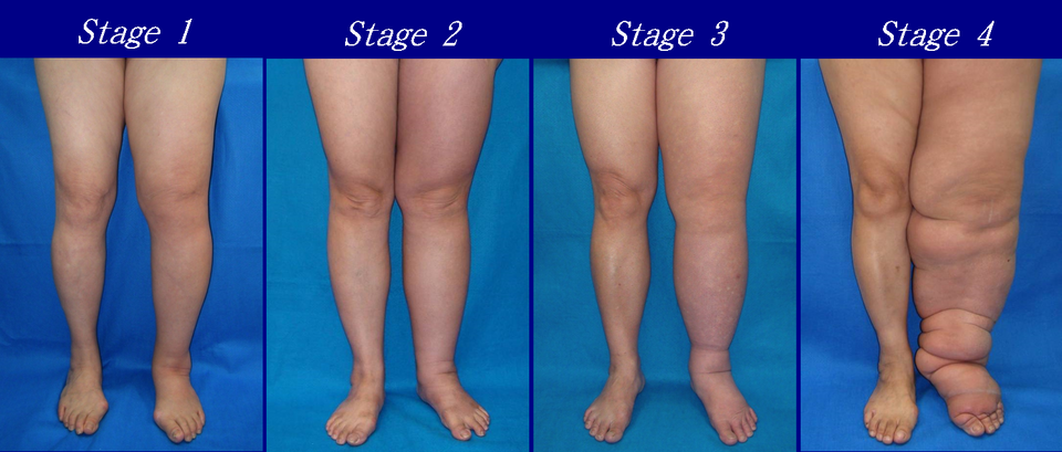
Edema: Swelling caused by excess fluid in the tissues. Lymphatic cause: Failure of the lymphatic system to drain interstitial fluid (e.g., lymph node removal during cancer surgery, leading to lymphedema).
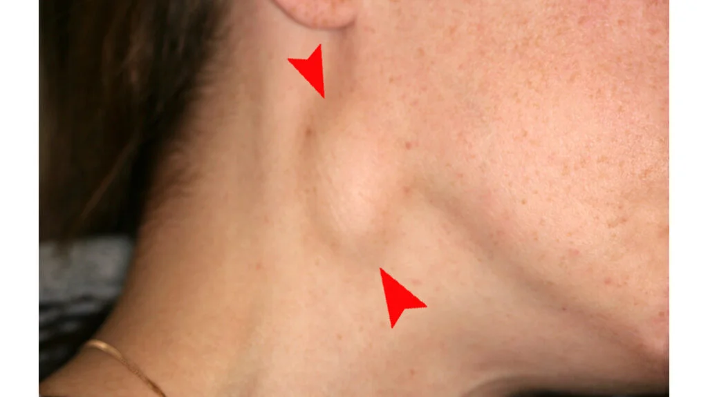
Lymph-adenopathy: Swollen or enlarged lymph nodes. Cause: Typically indicates an immune response to infection (e.g., strep throat, mono), or more seriously, cancer (e.g., lymphoma, metastasis).
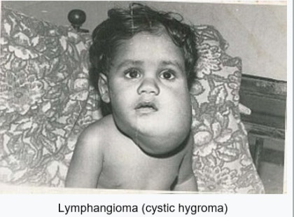
Lymphangioma: A benign malformation of the lymphatic system, often present at birth, resulting from abnormally formed lymphatic vessels.
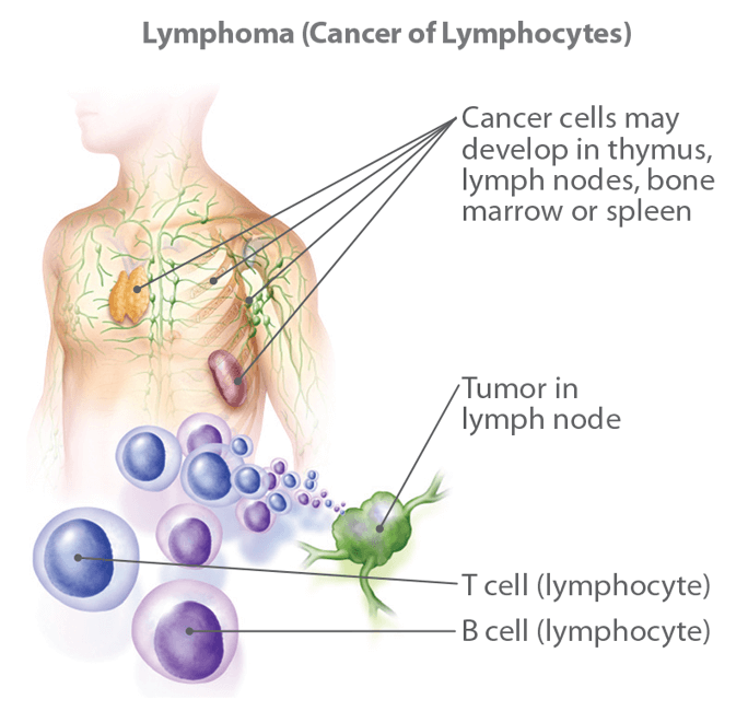
Lymphoma: A cancer of the lymphocytes themselves (e.g., Hodgkin's and Non-Hodgkin's lymphoma), causing uncontrolled growth of lymphocytes in lymph nodes and other tissues.
Lymph Flow & Metastasis: Cancer cells can break away from a primary tumor, enter lymphatic vessels, and travel to nearby lymph nodes. The lymph nodes are often the first site of spread, making them critical in cancer staging.
lymphedema
Etymology:
Lymph- (Latin: lympha): Meaning "clear water" or "goddess of water." This refers to the clear, water-like fluid.
-edema (Greek: oidēma): Meaning "swelling" or "a swelling tumor."
Edema: Swelling caused by excess fluid in the tissues. Lymphatic cause: Failure of the lymphatic system to drain interstitial fluid (e.g., lymph node removal during cancer surgery, leading to lymphedema).
Literal Translation: "Water swelling."
Clinical Meaning: The accumulation of lymph fluid in tissues, causing swelling, most often in the arms or legs. The name perfectly describes the condition: a swelling caused by clear lymphatic fluid.
Lymph-adeno-pathy
Lymph-adenopathy: Swollen or enlarged lymph nodes. Cause: Typically indicates an immune response to infection (e.g., strep throat, mono), or more seriously, cancer (e.g., lymphoma, metastasis).
Etymology:
Lymph- (Latin: lympha): "Clear water" (referring to lymph).
-adeno- (Greek: adēn): Meaning "gland." In this context, it refers to lymph nodes, which were historically called "lymph glands."
-pathy (Greek: pathos): Meaning "disease," "suffering," or "condition."
Literal Translation: "Disease of the lymph glands."
Clinical Meaning: Any disease or abnormality of the lymph nodes, most commonly presenting as swollen or enlarged nodes.
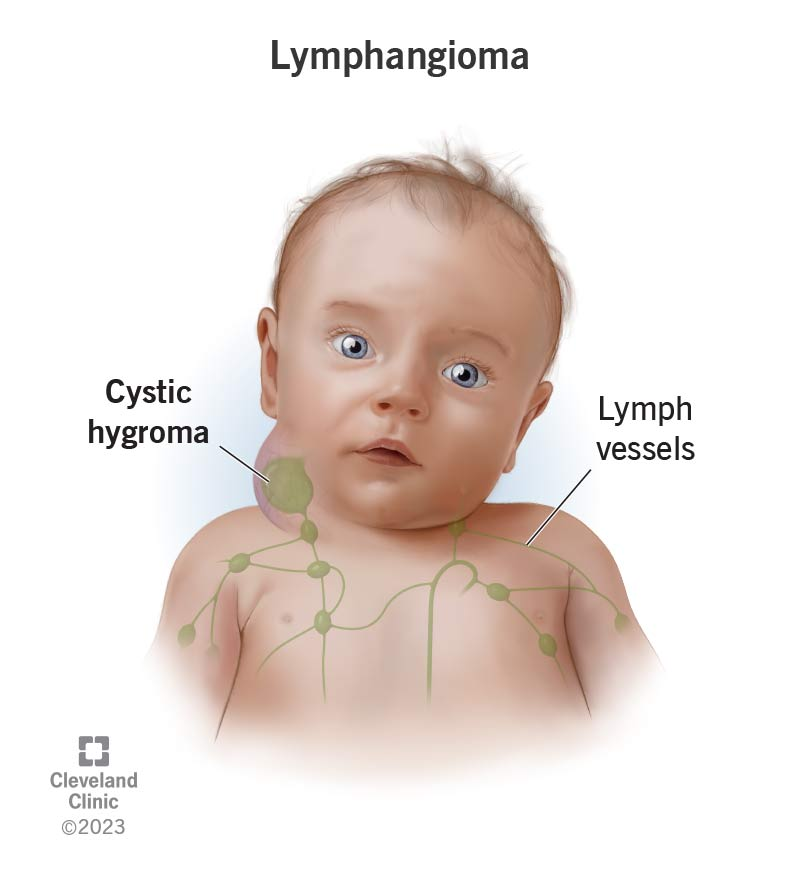
Lymph-angi-oma
A benign malformation of the lymphatic system, often present at birth, resulting from abnormally formed lymphatic vessels.
Etymology:
Lymph- (Latin: lympha): "Clear water."
-angio- (Greek: angeîon): Meaning "vessel" (as in a blood or lymphatic vessel).
-oma (Greek: -ōma): A suffix used to form nouns indicating a tumor or mass.
Literal Translation: "Tumor (swelling, non-cancerous) of the lymphatic vessels."
Clinical Meaning: A benign (non-cancerous) malformation of the lymphatic system, made up of fluid-filled spaces caused by abnormally formed lymphatic vessels. It is essentially a "tumor" in the original sense of the word—a swelling or mass
Lymph-oma
Etymology:
Lymph- (Latin: lympha): "Clear water."
-oma (Greek: -ōma): "Tumor" or "mass."
Literal Translation: "Tumor of the lymph."
Clinical Meaning: A malignant (cancerous) tumor that originates in the lymphocytes (a type of white blood cell) within the lymphatic system. Unlike lymphangioma, which is a mass of vessels, lymphoma is a cancerous mass of lymphoid cells (lymphocytes).
Lymphoma: A cancer of the lymphocytes themselves (e.g., Hodgkin's and Non-Hodgkin's lymphoma), causing uncontrolled growth of lymphocytes in lymph nodes and other tissues.
Lymph Flow & Metastasis (a way to spread cancer)
5. Lymph Flow & Metastasis
This is a combination of two concepts:
Lymph Flow:
Lymph: As above, from lympha, "clear water."
Flow: From Old English flōwan, meaning "to flow, stream, issue."
Metastasis:
Etymology: (Greek: metástasis)
Meta-: Meaning "change" or "beyond."
-stasis: Meaning "a standing" or "placement."
Literal Translation: "A change of place" or "displacement."
Clinical Meaning: The process by which cancer cells break away from the primary (original) tumor, travel through the lymphatic vessels (lymph flow) or blood vessels, and form new tumors (metastases) in other parts of the body. The path through the lymphatic system is a common route for metastasis, and the first place these cells often get trapped is in nearby lymph nodes.
Cancer cells can break away from a primary tumor, enter lymphatic vessels, and travel to nearby lymph nodes. The lymph nodes are often the first site of spread, making them critical in cancer staging.
what are the two subdivisions of the ANS?
Part 2: The Autonomic Nervous System (ANS)
1. Subdivisions of the ANS
The ANS is the involuntary division of the peripheral nervous system that controls glands, cardiac muscle, and smooth muscle. It has two primary, often antagonistic, subdivisions:
Sympathetic Nervous System (SNS): "Fight-or-Flight"
Prepares the body for emergency action, excitement, or exercise.
Parasympathetic Nervous System (PSNS): "Rest-and-Digest"
Promotes energy conservation, digestion, and elimination during rest.
what are the neurotransmitters and receptors used by the sympathetic nervous system?
The sympathetic nervous system (SNS) uses different neurotransmitters depending on the specific pathway and target organ.
sympathetic nervous system → acetylcholine to adrenal medulla nicotinic receptors → adrenal medulla makes epinephine (80%) and norepinephrine (20%) -→ send through entire body for sympathetic effect.
The primary neurotransmitters are Acetylcholine (ACh) and Norepinephrine (NE), but Epinephrine (Epi) is also crucial for its systemic effects.
Here’s a detailed breakdown:
1. At the Ganglion (Both Divisions= both parasympathetic and sympathetic)
Neurotransmitter: Acetylcholine (ACh) → nictonic receptros (N)
Receptor: Nicotinic Receptors (N)
Explanation: This is a universal step. All preganglionic neurons of the sympathetic nervous system (and the parasympathetic) release ACh to communicate with the postganglionic neuron in the ganglion. This synapse is always excitatory.
2. At the Target Organ (Most Common Pathway)
Neurotransmitter: No-repinephrine (NE)
Receptor: Adrenergic Receptors (α1, α2, β1, β2, β3)
Explanation: The vast majority of postganglionic sympathetic neurons release Norepinephrine (also called noradrenaline) onto their target organs. These target organs have adrenergic receptors, and the specific effect (excitatory or inhibitory) depends on the receptor type:
α1 receptors (vasoconstriction to reroute blood to areas that need it): leading to increased blood pressure.
a2 receptors (binds at a certain time): When the room (synapse) gets too hot (high NE from sympathetic nervous system), the thermostat (α₂ receptor) turns off the furnace (the neuron).
β1 receptors (heart): Increase heart rate and force of contraction.
β2 receptors (lungs & muscle): Cause bronchodilation (opening of airways) and vaso-dilation in skeletal muscle.
B3 receptors: lipid (fat) metabolism and bladder relaxation (increasing capacity to store urine)
3. Exception to the Rule: Sympathetic Cholinergic Fibers
Neurotransmitter: Acetylcholine (ACh)
Receptor: Muscarinic Receptors (M)
Explanation: A few specific post-ganglionic sympathetic neurons release ACh instead of NE. Their targets are:
Sweat Glands: To stimulate perspiration.
Some Blood Vessels: Specifically those supplying skeletal muscle (involved in thermoregulation).
4. The Adrenal Medulla Pathway (A Special Case)
Neurotransmitter: Acetylcholine (ACh)
Receptor: Nicotinic Receptors (N)
Explanation: The adrenal medulla is developmentally similar to a sympathetic ganglion. Preganglionic sympathetic neurons release ACh onto the adrenal medulla, stimulating it.
Hormones Released: Epinephrine (Epi) (~80%) and Norepinephrine (NE) (~20%)
Explanation: Instead of releasing a neurotransmitter at a specific synapse, the adrenal medulla releases these hormones directly into the bloodstream. They then act on adrenergic receptors throughout the entire body, creating a widespread, systemic "adrenaline rush" that complements the neural effects of the SNS.
what are the neurotransmitters and receptors used by the parasympathetic nervous system?
The parasympathetic nervous system (PSNS), known as the "rest and digest" system, uses a specific set of neurotransmitters and receptors to achieve its effects.
The key principle is that it uses acetylcholine (ACh) as its primary neurotransmitter at both synapses, but the type of receptor changes depending on the location.
Summary Table
Location | Neurotransmitter | Receptor Type | Effect |
|---|---|---|---|
Ganglion (Synapse between pre- and post-ganglionic neuron) | Acetylcholine (ACh) | Nicotinic Receptor (NN) | Stimulatory (Excites the post-ganglionic neuron) |
Target Organ (Synapse between post-ganglionic neuron and effector organ) | Acetylcholine (ACh) | Muscarinic Receptor (M) | Varies (Slows heart, stimulates digestion, etc.) M2 receptor is the HEART, M3 for EVERYTHING ELSE. |
1. Neurotransmitters
Acetylcholine (ACh): This is the sole neurotransmitter used by all neurons in the parasympathetic division where they synapse with another neuron or an organ.
Preganglionic Neurons: Release ACh at the ganglion to activate the postganglionic neuron.
Postganglionic Neurons: Release ACh at the target organ (e.g., heart, stomach, glands) to cause the final "rest and digest" effect.
2. Receptors
The type of receptor that receives ACh determines the response. There are two crucial types:
a) Nicotinic Receptors (nAChR)
Location: On the cell bodies of all post-ganglionic neurons (in the parasympathetic ganglia).
Mechanism: They are ligand-gated ion channels. When ACh binds, the channel opens, allowing ions to flow and causing rapid excitation of the post-ganglionic neuron.
Effect: The effect is always stimulatory. This is a relay switch—the message must always get passed on.
b) Muscarinic Receptors (mAChR)
Location: On the surface of all target organs (e.g., cardiac muscle, smooth muscle, glands).
Mechanism: They are G-protein coupled receptors (GPCRs). Their effects are slower and can be either stimulatory or inhibitory, depending on the specific organ and the signaling pathway activated.
Effect: Varies by organ. This is why ACh can have different effects in different parts of the body:
Heart (M₂ subtype): Inhibitory (Decreases heart rate and force of contraction).
Digestive Tract (M₃ subtype): Stimulatory (Increases motility, secretion, and relaxes sphincters).
Salivary & Gastric Glands (M₃ subtype): Stimulatory (Promotes salivation and gastric acid secretion).
Eye (Pupillary Sphincter Muscle) (M₃ subtype): Stimulatory (Causes constriction for near vision).
Lungs (Bronchial Smooth Muscle) (M₃ subtype): Stimulatory (Causes bronchoconstriction).
Why This Distinction is Important
The use of the same neurotransmitter (ACh) on two different receptor types (nicotinic vs. muscarinic) is what allows for precise control.
A drug that blocks nicotinic receptors (e.g., hexamethonium) would shut down all autonomic output (both sympathetic and parasympathetic) because it blocks the ganglia. This is rarely used.
A drug that blocks muscarinic receptors (e.g., atropine) will specifically block the parasympathetic effects on organs. This is very useful in medicine to:
Speed up a slow heart rate (by blocking the PSNS slowing effect).
Dilate the pupils for an eye exam.
Reduce excessive salivation and respiratory secretions during surgery.
In summary, the parasympathetic nervous system is a cholinergic system (uses ACh throughout) that acts on nicotinic receptors in ganglia and muscarinic receptors at organs to promote calming, restorative bodily functions.
components and basis for distinctions
origin of nerves in sympathetic nervous system (Thoraco-lumbar (T1-L2 spinal cord) AND parasympathetic nervous system (Cranio-sacral (Brainstem & S2-S4 spinal cord)
2. Components and Basis for Distinctions
The key anatomical distinctions are the origin of the nerves and the neurotransmitters used.
Feature | Sympathetic (SNS) | Parasympathetic (PSNS) |
|---|---|---|
Origin (CNS) | Thoraco-lumbar (T1-L2 spinal cord) | Cranio-sacral (Brainstem & S2-S4 spinal cord) |
Ganglion Location | Close to CNS (Para-vertebral chain & prevertebral ganglia) | In or near Target Organ (Terminal ganglia) |
Preganglionic Neurotransmitter | Acetylcholine (ACh) | Acetylcholine (ACh) |
Postganglionic Neurotransmitter | Norepinephrine (NE) (mostly) | Acetylcholine (ACh) |
General Effect | Widespread activation ("mass discharge") | Local, discrete effec |
1. Sympathetic: Ganglia "Close to CNS"
This means the ganglia are located very near the spinal column.
Paravertebral Chain Ganglia (Sympathetic Trunk): These are a linked chain of ganglia that lie immediately alongside the vertebral column (from the neck to the pelvis). They look like a string of beads next to the spine.
Prevertebral Ganglia: These are ganglia that lie anterior to the spine and close to the major abdominal arteries (e.g., celiac ganglion, superior mesenteric ganglion).
Why this matters:
Widespread Effect: Because the ganglion is far from the target organ, the postganglionic axon must be very long to reach its destination. A single preganglionic neuron can synapse with many postganglionic neurons in these central ganglia. This allows for mass activation—diverging the signal to multiple organs at once (e.g., heart, lungs, blood vessels), which is essential for the "fight-or-flight" response.
Analogy: Think of a central train station (the ganglion) located downtown. Trains (preganglionic axons) arrive from a nearby suburb (the spinal cord). From this central station, many long-distance trains (postganglionic axons) fan out to many different distant towns (target organs).
2. Parasympathetic: Ganglia "In or near Target Organ" (Terminal Ganglia)
This means the ganglia are located very far from the CNS, embedded in the walls or very close to the specific organ they control.
Terminal Ganglia: These are the final ganglia where the synapse occurs. They are microscopic and located within the tissue of the target organ itself. For example, they are in the wall of the heart, the wall of the bladder, or inside salivary glands.
Intramural Ganglia: A specific term meaning "within the wall," which describes terminal ganglia located inside the organ's structure.
Why this matters:
Localized, Discrete Effect: Because the ganglion is right at the organ, the postganglionic axon is extremely short. The signal is delivered to a single, specific organ without affecting others. This allows for precise, localized control (e.g., slowing the heart without affecting the digestive system), which is ideal for "rest-and-digest" functions.
Analogy: Think of a local delivery depot (the ganglion) built right in the middle of a neighborhood. A long-haul truck (preganglionic axon) drives from a distant city (the brainstem) to this local depot. From there, a very short, local delivery van (postganglionic axon) makes the final delivery to a single house (the specific cells of the target organ).
Summary Table
Feature | Sympathetic Ganglia | Parasympathetic Ganglia |
|---|---|---|
Location | Close to CNS (alongside or in front of spine) | In or near Target Organ (within the organ's wall) |
Another Name | Paravertebral or Prevertebral ganglia | Terminal or Intramural ganglia |
Resulting Axon Length | Long postganglionic axons | Short postganglionic axons |
Physiological Impact | Widespread, mass activation (divergence) | Discrete, localized control |
Effects on Organs
4. Effects on Organs
Organ | Sympathetic (SNS) Stimulation ("Fight-or-Flight") | Parasympathetic (PSNS) Stimulation ("Rest-and-Digest") |
|---|---|---|
Eye (Pupil) | Dilation (mydriasis) - for more light | Constriction (miosis) - for near vision |
Skin | Sweating (cholinergic fibers); Piloerection (goosebumps); Vasoconstriction | No significant innervation |
Heart | Increases heart rate (chronotropy) and force of contraction (inotropy) | Decreases heart rate and force of contraction |
Lungs (Bronchi) | Bronchodilation (increases air flow) | Bronchoconstriction |
Digestive Tract | Decreases motility and gland secretion; Sphincters contract | Increases motility and gland secretion; Sphincters relax |
Recognize the subdivisions of the autonomic division of the nervous system (ANS).
(sympathetic nervous system)
effect on heart
effect on blood pressure
effect on eyes
effect on lungs
effect on digestion
effect on liver
effect on adrenal gland
effect on blood flow to skin, digestive organs and skeletal muscles
1. Sympathetic Nervous System (SNS) (norepinephrine & norepinephrine)
Common Name: The "Fight-or-Flight" Division
Physiological Effects (What it does):
↑ Heart rate and force of contraction
↑ Blood pressure
Dilates pupils and bronchioles (in lungs)
Inhibits digestion (reduces salivation, slows peristalsis)
Stimulates glucose release from the liver (for energy)
Stimulates secretion of epinephrine (adrenaline) from adrenal glands
Shunts blood flow away from skin and digestive organs to skeletal muscles
the only thing minimized here is DIGESTION and BLOOD FLOW TO SKIN AND DIGESTIVE ORGANS to be given to SKELETAL MUSCLES.
Primary Function: To mobilize the body's resources during stressful or emergency situations. It prepares the body for intense physical activity.
Origin of Nerves: Neurons originate from the thoracic and lumbar regions of the spinal cord (T1 to L2), hence it is also called the thoracolumbar division.
Key Anatomical Feature: Has a long chain of ganglia (clusters of nerve cell bodies) located very close to the spinal cord, called the paravertebral ganglia or sympathetic chain.
what are the neurotransmitters the sympathetic nervous system uses?
The sympathetic nervous system (SNS) uses different neurotransmitters depending on the specific pathway and target organ.
sympathetic nervous system → acetylcholine to adrenal medulla nicotinic receptors → adrenal medulla makes epinephine (80%) and norepinephrine (20%) -→ send through entire body for sympathetic effect.
The primary neurotransmitters are Acetylcholine (ACh) and Norepinephrine (NE), but Epinephrine (Epi) is also crucial for its systemic effects.
Here’s a detailed breakdown:
1. At the Ganglion (Both Divisions= both parasympathetic and sympathetic)
Neurotransmitter: Acetylcholine (ACh) → nictonic receptros (N)
Receptor: Nicotinic Receptors (N)
Explanation: This is a universal step. All preganglionic neurons of the sympathetic nervous system (and the parasympathetic) release ACh to communicate with the postganglionic neuron in the ganglion. This synapse is always excitatory.
2. At the Target Organ (Most Common Pathway)
Neurotransmitter: No-repinephrine (NE)
Receptor: Adrenergic Receptors (α1, α2, β1, β2, β3)
Explanation: The vast majority of postganglionic sympathetic neurons release Norepinephrine (also called noradrenaline) onto their target organs. These target organs have adrenergic receptors, and the specific effect (excitatory or inhibitory) depends on the receptor type:
α1 receptors (vasoconstriction to reroute blood to areas that need it): leading to increased blood pressure.
a2 receptors (binds at a certain time): When the room (synapse) gets too hot (high NE from sympathetic nervous system), the thermostat (α₂ receptor) turns off the furnace (the neuron).
β1 receptors (heart): Increase heart rate and force of contraction.
β2 receptors (lungs & muscle): Cause bronchodilation (opening of airways) and vaso-dilation in skeletal muscle.
B3 receptors: lipid (fat) metabolism and bladder relaxation (increasing capacity to store urine)
3. Exception to the Rule: Sympathetic Cholinergic Fibers
Neurotransmitter: Acetylcholine (ACh)
Receptor: Muscarinic Receptors (M)
Explanation: A few specific post-ganglionic sympathetic neurons release ACh instead of NE. Their targets are:
Sweat Glands: To stimulate perspiration.
Some Blood Vessels: Specifically those supplying skeletal muscle (involved in thermoregulation).
4. The Adrenal Medulla Pathway (A Special Case)
Neurotransmitter: Acetylcholine (ACh)
Receptor: Nicotinic Receptors (N)
Explanation: The adrenal medulla is developmentally similar to a sympathetic ganglion. Preganglionic sympathetic neurons release ACh onto the adrenal medulla, stimulating it.
Hormones Released: Epinephrine (Epi) (~80%) and Norepinephrine (NE) (~20%)
Explanation: Instead of releasing a neurotransmitter at a specific synapse, the adrenal medulla releases these hormones directly into the bloodstream. They then act on adrenergic receptors throughout the entire body, creating a widespread, systemic "adrenaline rush" that complements the neural effects of the SNS.
Recognize the subdivisions of the autonomic division of the nervous system (ANS).
neurotransmitter?
effect on heart
effect on digestion
effect on pupils
effect on bronchioles
effect on urination and defacation
effect on sexual arousal
2. Parasympathetic Nervous System (PNS)
Common Name: The "Rest-and-Digest" or "Feed-and-Breed" Division
Primary Function: To conserve energy and promote bodily functions that occur at rest. It is responsible for the body's "housekeeping" activities.
Origin of Nerves: Neurons originate from the brainstem (cranial nerves, e.g., Vagus Nerve (CN X)) and the sacral region (S2-S4) of the spinal cord, hence it is also called the craniosacral division.
Key Anatomical Feature: Its ganglia are typically located very close to or within the target organ itself (terminal ganglia).
Neurotransmitter: The primary neurotransmitter released onto target organs is acetylcholine (ACh).
Physiological Effects (What it does):
↓ Heart rate
Stimulates digestion (increases salivation, intestinal peristalsis)
Constricts pupils and bronchioles
Stimulates urination and defecation
Stimulates sexual arousal
Recognize the subdivisions of the autonomic division of the nervous system (ANS).
3. Enteric Nervous System (ENS)
Common Name: The "Second Brain" of the Gut
Primary Function: An extensive, self-contained network of neurons that independently governs the function of the gastrointestinal tract.
Location: It is embedded in the lining of the esophagus, stomach, intestines, pancreas, and gallbladder.
Relationship to ANS: While it can operate autonomously, it is heavily influenced and regulated by the sympathetic and parasympathetic divisions. The Sympathetic nervous system generally inhibits digestive activity, while the Parasympathetic nervous system (via the Vagus nerve) stimulates it.
Physiological Effects (What it does):
Controls motility (peristalsis) of the digestive tract
Regulates secretion of enzymes and acid
Manages local blood flow within the gut
Monitors the physiological condition of the gastrointestinal tract
Recognize the subdivisions of the autonomic division of the nervous system (ANS).
Summary Table: A Side-by-Side Comparison
Feature | Sympathetic (SNS) | Parasympathetic (PNS) |
|---|---|---|
Overall Function | Fight-or-Flight (expends energy) | Rest-and-Digest (conserves energy) |
Origin (CNS) | Thoracolumbar (T1-L2) | Craniosacral (Brainstem & S2-S4) |
Ganglion Location | Close to spinal cord (Chain ganglia) | Near or within target organ |
Fiber Length | Short preganglionic, Long postganglionic | Long preganglionic, Short postganglionic |
Key Neurotransmitter | Norepinephrine (at target organ) | Acetylcholine (at target organ) |
Effect on Body | Widespread, systemic effects | Localized, discrete effects |
Example Actions | Increases heart rate, dilates pupils | Slows heart rate, stimulates digestion |
Recognize the subdivisions of the autonomic division of the nervous system (ANS).
In essence, the Sympathetic and Parasympathetic systems are complementary, often having antagonistic (opposing) effects on the same organs to provide precise control, much like accelerating and braking in a car. The Enteric system is a specialized, semi-independent network that manages the complex process of digestion under their influence.
Identify the components of both divisions of ANS and the basis for these distinctions.
Building on the recognition of the subdivisions, here is a detailed identification of their components and the basis for these distinctions.
The two primary divisions of the Autonomic Nervous System (ANS)—the Sympathetic and Parasympathetic systems—are distinguished by their anatomy, neurotransmitters, and functional effects. A third division, the Enteric Nervous System, is often considered a semi-independent part of the ANS.
Identify the components of both divisions of ANS and the basis for these distinctions.
Feature | Sympathetic Division | Parasympathetic Division |
|---|---|---|
Origin in CNS | Thoracolumbar outflow. Preganglionic neurons originate from the lateral gray horn of the spinal cord segments T1 to L2. | Craniosacral outflow. Preganglionic neurons originate in the brainstem (cranial nerves III, VII, IX, X) and the sacral spinal cord (segments S2-S4). |
Location of Ganglia | Ganglia are close to the CNS and far from the target organ. They form two chains along the vertebral column (paravertebral ganglia/chain) or a few prevertebral ganglia (e.g., celiac ganglion). | Ganglia are very close to or embedded within the wall of the target organ itself (terminal or intramural ganglia). |
Length of Neurons | Short pre-ganglionic axons (as the ganglion is nearby). Long post-ganglionic axons (must travel from the chain to the organ). | Long pre-ganglionic axons (must travel all the way to the organ). Short post-ganglionic axons (only need to innervate tissue right next to the ganglion). |
Divergence | High divergence. One preganglionic neuron synapses with many postganglionic neurons. This allows for a widespread, mass activation. | Low divergence. One preganglionic neuron synapses with very few postganglionic neurons. This allows for discrete, localized effects. |
Differentiate the location of the cell body and tract of axon of the neurons in the sympathetic and parasympathetic system.
This is a fundamental distinction between the two systems. The key is to follow the path of a single nerve signal from the spinal cord to the target organ.
Core Concept: Two-Neuron Chain
In both the Sympathetic (SNS) and Parasympathetic (PNS) systems, the pathway from the CNS to the target organ consists of two neurons:
Preganglionic Neuron: Cell body in the CNS. Its axon (preganglionic axon) travels to a ganglion.
pre-ganglionic because it travels to the ganglion
Postganglionic Neuron: Cell body in the ganglion. Its axon (postganglionic axon) travels to the target organ.
postganglionic because it travels to the target organ.
The differences lie in where these cell bodies are located and the length of their axons.
AUTONOMIC NERVOUS SYSTEM SELF DIRECTED LEARNING VIDEO)