A&P 1 Cell Anatomy
1/35
There's no tags or description
Looks like no tags are added yet.
Name | Mastery | Learn | Test | Matching | Spaced | Call with Kai |
|---|
No analytics yet
Send a link to your students to track their progress
36 Terms
Interstitial Fluid
The fluid that surrounds cells. It contains nutrients such as glucose, salt, fatty acids, and minerals such as Ca, Mg, and K. As such, it provides a means of delivering materials to the cells, intercellualr communication, and removal of metabolic waste.
What does ECF stand for? How is it different than interstitial fluid?
Extracellular fluid
Extracelluar fluid includes both interstitial fluid (the fluid that surrounds and "bathes" the cells); and blood plasma, or intravascular fluid (the fluid within the blood vessels).
Interstitial fluid composes about 2/3 of the extracellular fluid, adn blood plasma about 1/3.
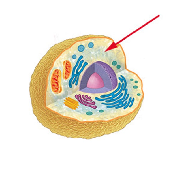
What is Cytosol? What does ICF stand for?
Cytosol is also called intracelluar fluid (ICF) or cytoplasmic matrix and is the viscous, syruplike fluid of the cytoplasm. It has a high water content and contains many dissolved macromolecules (carbohydrates, lipids, proteins).
It functions as the site of multiple cell processes such as signal transduction within the cell.
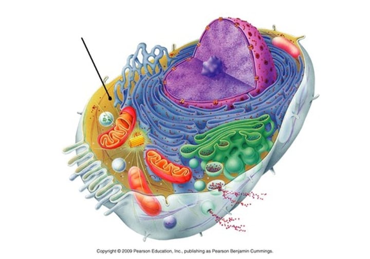
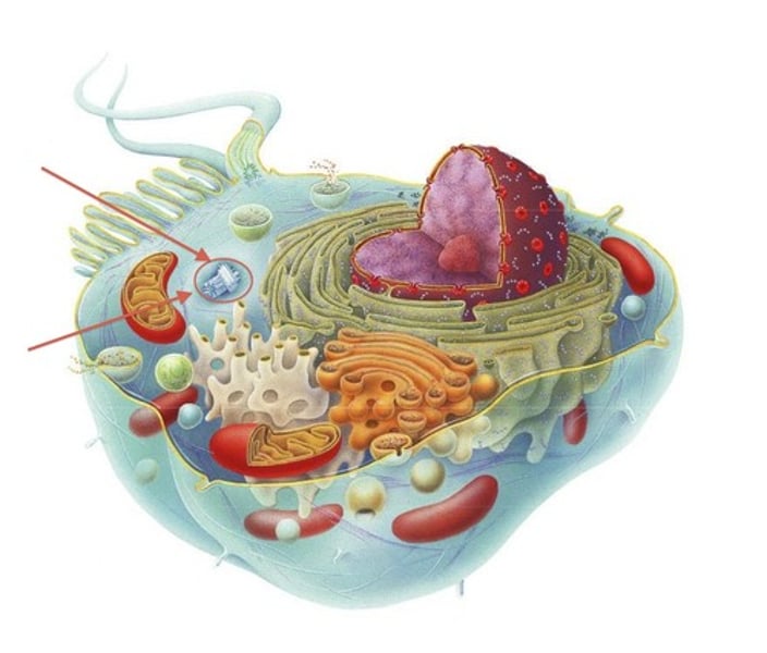
Cytoplasm
A general term for all cellular contents located between the plasma membrane and the nucleus. The three primary componenets are cytosol, organelles, and inclusions.
It has many different functions. For example, it gives a cell its shape and keeps organelles in their place.
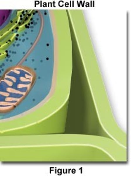
Cell wall
The cell wall is located outside the cell membrane of plant cells, bacterial cells, fungal cells, and some protozoan cells. Animal cells do not have cell walls.
The cell wall functions to protect the cell and to allow things in and out of the cell. In bacteria, it can help the cell attach to a host.
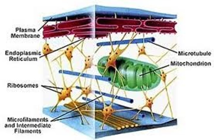
Cytoskeleton
Most eukaryotic cells contain a complex network of protein fibers called the cytoskeleton. it forms a framework for the movement of organelles around the cytoplasm - most of the organelles are attached to the cytoskeleton. It consists of protein microfilaments, intermediate filaments, and microtubules.
It determines cell shape, helps organelles move within the cell, and move chromosomes to the daughter cell in cell division.
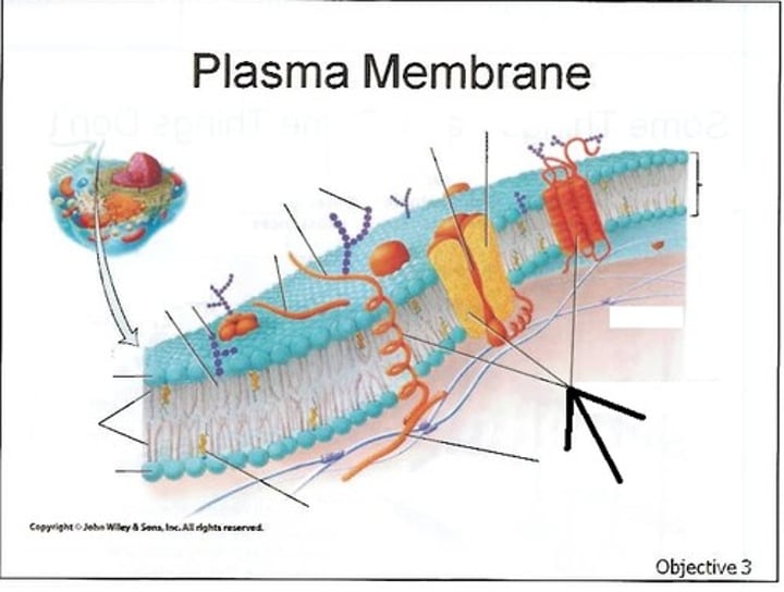
Cell membrane
Also called the plasma membrane. It forms the outer barrier separating the internal contents from the external environment of the cell. Modified extensions of the plasma membrane include cilia, a flagellum, and mircrovilli.
It is selectively permeable to ions and organic molecules and controls the movement of substances in and out of cells.
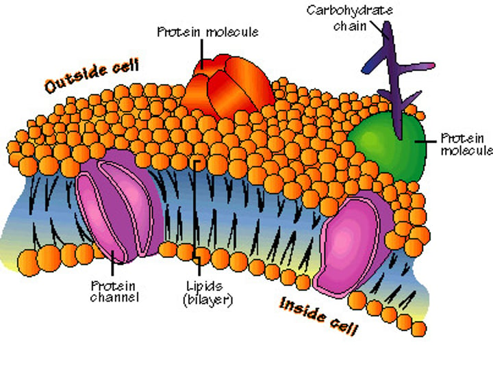
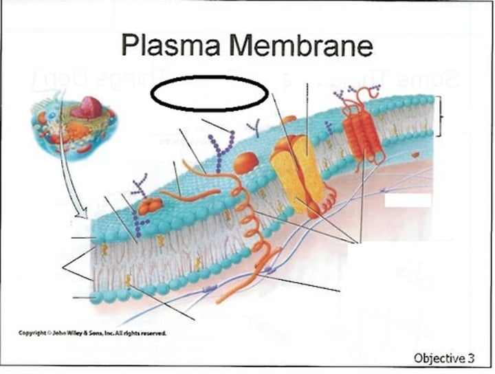
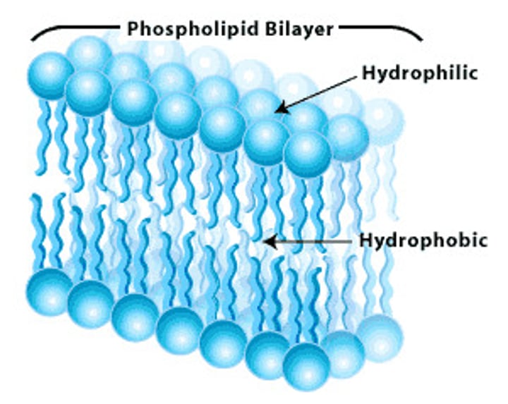
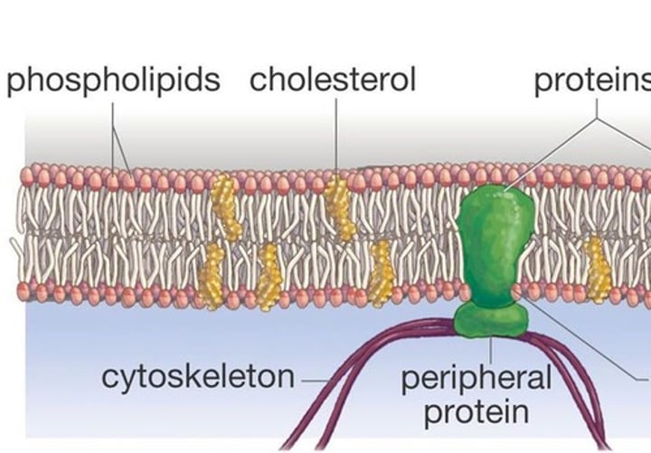
Phospholipid bilayer
The plasma membrane is composed of a phospholipid bilayer with polar, hydrophilic (water loving) heads and non-polar, hydrophobic (water fearing) tails. The heads point outward, and the tails point inward. Interspersed in the phospholipids are proteins.
Cholesterol
Cholesterol (as it pertains to a cell) is also a component of cell membranes/plasma membranes. It is scattered within the hydrophobic regions of the bilayer.
It strengthens the membrane and stabilizes it at termperature extremes.
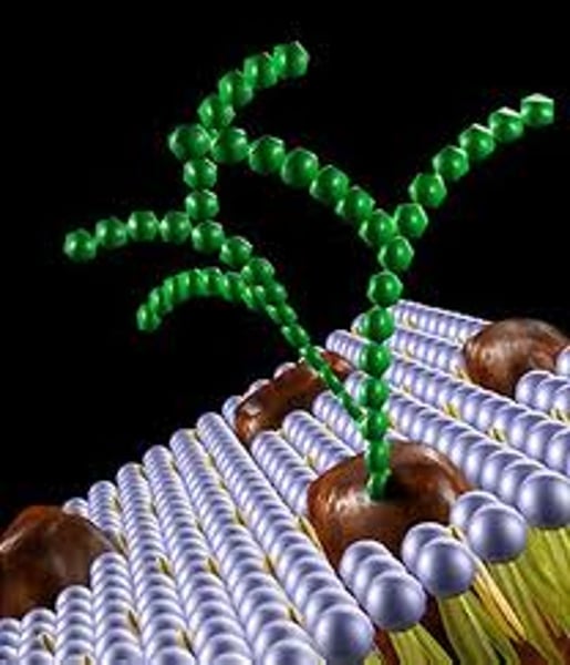
Glycoproteins
Many integral proteins (proteins embedded within and extending across the bilayer) are glycoproteins that have carbohydrates exposed to the interstitial fluid.
They provide many functions. They give structural support, help to form connective tissues, facilitate digestion by producing secretions/mucous in the gastrointestinal tract.
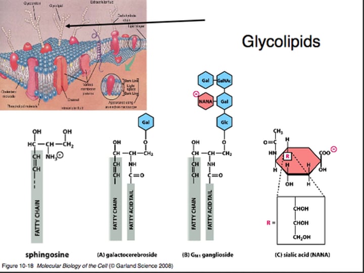
Glycolipids
Lipids with attached carbohydrate groups. They are only on the outer phospholipid layer of the membrane, and are exposed to the interstitial fluid.
Together the CHO portion of the glycolipids and the glycoproteins form the glycocalyx (the sugar coating on the cell's surface).
Glycolipids provide energy and also serve as markers for cellular recognition.
Channel proteins
These are integral proteins and are one of three types of transport proteins (the other two are carriers and pumps).
In some cases the channel proteins simply act as a passive pore. Molecules will randomly move through the opening in a process called diffusion, requiring no energy. Molecules move from an area of high concentration to an area of low concentration.

Transmembrane proteins
A class of integral proteins. They commonly form a helix or coil.
These proteins function as a means of signaling to the cell what the external environment contains. They also help control the exchange of materials across the membrane.
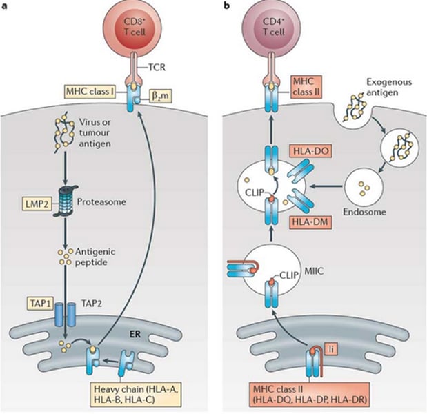
Peripheral proteins: MHC-I proteins and MHC-II proteins
MHC (Major HistoCompatibility) Class I and Class II proteins are glycoproteins. They are continously synthesized by the rough endiplasmic reticulum, insterted into the ER and shipped and modified by the endomembrane system. Then they're embedded in the plasma membrane.
Their main function is to bind to peptide fragments derived from pathogens and display them on the cell surface for recognition by the appropriate T-cells.
MHC-I's have beta-2 subunits so can only be recognized by CD8 co-receptors.
MHC-II's have no beta-2 subunits so can be recognized by CD4 co-receptors.
In this way MHC molecules chaperone which type of lymphocytes (WBC) may bind to the given antigen with high affinity.
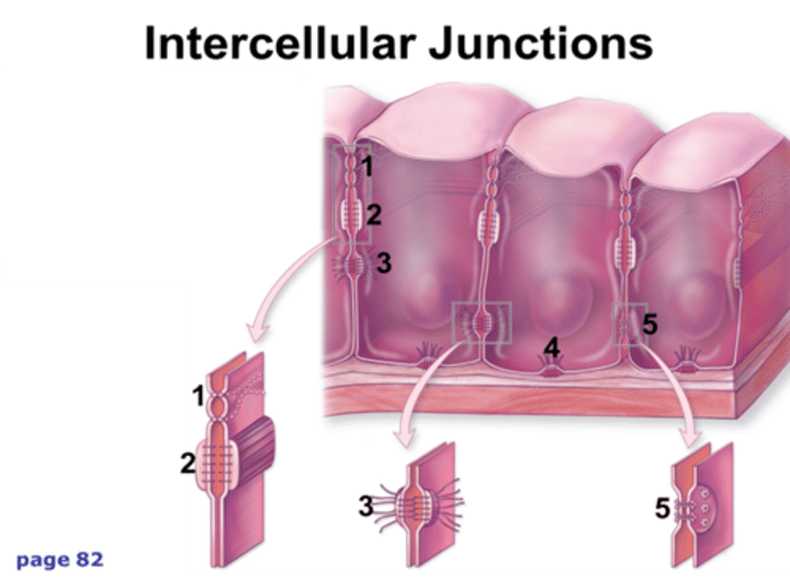
Tight Junctions
Also called a zonula occludens. It encircles certain types of cells (like epithelial cells) near their exposed apical (upper) surface and completely attaches each cell to its neighbors. Plasma membrane proteins among neighboring cells fuse, so the apical surfaces of the cells are tightly connected everywhere around the cell.
This seals off the intercellular space and prevents substances from passing between the epithelial cells and forces materials to move through, rather than between cells.
Example: in the small intestine, tight junctions prevent corrosive digestive enzymes from moving between cells and damaging internal body structures. They also prevent leakage of urine through the urinary bladder wall.
(#1 and 2 in the image are showing a tight junction, #3 is a desmosome, and #5 is a gap junction)
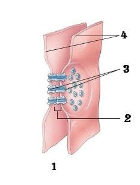
Gap junctions
These are formed across the intercellular space between neighboring cells. This gap (2 nanometers in length) is bridged by six tansmembrane proteins, called connexons that form tiny, fluid-filled tunnels or pores.
Gap junctions provide a direct passageway for substances (such as ions, glucose, amino acids, and other small solutes)to travel between neighboring cells.
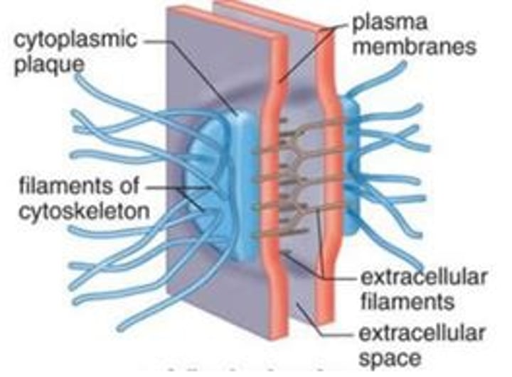
Desmosomes
Also called macula adherens (adhering spot). These are like snaps between adjacent cells. Each is a small region that holds cells together and provides resistance to mechanical stress at a single point. The neighboring cells are separated by a small space that is spanned by a fine web of protein filaments. Each cell contributes half of the the complete desmosome.
Cells of tissues exposed to stress, such as the external layer of the skin and cardiac muscle, contain desmosomes. Hemidesmosomes (half of a desmosome) anchor the basal layer of cells of the epidermis to the underlying components.
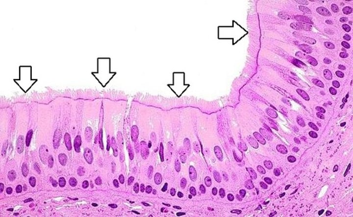
Cilia
Singular is cilium. Cilia are projections extending from the cell. They contain both cytoplasm and supportive microtubule proteins, and they are enclosed by the plasma membrane. Cilia are usually found in large numers on the exposed surfaces of specific cells such as those that line portions of the respiratory passageways.
The beating of cilia moves the mucus formed from mucin and any adherent substances along the cell surface (in the respiratory passageways it moves it to the throat, where it can be expelled from the respiratory system).
*Can be seen with the microscope at the cellular level.
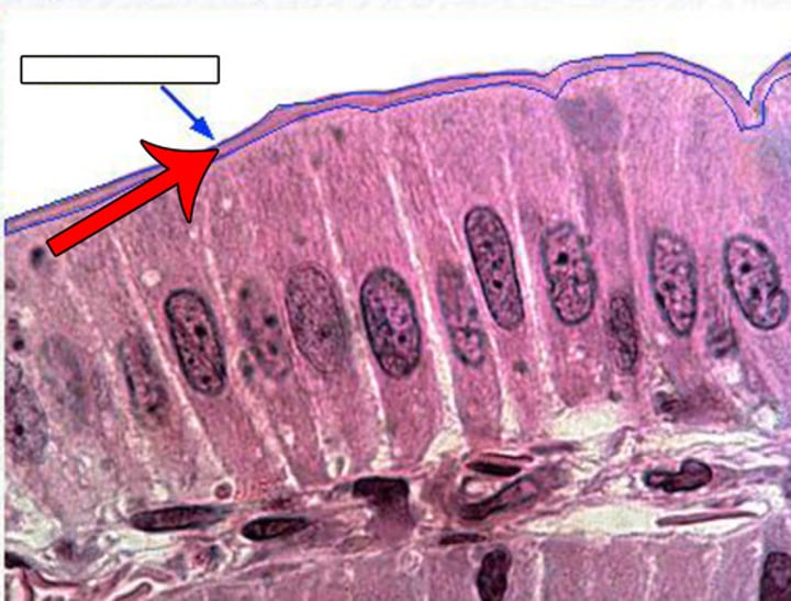
Microvilli
Singular is microvillus. Microvilli are thin, microscopic membrane extenstions from tthe surface of the plasma membrane. They are much smaller than cilia, much more densely packed together, and lack powered movement.
These projections form a more extensive plasma membrane surface for molecules to travel across to faciliate more efficent membrane transport.
Cells with microvilli occur throughout the small intestine, where increased surface area is needed to absorb digested nutrients.
*Can't really be seen at the cellular level of microscopic images. It looks just like a solid mass adjacent to the apical surface of cells and is called a brush border.
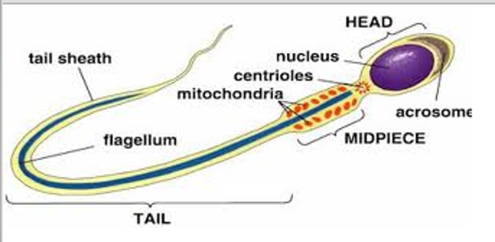
Flagella
Singular is flagellum. Flagella are similar to cilia in basic structure, however they are longer and usually appear alone.
The function of a flagellum is to help propel an entire cell. In humans, the only example of a cell with a a flagellum is sperm, which needs to move through the female reproductive tract to reach the ovum.
Nucleus
The nucleus is the largest structure in the cell, typically averaging 5-7 micrometers in diameter. It is often called the cell's control center A cell typically has one nucleus, however skeletal muscle cells have many nuclei.
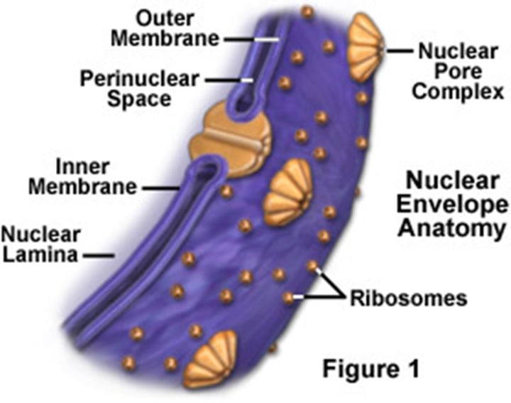
Nuclear envelope
The nucleus is enclosed in a double membrane that is called the nuclear envelope. It separates the cytoplasm from the nucleoplasm (the fluid within the nucleus).
The nuclear envelope controls the movement of materials between the nucleus and the surrounding cytoplasm. Each layer of the nuclear envelope is a phospholipid bilayer, similar to the plasma membrane.
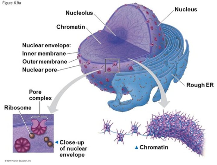
Nuclear pores
Channel-like open passageways that penetrate fused regions within the double membrane throughout the entire nuclear envelope.
They allow the passage of large particles both into the nucleus (like proteins) and out of the nucleus (like mRNA). Ions and water-soluble molecules also pass through nuclear pores.
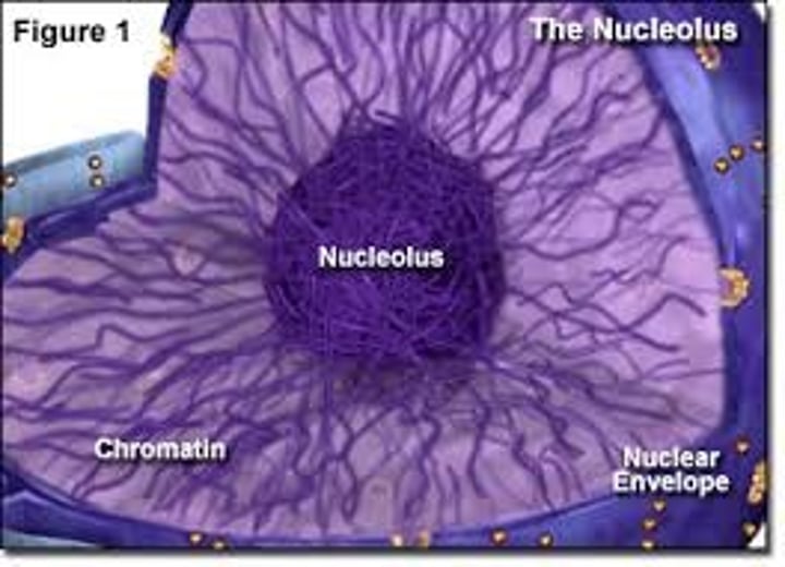
Nucleolus
The cell typically contains one dark-staining, usually spherical, body called a nucleolus (plural nucleoli). It is not membrane bound. It is composed of protein and RNA and is responsible for producing the small and large subunits of ribosomes.
Nucleoplasm
The fluid within the nucleus. Composed primarily of water, dissolved ions, and a complex mixture of molecules.
Its primary function is to act as a suspension medium for the organelles of the nucleus. other functions include the maintenance of nuclear shape and structure, and the transportation of ions, molecules, and other substances.
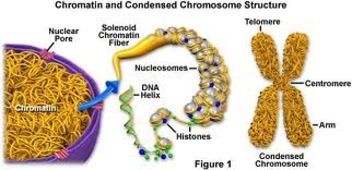
Chromatin
When a cell is not dividing, the DNA and its associated proteins are in the form of a finely filamented mass called chromatin, which resembles an unrolled spool of thread.

Chromosomes (what is the difference between this and chromatin? What are both made of?
The tightly coiled chromatin is what we see as chromomsomes during cell division. Both chromatin and chromosomes are made of DNA and proteins.
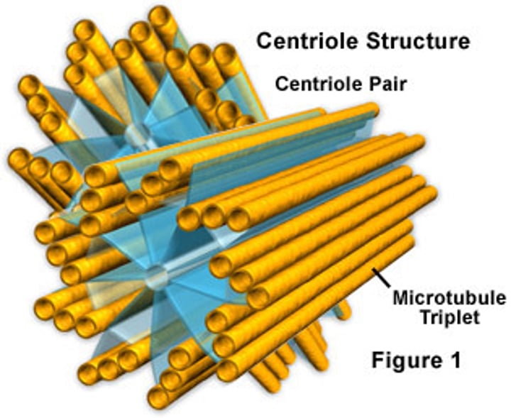
Centrosome
It is typically in close proximity to the nucleus and contains a pair or perpendicularly oriented cylindrical centrioles surrounded by protein that is amorphous (without distinctive shape).
The centrosome (or microtubule organizing center), is an area in the cell where microtubles are produced.
Centriole
Centrioles are made of microtubules and exist in perpenicular pairs within centrosomes.
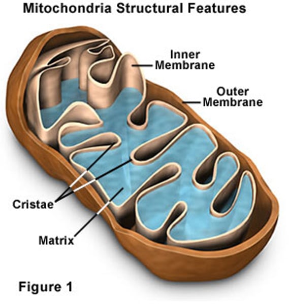
Mitochondria
Singular is mitochondrion. They are oblong-shaped organelles with a double membrane and contain a small, unique circular fragment of DNA that has genes for producing mitochondrial proteins.
Mitochondria engage in aerobic cellular respiration to complete the digestion of glucose and other fuel molecules, such as fatty acids, for the tranfer of energy to synthesize ATP molecules, the cell's energy currency.
Mitochondria are also called the "powerhouses" of the cell.
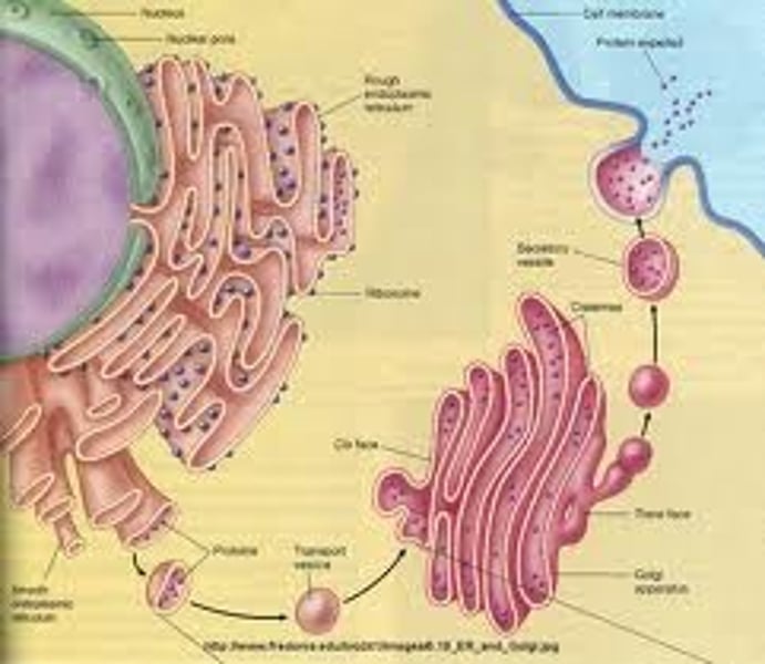
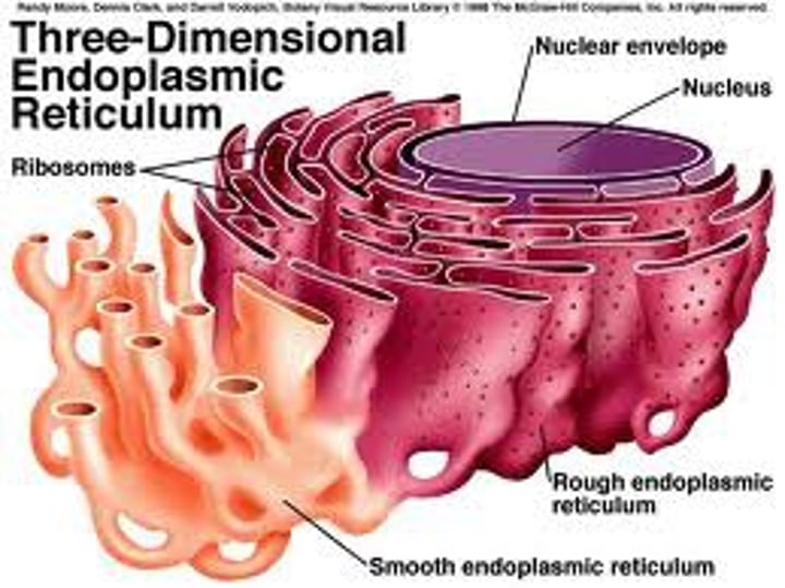
Rough endoplasmic reticulum
The endoplasmic reticulum is an extensive interconnected membrane network that varies in shape (ex. parallel sheets, sacs, or tubules) and separates fluid within the membranous structure from the cytosol. It typically extends form the nuclear envelope to the plasma membrane. Endoplasmic reticulum with ribosomes attached to it is called ROUGH endoplasmic reticulum, or rough ER.
The ribosomes of the rough ER produce, modify, package, and store proteins.
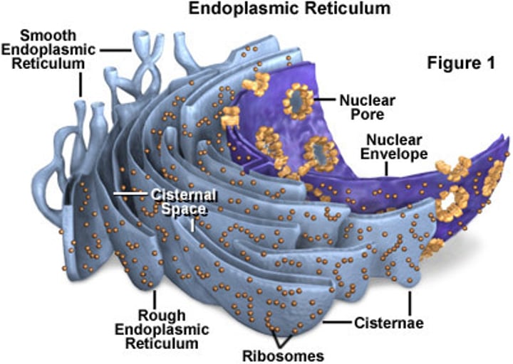
Smooth endoplasmic reticulum
Smooth ER is ER that doesn't have attached ribosomes. It is however continuous with the rough ER. It resembles multiple interconnected branches of tubules.
The smooth ER carries out diverse metabolic processes that vary by cell type. Its functions include synthesis, transport, and storage of different types of lipids, carbohydrate metabolism, and detoxification of drugs, alcohol, and poisons.
Free ribosomes
Both free and fixed/bound ribosomes are non-membrane bound organelles containing protein and ribonucleic acid that are arranged into both a large and small subunit. Free ribosomes are suspended within the cytosol.
In general, all other proteins that function within the cell that are not made by fixed/bound ribosomes are made by free ribosomes.

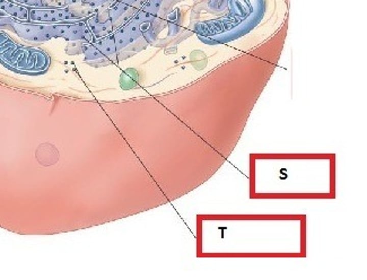
Fixed ribosomes
These are attached to the external surface of the ER membrane to form rough ER
They are used to synthesize proteins that are destined for export out of the cell, to become an integral part of the plasma membrane, or to serve as enzymes within lysosomes. (Free ribosomes synthesize all other types of proteins).
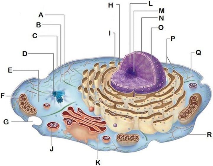
*in the image they are labeled "S". Free ribosomes are labeled "T" in the image.
Golgi apparatus/Golgi bodies/Golgi complex
The golgi apparatus is composed of about 5 elongated, flattened saclike membranous structrues called cisternae. It exhibits a distinct polarity. The two poles are called the cis-face and the trans-face. The cis-face is closer in proximity to the ER and the diameter of its flattened sac is larger compare to the trans-face. The cis-face is the receiving region and the trans-face is the shipping region.
One of the primary functions of the GA is to modify, package, and sort proteins that are made in the ER.

Golgi Vesicles
A vesicle is a small organelle within a cell, consisting of fluid enclosed by a lipid bilyaer membrane.
It stores and transports substances throughout the cell.
Examples are lysosomes (clean-up organelles) and peroxisomes (pinched off rough ER).