OTHY102 Weeks 1-5
 Call Kai
 Learn
Learn Practice Test
 Spaced Repetition
 Match
Match Flashcards
 Knowt Play
1/237

Name | Mastery | Learn | Test | Matching | Spaced | Call with Kai | Chat |
|---|
No analytics yet
Send a link to your students to track their progress
238 Terms
What are the five major peripheral nerves of the upper limb?
The five major peripheral nerves of the upper limbs are the musculocutaneous, nerve, median nerve, ulnar nerve, radial nerve and the axillary nerve
Which nerve primarily supplies the biceps brachii, brachialis and coracobrachialis (anterior compartment of the arm)?
musculocutaneous nerve
Which nerve supplies the triceps brachii and anconeus (posterior compartment of the arm)?
radial nerve
Which nerve supplies the deltoid and teres minor?
axillary nerve
Which nerve primarily supplies the triceps brachii and anconeus (posterior compartment of the arm) and the extensor muscles of the forearm (posterior compartment of the forearm)?
radial nerve
Which nerve supplies most of the flexor muscles of the forearm (anterior compartment of the forearm)?
median nerve
Which nerve supplies most of the intrinsic muscles of the hand?
ulnar nerve
What is the difference between intrinsic and extrinsic muscles?
Intrinsic muscles originate and insert within the body part they move.
Extrinsic muscles originate outside the body part they move and insert within it.
What is the difference between intrinsic and extrinsic muscles of the hand?
Intrinsic hand muscles: originate and insert within the hand.
Extrinsic hand muscles: originate in the forearm but their tendons extend into the hand to produce movement.
Which nerve supplies the deltoid muscle?
axillary nerve
Which nerve supplies the biceps brachii?
musculocutaneous nerve
Which nerve supplies the triceps brachii?
radial nerve
Which nerve passes through the carpal tunnel?
median nerve
Which major nerve passes posterior to the medial epicondyle of the humerus (the “funny bone” area)?
ulnar nerve
Which nerve is particularly vulnerable at the surgical neck of the humerus?
axillary nerve
Which nerve is particularly vulnerable in a midshaft fracture of the humerus?
radial nerve?
Which nerve provides sensation over the lateral shoulder (“regimental badge” area)?
axillary nerve
Which nerve provides cutaneous sensation to the lateral forearm?
Lateral cutaneous nerve of the forearm (terminal continuation of the musculocutaneous nerve).
Which nerve provides sensation to much of the posterior arm, posterior forearm and dorsolateral hand?
radial nerve
Which nerve provides sensation to the lateral palm and palmar surface of the lateral 3½ digits?
median nerve
Which nerve provides sensation to the medial 1½ digits of the hand?
ulnar nerve

Identify each labelled structure in the image (Scapula Anterior View).
A. Acromion
B. Coracoid Process
C. Glenoid Cavity
D. Infraglenoid Tubercle
E. Lateral Border
F. Medial Border
G. Subscapular Fossa
H. Suprascapula Notch
I. Inferior angle
J. Superior angle
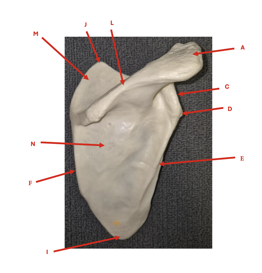
Identify each labelled structure in the image (Scapula Posterior View).
A. Acromion
C. Glenoid Cavity
D. Infraglenoid Tubercle
E. Lateral Border
F. Medial Border
I. Inferior Angle
J. Superior Angle
L. Spine of Scapula
M. Supraspinous Fossa
N. Infraspinous Fossa
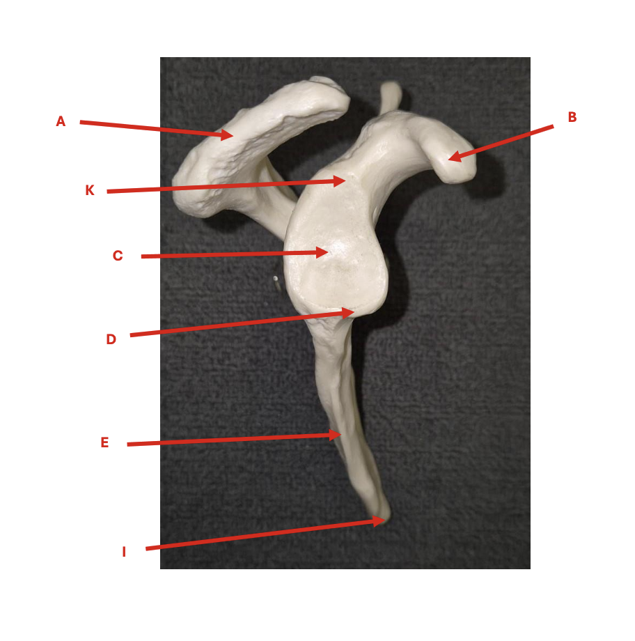
Identify each labelled structure in the image (Scapula Lateral View).
A. Acromion
B. Coracoid
C. Glenoid Cavity
D. Infraglenoid Tubercle
E. Lateral Border
I. Inferior Angle
K. Supraglenoid Tubercle

Identify each labelled structure in the image (Clavicle Superior View).
A. Sternal end
B. Acromial end
C. Curvatures

Identify each labelled structure in the image (Clavicle Inferior View)
A. Sternal end
B. Acromial end
C. Curvatures
Identify each labelled structure in the image and state the view of each humerus
Left: Anterior view
Right: Posterior view
A. Head of humerus
B. Greater Tubercle
C. Lesser Tubercle
D. Intertubecular Groove
E. Deltoid tuberosity
F. Lateral Supracondylar Ridge
G. Medial Supracondylar Ridge
H. Radial Fossa
I. Coronoid Fossa
J. Lateral Epicondyle
K. Medial Epicondyle
L. Capitulum
M. Trochlea
N. Olecranon Fossa
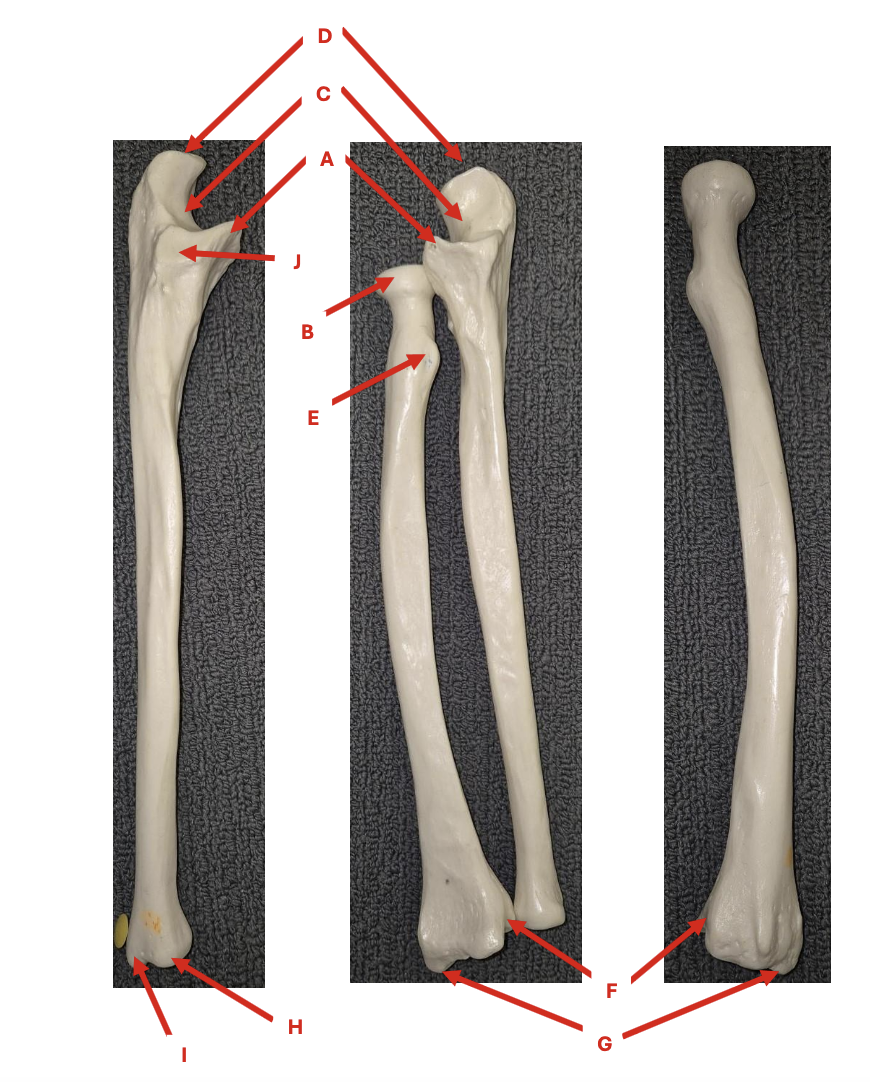
Identify each labelled structure in the image and state the view of each ulna and radius
Left: Ulna, anterior view
Middle: Ulna and radius, anterior view
Right: Radius, posterior view
A. Coronoid process
B. Radial head
C. Trochlear notch
D. Olecranon process
E. Radial tuberosity
F. Ulnar notch of radius
G. Styloid process of radius
H. Ulnar head
I. Styloid process of ulnar
J. Radial Notch of ulnar

Identify each labelled structure in the image (Hand)
A. Distal Phalange
B. Middle phalange
C. Proximal phalange
D. 1st distal phalange
E. 1st Proximal phalange
F. 1st metacarpal
G. Trapezoid
H. Trapezium
I. Scaphoid
J. Lunate
K. Pisiform
L. Triquetrum
M. Hamate
N. Capitate

Identify each labelled structure in the image (Shoulder Posterior View)
A. Acromioclavicular ligament
D. Coracoclavicular ligament (conoid)
G. Supraspinatus tendon

Identify each labelled structure in the image (Shoulder Anterior View)
A. Acromioclavicular ligament
B. Coracoacromial ligament
C. Coracoclavicular ligament (trapezoid)
D. Coracoclavicular ligament (conoid)
E. Long head of Biceps (tendon)
F. Coracohumeral ligament
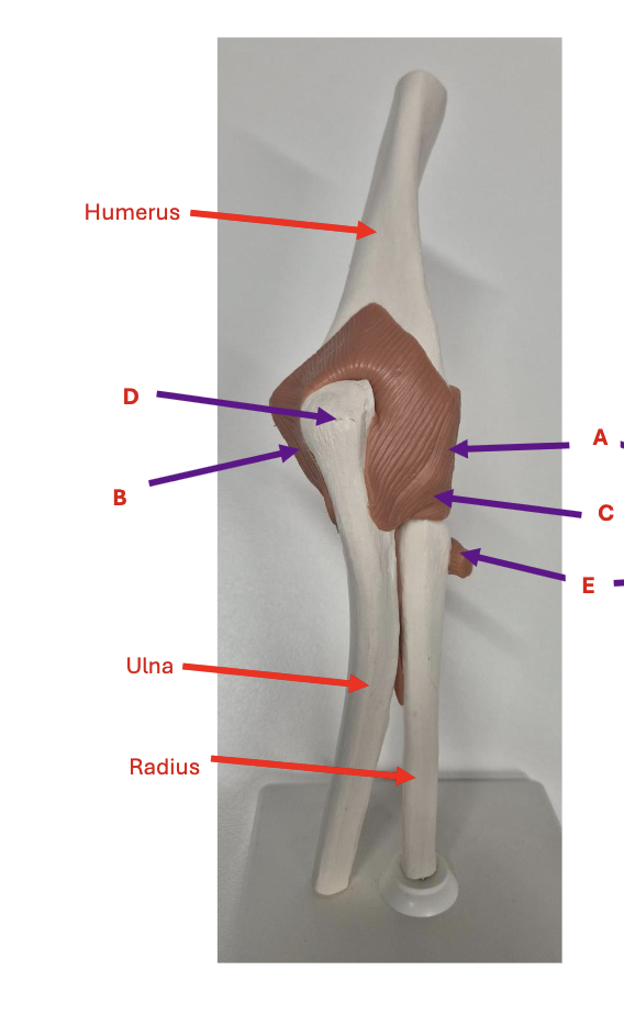
Identify each labelled structure in the image (Elbow Anterior View)
A – radial collateral ligament
B – ulnar collateral ligament
C – annular ligament
D – olecranon process
E – Tendon of Biceps

Identify each labelled structure in the image (Elbow Posterior View)
A – radial collateral ligament
B – ulnar collateral ligament
C – annular ligament
D – olecranon process
E – Tendon of Biceps

Identify each labelled structure in the image (Hand Anterior View)
A – ulna
B – radius
C – radiocarpal joint
D – ulnar collateral ligament
E – radial collateral ligament
F – 1st CMC joint (carpometacarpal)
G – 1st MCP joint (metacarpophalangeal)
H – 1st interphalangeal joint I – 5th MCP joint
J – 5th PIP joint (proximal interphalangeal)
K – 5th DIP joint (distal interphalangeal)
L – hook of hamate
M - pisiform
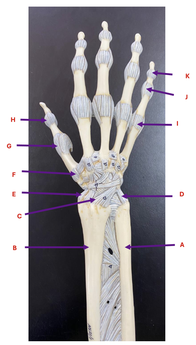
Identify each labelled structure in the image (Hand Posterior View)
A – ulna
B – radius
C – radiocarpal joint
D – ulnar collateral ligament
E – radial collateral ligament
F – 1st CMC joint (carpometacarpal)
G – 1st MCP joint (metacarpophalangeal)
H – 1st interphalangeal joint I – 5th MCP joint
J – 5th PIP joint (proximal interphalangeal)
K – 5th DIP joint (distal interphalangeal)
L – hook of hamate
M - pisiform
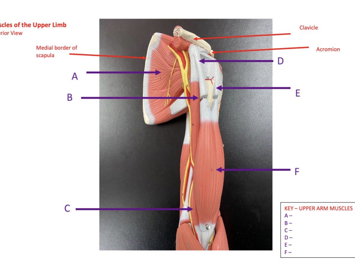
Identify each labelled structure in the image (Upper Arm Muscles Anterior View)
A – Subscapularis
B – Coracobrachialis
C – Brachialis
D – Biceps brachii – short head tendon
E – Biceps brachii – long head tendon
F – Biceps brachii – muscle belly

Identify each labelled structure in the image (Upper Arm Muscles Anterior View)
A – Subscapularis
B – Coracobrachialis
C – Brachialis
D – Biceps brachii – short head tendon
E – Biceps brachii – long head tendon
F – Biceps brachii – muscle belly

Identify each labelled structure in the image (Upper Arm Muscles Posterior View)
G – Supraspinatus
H – Infraspinatus
I – Teres minor
J – Teres major
K – Triceps brachii (lateral head)
L – Triceps (long head)

Identify each labelled structure in the image (Upper Arm Muscles Posterior View)
C – Brachialis
F – Biceps brachii – muscle belly
G – Supraspinatus
H – Infraspinatus
I – Teres minor
J – Teres major
K – Triceps brachii (lateral head)
L – Triceps (long head)
M – Tendon of Triceps
N – posterior deltoid
O – middle deltoid

Identify each labelled structure in the image (Muscles of the Forearm Anterior View)
A – pronator teres
B – flexor carpi radialis
C – palmaris longus
D – flexor carpi ulnaris
E – flexor retinaculum
F – brachioradialis
G – extensor carpi radialis longus

Identify each labelled structure in the image (Muscles of the Forearm Posterior View)
F – brachioradialis
G – extensor carpi radialis longus
H – extensor carpi radialis brevis
I – extensor carpi ulnaris
J – extensor digitorum
K – extensor digiti minimi
L – extensor retinaculum
M – anconeus
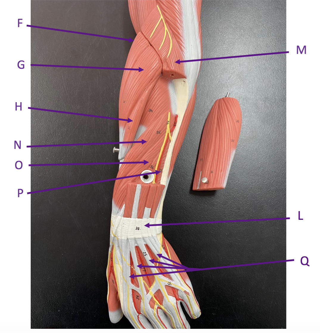
Identify each labelled structure in the image (Muscles of the Forearm Posterior View)
F – brachioradialis
G – extensor carpi radialis longus
H – extensor carpi radialis brevis
L – extensor retinaculum
M – anconeus
N – abductor pollicis longus
O – extensor pollicis longus
P – extensor indicis
Q – tendons of extensor digitorum
What are the major groups of intrinsic muscles of the hand?
Thenar muscles, hypothenar muscles, lumbricals and interossei.
What are the three thenar muscles?
Abductor pollicis brevis, flexor pollicis brevis and opponens pollicis.
What is the primary function of abductor pollicis brevis?
ABduction of the thumb
What is the primary function of flexor pollicis brevis?
Flexion of the thumb
What is the primary function of opponens pollicis?
Opposition of the thumb
What is the primary action of adductor pollicis?
ADduction of the thumb.
What are the three hypothenar muscles?
Abductor digiti minimi, flexor digiti minimi brevis and opponens digiti minimi.
What is the primary function of abductor digiti minimi?
ABduction of the little finger
What is the primary function of flexor digiti minimi brevis?
Flexion of the little finger
What is the primary function of opponens digiti minimi?
Opposition of the little finger
How many lumbricals are in the hand?
Four
What do the lumbricals do?
Flex the MCP joints and extend the IP joints of digits 2–5
How many dorsal interossei are in the hand?
Four
What is the primary action of the dorsal interossei?
ABduct digits 2–4 away from the middle finger
How many palmar interossei are in the hand?
Three
What is the primary action of the palmar interossei?
ADduct digits 2, 4 and 5 toward the middle finger.
What mnemonic distinguishes the actions of the dorsal and palmar interossei?
DAB = Dorsal ABduct. PAD = Palmar ADduct.
What is a retinaculum?
A band of connective tissue that holds tendons close to the underlying bones and prevents bowstringing during movement.
Where is the flexor retinaculum located?
On the anterior (palmar) side of the wrist.
What is the main function of the flexor retinaculum?
Holds the flexor tendons close to the wrist and forms the roof of the carpal tunnel.
Where is the extensor retinaculum located?
On the posterior (dorsal) side of the wrist.
What is the main function of the extensor retinaculum?
Holds the extensor tendons close to the wrist and prevents bowstringing during extension.
What is the palmar aponeurosis?
A thick triangular sheet of connective tissue in the palm.
What is the main function of the palmar aponeurosis?
Protects underlying structures and helps anchor the skin of the palm.
What is the extensor expansion (extensor hood)?
A connective-tissue expansion on the dorsal surface of the fingers that receives tendons from the extensor and intrinsic hand muscles.
What is the function of the extensor expansion?
Coordinates extension of the fingers, particularly at the interphalangeal joints.
What are synovial tendon sheaths?
Fluid-filled sheaths surrounding tendons that reduce friction as the tendons move.
What is the purpose of the flexor tendon sheaths in the hand?
They allow the flexor tendons to glide smoothly during finger movement.
What is the digital sweep of the fingers?
During finger flexion, the fingertips move toward the scaphoid tubercle.
What is the resting position of the hand?
Wrist slightly extended, fingers slightly flexed, and thumb slightly abducted and flexed.
What is the functional position of the hand?
Wrist extended, MCP joints flexed, IP joints slightly flexed, and thumb positioned for opposition.
Why is wrist extension important for grip?
Wrist extension places the finger flexors in a more effective position to generate grip force.
What are the two broad categories of grip?
Power grip and precision grip.
What is a power grip?
A grip in which an object is held against the palm by the flexed fingers and thumb to generate force.
What is a precision grip?
A grip in which an object is controlled mainly between the thumb and fingertips for fine manipulation.
What is an example of a power grip?
Holding an object such as a hammer or cylindrical handle.
What is an example of a precision grip?
Holding a small object such as a pen or key between the thumb and fingers.
Which digit forms the reference axis for abduction and adduction of the fingers?
Digit 3 (middle finger).
What is finger abduction?
Movement of the fingers away from digit 3.
What is finger adduction?
Movement of the fingers toward digit 3.
What is thumb opposition?
Movement of the thumb across the palm so its pad can contact the pads of the other digits.
What is thumb reposition?
Movement of the thumb from opposition back toward its anatomical position.
Why are the movement planes of the thumb different from those of the fingers?
The thumb is rotated approximately 90° relative to the fingers, changing the planes in which its movements occur.
In which plane does thumb flexion and extension occur?
Frontal (coronal) plane.
In which plane does thumb abduction and adduction occur?
Sagittal plane.
What are the thenar and hypothenar eminences?
Thenar eminence: muscular prominence at the base of the thumb. Hypothenar eminence: muscular prominence at the base of the little finger.
What are the three main types of power grip?
Cylindrical, spherical and hook grips.
What are the main types of precision grip covered in the lecture?
Lateral pinch/prehension, pincer, pad-to-pad (pinch), and lumbrical/plate grip.
What structures help open the hand during release of grip?
Extrinsic finger extensors, thumb abductors, thumb extensors, intertendinous connections and the dorsal digital expansion.
What are intertendinous connections?
Fibrous connections between extensor digitorum tendons near the MCP joints.
What is the functional role of intertendinous connections?
They limit completely independent finger extension and help the fingers open together during hand release.
What are the three groups of bones in the hand?
Carpals, metacarpals and phalanges.
What are the carpal bones in the proximal row, lateral to medial?
Scaphoid, lunate, triquetrum, pisiform.
What are the carpal bones in the distal row, lateral to medial?
Trapezium, trapezoid, capitate, hamate.
Which carpal bones have important palpable projections?
Scaphoid — tubercle
trapezium — tubercle
hamate — hook.
What are the three main parts of a metacarpal or phalanx?
Base, shaft and head.
What type of joint is the radiocarpal (wrist) joint?
Synovial, biaxial, condyloid (ellipsoid).
What movements occur at the wrist?
Flexion, extension, abduction, adduction and circumduction.