MRI Spine Anatomy
1/74
Earn XP
Description and Tags
Questions
Name | Mastery | Learn | Test | Matching | Spaced |
|---|
No study sessions yet.
75 Terms

Letter G in Image 37 is pointing to:
A. Spinal canal
B. Abdominal aorta
C. Intervertebral disc
D. Spinous process
A. Spinal canal

Image 52 is a ____ weighted image acquired in the ____ scan plane.
A. T1; Axial
B. T1; Sagittal
C. T2; Axial
D. T2; Coronal
E. STIR; Axial
C. T2; Axial

Letter A in Image 37 is pointing to:
A. Inferior vena cava
B. Azygos vein
C. Abdominal aorta
D. Psoas muscle
C. Abdominal aorta

Letter H in Image 55 is pointing to:
A. 6th thoracic vertebrae
B. 7th thoracic vertebrae
C. 8th Thoracic vertebrae
D. 9th thoracic vertebrae
E. 10th thoracic vertebrae
B. 7th thoracic vertebrae

Letter G in Image 19 is pointing to:
A. Ventral nerve root
B. Dorsal nerve root
C. Vertebral body
D. Spinous process
E. Cerebrospinal fluid (CSF)
D. Spinous process

Letter B in Image 36 is pointing to:
A. Body of L1
B. Body of L3
C. Body of L5
D. Body of S1
D. Body of S1

Letter A in Image 53 is pointing to:
A. Ligamentum flavum
B. Spinous ligament
C. Spinal cord
D. Cerebrospinal fluid
E. spinous process
C. Spinal cord

Letter E in Image 37 is pointing to:
A. Facet joint
B. Lamina
C. Transverse process
D. Spinous process
B. Lamina

Letter B in Image 132 is pointing to:
A. Lamina
B. Pedicle
C. Facet joint
D. Spinal canal
E. Spinous process
B. Pedicle

Image 36 is a ___ weighted image acquired in the _____ scan plane.
A. T1; Axial
B. T1; Sagittal
C. T2; Axial
D. T2; Sagittal
E. STIR; Sagittal
D. T2; Sagittal

Letter J in Image 132 is pointing to:
A. Lamina
B. Pedicle
C. Facet joint
D. Zygapophyseal joint
E. Nerve roots
A. Lamina

Letter D in Image 53 is pointing to:
A. Ligamentum flavum
B. Spinous ligament
C. Spinal cord
D. Cerebrospinal fluid
E. Spinous process
D. Cerebrospinal fluid
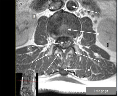
Image 37 was acquired at what anatomical position?
A. L1-2 disc space
B. L2-3 disc space
C. L4-5 disc space
D. L5-S1 disc space
B. L2-3 disc space

Letter B in Image 53 is pointing to:
A. Ligamentum flavum
B. Spinous ligament
C. Spinal cord
D. Cerebrospinal fluid
E. Spinous process
A. Ligamentum flavum

Letter J in Image 18 is pointing to:
A. Anterior longitudinal ligament (ALL)
B. Posterior longitudinal ligament (PLL)
C. Spinous ligament
D. Venous plexus
A. Anterior longitudinal ligament (ALL)

Letter B in Image 18 is pointing to:
A. Posterior arch of C1
B. Cerebellum
C. Spinal cord
D. Fourth ventricle
E. Cerebrospinal fluid (CSF)
B. Cerebellum

Letter B in Image 19 is pointing to:
A. Ventral nerve root
B. Dorsal nerve root
C. Spinal cord
D. Transverse process
E. Cerebrospinal fluid (CSF)
C. Spinal cord

Letter H in Image 18 is pointing to:
A. Anterior longitudinal ligament (ALL)
B. Posterior longitudinal ligament (PLL)
C. Spinous ligament
D. Venous plexus
B. Posterior longitudinal ligament (PLL)

Letter J in Image 55 is pointing to:
A. Abdominal aorta
B. Vertebral body
C. Suprasternal notch
D. Spinous process
E. Xiphoid process
C. Suprasternal notch

Letter F in Image 55 is pointing to:
A. Vertebral body
B. Spinal cord
C. Intervertebral disk
D. Spinous process
E. Cerebrospinal fluid (CSF)
C. Intervertebral disk
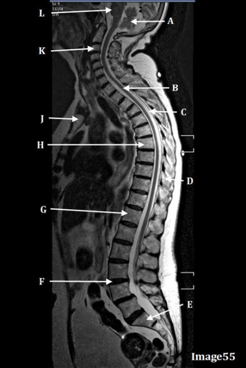
Letter B in Image 55 is pointing to:
A. Medulla oblongata
B. Spinal cord
C. Cerebrospinal fluid (CSF)
D. Spinous process
E. Cerebellum
B. Spinal cord

Letter B in Image 52 is pointing to:
A. Spinous process
B. Transverse process
C. Spinal cord
D. Cerebrospinal fluid
E. Azygos vein
C. Spinal cord

Letter C in Image 52 is pointing to:
A. Spinous process
B. Transverse process
C. Spinal cord
D. Cerebrospinal fluid
E. Azygos vein
A. Spinous process
Letter F in Image 132 is pointing to:
A. Lamina
B. Pedicle
C. Facet joint
D. Zygapophyseal joint
E. C and/or D
E. C and/or D (Facet joint and/or Zygapophyseal joint)

Letter G in Image 18 is pointing to:
A. Anterior longitudinal ligament (ALL)
B. Posterior longitudinal ligament (PLL)
C. Spinous ligament
D. Venous plexus
D. Venous plexus

Letter A in Image 132 is pointing to:
A. L3 vertebral body
B. L5 vertebral body
C. L3-4 intervertebral disc
D. L4-5 intervertebral disc
E. L5-S1 intervertebral disc
D. L4-5 intervertebral disc

Letter C in Image 55 is pointing to:
A. Medulla oblongata
B. Spinal cord
C. Cerebrospinal fluid (CSF)
D. Spinous process
E. Cerebellum
C. Cerebrospinal fluid (CSF)

Letter G in Image 55 is pointing to:
A. 10th thoracic vertebrae
B. 11th thoracic vertebrae
C. 12th Thoracic vertebrae
D. 1st lumbar vertebrae
E. 2nd lumbar vertebrae
C. 12th Thoracic vertebrae

Letter B in Image 37 is pointing to:
A. Facet joint
B. Intervertebral disc
C. Transverse process
D. Spinous process
E. Spinal cord
B. Intervertebral disc

Letter K in Image 55 is pointing to:
A. Foramen magnum
B. 1st cervical vertebrae
C. 2nd cervical vertebrae
D. 3rd cervical vertebrae
E. 4th cervical vertebrae
D. 3rd cervical vertebrae

Letter D in Image 55 is pointing to:
A. Medulla oblongata
B. Spinal cord
C. Cerebrospinal fluid (CSF)
D. Spinous process
E. Cerebellum
D. Spinous process

Letter D in Image 52 is pointing to:
A. Ligamentum flavum
B. Lamina
C. Azygos vein
D. Spinous process
E. Posterior longitudinal ligament
B. Lamina

Letter C in Image 37 is pointing to:
A. Facet joint
B. Intervertebral disc
C. Transverse process
D. Spinous process
E. Spinal cord
A. Facet joint

Letter D in Image 37 is pointing to:
A. Facet joint
B. Intervertebral disc
C. Transverse process
D. Spinous process
E. Spinal cord
D. Spinous process

Letter D in Image 132 is pointing to:
A. Lamina
B. Pedicle
C. Facet joint
D. Cerebrospinal fluid
E. Nerve roots
F. Abdominal aorta
D. Cerebrospinal fluid

Letter E in Image 18 is pointing to:
A. Posterior arch of C1
B. Cerebellum
C. Spinal cord
D. Fourth ventricle
E. Cerebrospinal fluid (CSF)
E. Cerebrospinal fluid

Letter E in Image 55 is pointing to:
A. S1 vertebral body
B. L5 vertebral body
C. Intervertebral disk
D. L4 vertebral body
E. S2 vertebral body
A. S1 vertebral body
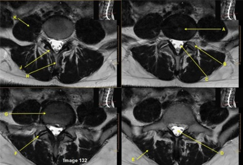
Letter H in Image 132 is pointing to:
A. Lamina
B. Pedicle
C. Facet joint
D. Spinal canal
E. Spinous process
E. Spinous process

Letter E in Image 132 is pointing to:
A. Infraspinatus muscle
B. Psoas muscle
C. Erector spinae muscle
D. Abdominus rectus muscle
C. Erector spinae muscle

Letter D in Image 17 is pointing to:
A. Vertebral body
B. Intervertebral disc space
C. Spinal cord
D. Spinous process
A. Vertebral body

Letter A in Image 55 is pointing to:
A. Medulla oblongata
B. Spinal cord
C. Cerebrospinal fluid (CSF)
D. Spinous process
E. Cerebellum
E. Cerebellum

Letter A in Image 36 is pointing to:
A. L1-2 HNP
B. L2-3 HNP
C. L4-5 HNP
D. L5-S1 HNP
C. L4-5 HNP

Letter D in Image 36 is pointing to:
A. L1-2 disc space
B. L2-3 disc space
C. L4-5 disc space
D. L5-S1 disc space
B. L2-3 disc space

Letter F in Image 53 is pointing to:
A. Sternum
B. Vertebral body
C. Suprasternal notch
D. Intervertebral disc space
E. Xiphoid process
A. Sternum

Letter A in Image 19 is pointing to:
A. Ventral nerve root
B. Dorsal nerve root
C. Spinal cord
D. Transverse process
E. Cerebrospinal fluid (CSF)
E. Cerebrospinal fluid (CSF)

Letter H in Image 19 is pointing to:
A. Transverse process
B. Lamina
C. Dorsal nerve root
D. Ventral nerve root
E. Spinous process
F. Vertebral body
A. Transverse process

Letter A in Image 52 is pointing to:
A. Esophagus
B. Inferior vena cava
C. Azygos vein
D. Descending thoracic aorta
E. Spinal cord
D. Descending thoracic aorta

Letter G in Image 132 is pointing to:
A. L3 vertebral body
B. L5 vertebral body
C. L3-4 intervertebral disc
D. Zygapophyseal joint
E. L5-S1 intervertebral disc
B. L5 vertebral body

Letter D in Image 19 is pointing to:
A. Ventral nerve root
B. Dorsal nerve root
C. Spinous ligament
D. Transverse process
E. Cerebrospinal fluid (CSF)
B. Dorsal nerve root

Letter C in Image 132 is pointing to:
A. Lamina
B. Pedicle
C. Facet joint
D. Zygapophyseal joint
E. Nerve roots
E. Nerve roots

Letter H in Image 37 is pointing to:
A. Trachea
B. Abdominal aorta
C. Interior vena cava
D. Right external iliac artery
C. Inferior vena cava
The ________ is posterior to the vertebral body and consists of two pedicles and two laminae.
A. Vertebral notch
B. Vertebral arch
C. Transverse foramen
D. Spinous process
B. Vertebral arch

Letter E in Image 19 is pointing to:
A. Ventral nerve root
B. Dorsal nerve root
C. Spinal cord
D. Transverse process
E. Cerebrospinal fluid (CSF)
A. Ventral nerve root
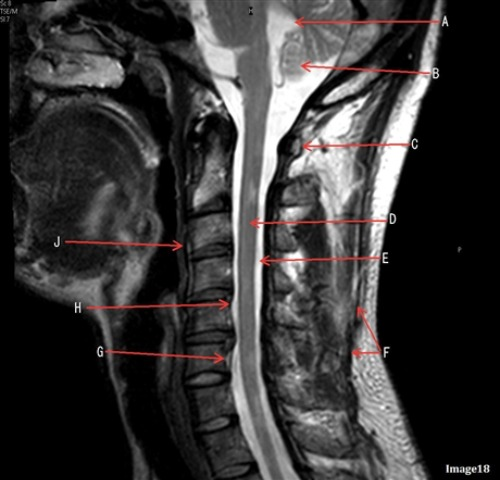
Letter A in Image 18 is pointing to:
A. Posterior arch of C1
B. Cerebellum
C. Spinal cord
D. Fourth ventricle
E. Cerebrospinal fluid (CSF)
D. Fourth ventricle

Letter A in Image 17 is pointing to:
A. Vertebral body
B. Intervertebral disc space
C. Spinal cord
D. Spinous process
D. Spinous process

Letter L in Image 55 is pointing to:
A. Vertebral body
B. Pons
C. Spinal cord
D. Fourth ventricle
E. Cerebellum
B. Pons
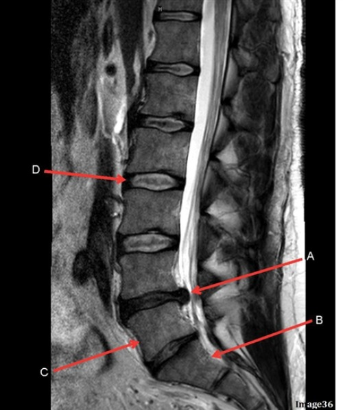
Letter C in Image 36 is pointing to:
A. Body of L1
B. Body of L3
C. Body of L5
D. Body of S1
C. Body of L5
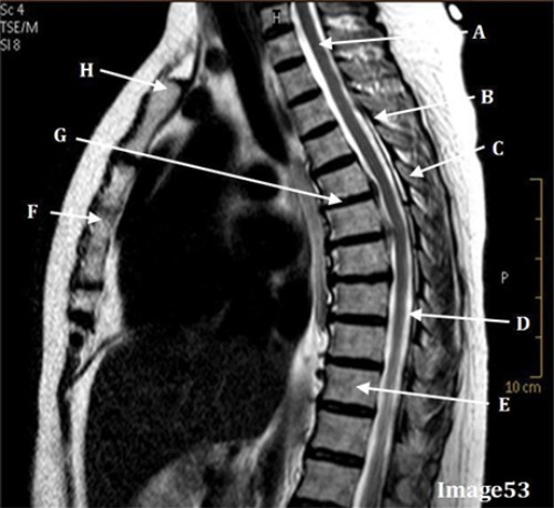
Image 53 is a ____ weighted image acquired in the ____ scan plane.
A. T1; Axial
B. T1; Sagittal
C. T2; Axial
D. T2; Sagittal
E. STIR; Sagittal
D. T2; Sagittal
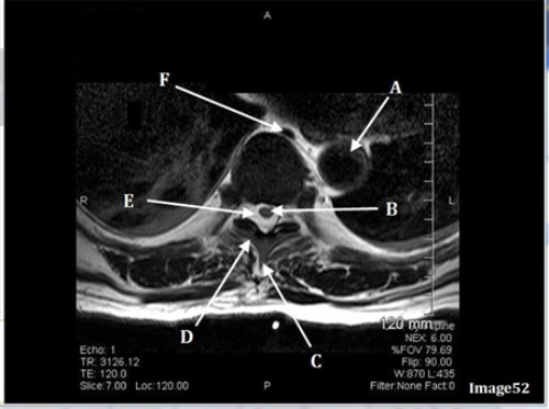
Letter F in Image 52 is pointing to:
A. Abdominal aorta
B. Inferior vena cava
C. Spinal cord
D. Cerebrospinal fluid
E. Azygos vein
E. Azygos vein

Letter H in Image 53 is pointing to:
A. Sternum
B. Vertebral body
C. Suprasternal notch
D. Intervertebral disc space
E. Xiphoid process
C. Suprasternal notch

Letter D in Image 18 is pointing to:
A. Posterior arch of C1
B. Cerebellum
C. Spinal cord
D. Fourth ventricle
E. Cerebrospinal fluid (CSF)
C. Spinal cord

Letter K in Image 132 is pointing to:
A. Infraspinatus muscle
B. Psoas muscle
C. Erector Spinae muscle
D. Abdominus rectus muscle
B. Psoas muscle
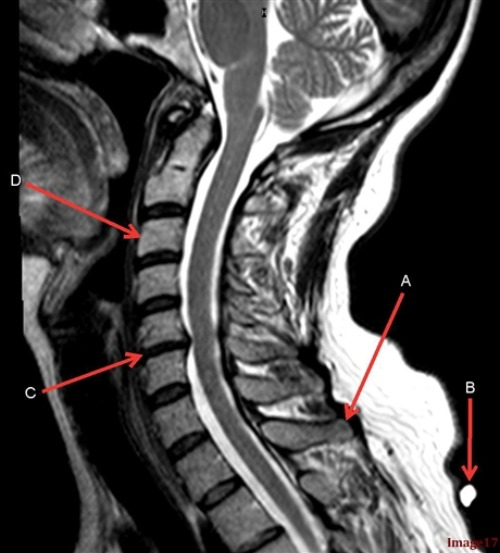
Letter C in Image 17 is pointing to:
A. Vertebral body
B. Intervertebral disc space
C. Spinal cord
D. Spinous process
B. Intervertebral disc space

Letter C in Image 53 is pointing to:
A. Ligamentum flavum
B. Spinous ligament
C. Spinal cord
D. Cerebrospinal fluid
E. Spinous process
E. Spinous process
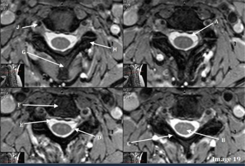
Letter C in Image 19 is pointing to:
A. transverse process
B. Lamina
C. Dorsal nerve root
D. Ventral nerve root
E. Spinous process
B. Lamina

Letter J in Image 19 is pointing to:
A. Jugular vein
B. Iliac artery
C. Vertebral artery
D. Cerebrospinal fluid
E. Fourth ventricle
C. Vertebral artery

Letter E in Image 53 is pointing to:
A. Ligamentum flavum
B. Vertebral body
C. Spinal cord
D. Intervertebral disc space
E. Spinous process
B. Vertebral body
which spinal ligament is vertically oriented, connecting the dorsal tips of the spinous processes?
A. Ligamentum flavum
B. Supraspinous ligament
C. Interspinous ligament
D. Intertransverse ligament
B. Supraspinous ligament

Letter F in Image 37 is pointing to:
A. Spinous process
B. Superior articular process
C. Lamina
D. Transverse process
B. Superior articular process

Letter C in Image 18 is pointing to:
A. Posterior arch of C1
B. Cerebellum
C. Spinal cord
D. Fourth ventricle
E. Cerebrospinal fluid (CSF)
A. Posterior arch of C1

Letter E in Image 52 is pointing to:
A, Spinous process
B. Transverse process
C. Spinal cord
D. Cerebrospinal fluid
E. Azygos vein
D. Cerebrospinal fluid

Letter F in Image 18 is pointing to:
A. Anterior longitudinal ligament (ALL)
B. Posterior longitudinal ligament (PLL)
C. Spinous ligament
D. Venous plexus
C. Spinous ligament
What is the intervertebral disc’s outer ring of concentric fibrous layer termed
A. nucleus pulposus
B. Zygapophyseal joint
C. Centrum
D. Anulus fibrosis
D. Anulus fibrosis

Letter F in Image 19 is pointing to:
A. Ventral nerve root
B. Dorsal nerve root
C. Vertebral body
D. Spinous process
E. Cerebrospinal fluid (CSF)
C. Vertebral body

Letter G in Image 53 is pointing to:
A. Sternum
B. Vertebral body
C. Suprasternal notch
D. Intervertebral disc space
E. Xiphoid process
D. Intervertebral disc space