Biology topic 2 part 2- Genes and health
1/79
There's no tags or description
Looks like no tags are added yet.
Name | Mastery | Learn | Test | Matching | Spaced | Call with Kai |
|---|
No analytics yet
Send a link to your students to track their progress
80 Terms
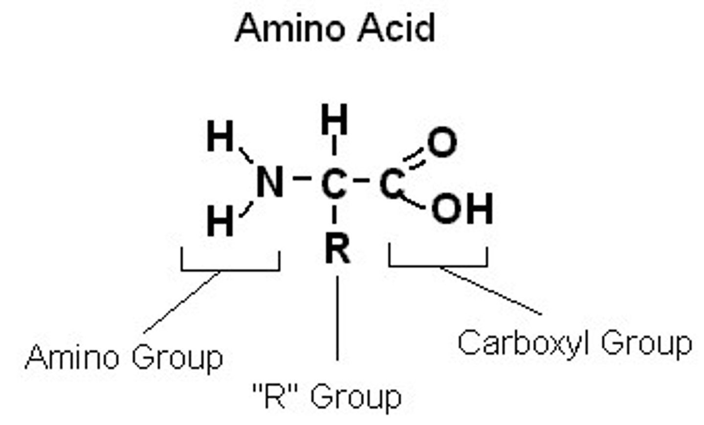
Amino acid structure
Central carbon atom
Amino group
Carboxyl group
Single hydrogen
Variable R group
How can R groups on amino acids differ
Size, polarity and charge
What are the two main shapes of the tertiary structure
alpha helix and beta plated sheets
What is the secondary structure of a protein?
The curling or folding of the polypeptide chain into alpha helices and beta pleated sheets due to the formation of hydrogen bonds
Tertiary structure
The third level of protein structure; the overall, three-dimensional shape of a polypeptide due to interactions of the R groups of the amino acids making up the chain.
What bonds are formed in the tertiary structure
Ionic bonds between oppositely charged R groups in different amino acids in the polypeptide chain.
Disulphide bridges between R groups containing Sulphur. Hydrogen bonds between slightly charged H and slightly charge O/N (delta negative) in different amino acids.
Quaternary structure
The specific 3D shape of a protein that is determined by multiple polypeptide chains and/or prosthetic groups bonded together
What is a globular protein
A protein with a spherical shape that is soluble in water and typically have metabolic roles
Why are globular proteins soluble in water
The hydrophilic groups are on the outside, so they are soluble in water.
What are the roles of globular proteins
- Transport proteins
- Hormones
- Enzymes
What are fibrous proteins
Structural proteins. The long chains run parallel to one another and are linked by cross bridges and so form very stable molecules.
How does the induced fit model work
The active site forms as the enzyme and substrate interact. The enzyme is flexible and can mould itself around its substrate. As the enzyme changes shape, it puts a strain on the substrate, which distorts a particular bond or bonds in the substrate and lowers the activation energy needed to break the bond.
What is a conjugated protein
One that gets part of their function from a prosthetic group.
What is the phospholipid bilayer
a two-layered arrangement of phosphate and lipid molecules that form a cell membrane, the hydrophobic lipid ends facing inward and the hydrophilic phosphate ends facing outward.
What 3 arrangements can phospholipids have in water
Phospholipid bilayer, spherical (micelles) and a monolayer on the surface
Why is the phosphate head in phospholipids hydrophilic
It is polar, so attracts other polar molecules, like water
Why is a bilayer the favoured structure of phospholipids in water
The two fatty acids are too large to fit into the interior of a micelle
What is the fluid mosaic model
states that a membrane is a fluid structure with a "mosaic" of various proteins embedded in it
What is the cell surface membrane made up of
Phospholipid bilayer with proteins, cholesterol, glycoproteins and glycolipids embedded throughout the structure
What are glycoproteins and glycolipids
Proteins and lipids with polysaccharides attached
What happens when there is an increased amount of unsaturated fatty acids in the cell membrane
It becomes more fluid because the kinks in the hydrocarbon chains prevent them from packing closely together, so more movement is possible
Roles of glycoproteins and glycolipids
Involved in cell recognition, cell signalling and cell attachment
Role of cholesterol in cell membranes
Controls membrane fluidity. The more cholesterol, the less fluid the membrane is and the less permeable. It is very important for keeping membranes stable at body temperature, without it cells would burst open.
Why is it important for a membrane to be fluid
- Diffusion of substances across a membrane
- Fusion of membranes e.g. exocytosis
- Membranes to move and change shape e.g. phagocytosis
If lipids in the cell membrane have longer tails, does this increase or decrease membrane fluidity
Decrease because they have higher melting point
How does cholesterol affect membrane fluidity
- At body temperature, it interacts with the tails, decreasing fluidity
- At low temperatures it prevents it from solidifying
- At normal body temperature, a low level of cholesterol can make the membrane too fluid, causing it to burst
What are integral proteins
Proteins that penetrate the hydrophobic core.
What are peripheral proteins
bound to the surface of the membrane or bound to an integral protein
Peripheral protein roles
- Peripheral proteins on the extracellular side of the membrane act as receptors for hormones or neurotransmitters, or are involved in cell recognition. Many are glycoproteins.
- Peripheral proteins on the cytosolic side of the membrane are involved in cell signalling or chemical reactions. They can dissociate from the membrane and move into the cytoplasm.
Integral proteins roles
Can transport molecules or ions across the membrane or have an enzymatic function.
Facilitated diffusion
Hydrophilic molecules and ions that are larger cannot diffuse through the phospholipid bilayer, as they are insoluble in lipids, so cannot move through the barrier. They can diffuse through the cell membrane either via channel proteins or carrier proteins
Channel proteins
Water filled pores that span the membrane that allow specific water soluble ions to pass through. The ions bind with the protein causing it to change shape in a way that closes it to one side of the membrane and opens it to the other
Carrier proteins
The ion or molecule binds to a specific point on the carrier protein, which changes the shape, allowing the ion or molecule to move across the membrane
Osmosis
Movement of water molecules from a solution with a lower concentration of solute, to a solution with a higher concentration of solute through a partially permeable membrane
What is meant by isotonic
When two solutions have the same water potential - therefore there is no net movement of water molecules
Active transport
The carrier proteins span the plasma membrane and bind to the molecule or ion to be transported on one side of it.
On the inside of the cell/organelle, ATP binds to the protein, causing it to split into ADP and a phosphate molecule. This releases energy. As a result the protein molecule changes shape and opens to the opposite side of the membrane.
The phosphate molecule is released from the protein which causes the protein to revert to its original shape, ready for the process to be repeated. The phosphate molecule then recombines with the ADP to form ATP during respiration.
6 main functions of membrane proteins
- Transport
- Enzymic activity
- Signal transduction
- Cell-cell recognition
- Intercellular joining
- Attachment to the cytoskeleton and extracellular matrix
enzymic membrane proteins
A protein built into the membrane may be an enzyme with its active site exposed to substances in the adjacent solution. In some cases, several enzymes in a membrane are organized as
a team that carries out sequential steps of a metabolic pathway.
Signal transduction membrane proteins
A membrane protein may have a binding site with a specific shape that fits the shape of a chemical messenger, such as a hormone. The external messenger (signal) may cause a conformational change in the protein (receptor) that relays the message to the inside of the cell.
Cell-cell recognition membrane proteins
Some glyco-proteins serve as identification tags that are specifically recognized by other cells.
Intercellular joining
Membrane proteins of adjacent cells may hook together in various kinds of junctions, such as gap junctions or tight junctions
Attachment to the cytoskeleton and extracellular matrix
Microfilaments or other elements of the cytoskeleton may be bonded to membrane proteins, a function that helps maintain cell shape and stabilizes the location of certain membrane proteins. Proteins that adhere to the ECM can coordinate extracellular and intracellular changes
When are exocytosis and endocytosis needed
When large quantities of molecules or large molecules need to be transported across a membrane
Exocytosis
Used in the release of substances across a cell surface membrane, usually proteins or polysaccharides. Vesicles fuse with the cell membrane and the contents are released.
Endocytosis
Substances are taken in by the cell by the creation of vesicles from the cell surface membrane. Part of the cell membrane engulfs the material to be transported. In some cases the substance to be absorbed attaches to a receptor in the cell membrane and is then absorbed by endocytosis.
What is a vesicle
A small, membrane bound sac
Process of inspiration
Diaphragm contracts so it flattens and lowers, external intercostal muscles contract, ribs moved up and out, volume of thorax increases, pressure in thorax reduced so it is lower than air pressure, air drawn through the gaseous exchange system to equalise the pressure gradient
Process of expiration
The internal intercostal muscles contract while the external intercostal muscles relax, the ribs move downwards and inwards, decreasing the volume of the thorax. The diaphragm muscles relax decreasing the volume of the thorax. The decreased volume of the thorax increases the pressure in the lungs. The pressure is now greater than that of the atmosphere, and so air is forced out of the lungs.
Role of goblet cells in the tubes of the gas exchange system
continuously produce mucus
Role of cilia in gas exchange
continuously remove mucus that cover the epithelial cells lining the tubes of the gas exchange system
Epithelial cells
skin cells that cover the outside of the body and line the internal surfaces of organs. Work together as epithelium (a tissue), which consists of one or more layers of cells sitting on a basement membrane
What is the basement membrane
The layer that anchors epithelial tissue to connective tissues. It is made of protein fibres in a jelly-like protein-carbohydrate matrix.
Basal membrane
Membrane surface of epithelial cells that face the basement membrane
Apical membrane
Membrane surface of epithelial cells that faces away from the basement membrane
Types of epithelia
squamous and columnar
Squamous epithelia
Found in the lining of the alveoli and capillaries, very thin (less than 0.2 micrometers)
Columnar epithelia
Found in the small intestine and in gas exchange tubes. Epithelial cells extend out from the basement membrane.
What does pseudostratified mean
false stratified; all cells touch basement membrane
Alveoli properties
One cell thin, large surface area, covered in capillaries, moist,
Properties that effect rate of gas exchange
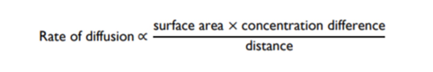
Surface area, concentration gradient and thickness of the exchange surface
Ficks law
law stating that the net diffusion rate of a gas across a fluid membrane is proportional to the difference in partial pressure, proportional to the area of the membrane, and inversely proportional to the thickness of the membrane
Gills
Series of bony gill arches each with two stacks of gill filaments. They have protruding rows of very thin lamellae, which each consist of a network of capillaries covered by a single layer of epithelial cells
Why do gills have only a single layer of epithelial cells
To allow for a short diffusion distance
What is the name of the bony plate that protects the gills
operculum
How do fish ventilate their gills
-The pumping action of their buccal cavity and operculum creates a pressure gradient that moves water over their gills
-Fast swimming fish just swim with their mouth open to breathe
What is countercurrent flow
When the blood flows in the opposite direction to the water
Why is countercurrent flow essential in fish
It maintains a high concentration gradient and ensures maximum diffusion of oxygen into the blood from the water. Parallel flow would result in both the blood and the water having equal amounts of oxygen.
Gas exchange in insects
Tracheal system
- Gas enters through the spiracles, which then passes into the trachea, which branches into tracheoles.
- The tracheoles contain fluid which seep into them from surrounding cells when the insect is resting
- When active, the muscles draw up the tracheal fluid which provides them with oxygen for respiration.
- The pressure in the tracheoles lower, drawing in more air through the spiricals. SA available for oxygen to diffuse through the tracheal walls directly will increase
Why does an insects exoskeleton allow for efficient gas exchange
It is thick and waxy, so not a good substance for things to diffuse through
How do larger insects ventilate their tracheal systems
- Air sacs can be squeezed by flight muscles to push air in and out
- Flight muscles can alter the volume of the insects thorax
- Some insects have a specialised breathing mechanism. As they expand their abdomen, they close spiracles at the back end of their body. When they contract their abdomen, they open up the spiracles at the rear and close those at the front.
Overton
Discovered that membranes are made of lipids. Discovered similarities between lipids and membranes and that lipid-soluble molecules moved through cells much faster than others
Langmuir
Experimented with the interaction between oil and water. Proposed that lipids form a monolayer on water
Gorter and Grendel
Extracted lipids from red blood cells and found that the surface area of the lipids was twice the surface area of the cell, so proposed membranes are made up of a phospholipid bilayer
Davson and Danielli
A protein-lipid sandwich. Proteins coat surface. Do not permeate the bilayer.
Problems with the Davson and Danielli model
1) the amount and types of membrane proteins largely differed between cells
2) It was unclear how the proteins would allow the cell to change shape without breaking bonds
3) Membrane proteins are largely hydrophobic, so would not be found where the model positioned them
Branton
Used freeze fracturing to split cell membranes between the two lipid layers. he p-face (looking down on the inner layer) showed many bumps that were identified as proteins. This was evidence against the Davson and Danielli model
Who proposed the fluid mosaic model
Singer and Nicholson
Investigation of cell membrane structure
1) Using a scalpel, cut beetroot into 5 equal pieces and rinse off any pigment released
2) Using a measuring cylinder, measure 5cm^3 of water into 5 different test tubes
3) Place the test tubes into water baths of different temperatures (10-50 degrees) and leave for 5 mins to adjust
4) Place the 5 beetroot pieces into the 5 different test tubes and leave them for the same length of time
5) Remove the beetroot from the tubes
6) Turn on the colorimeter and allow it to stabalise for 5 minutes. Set up the colorimeter using a blue filter (470nm) and calibrate it to zero using pure water
7) Use a pipette to transfer a sample of the liquid from the first beetroot sample to a clean cuvette, filling it about 3/4 full
8) Put the colourimeter and measure the absorbance
9) repeat steps 7-8 for the other samples
10) The higher the absorbance, the less light is passing through, so the more pigment that was released, meaning a more permeable membrane
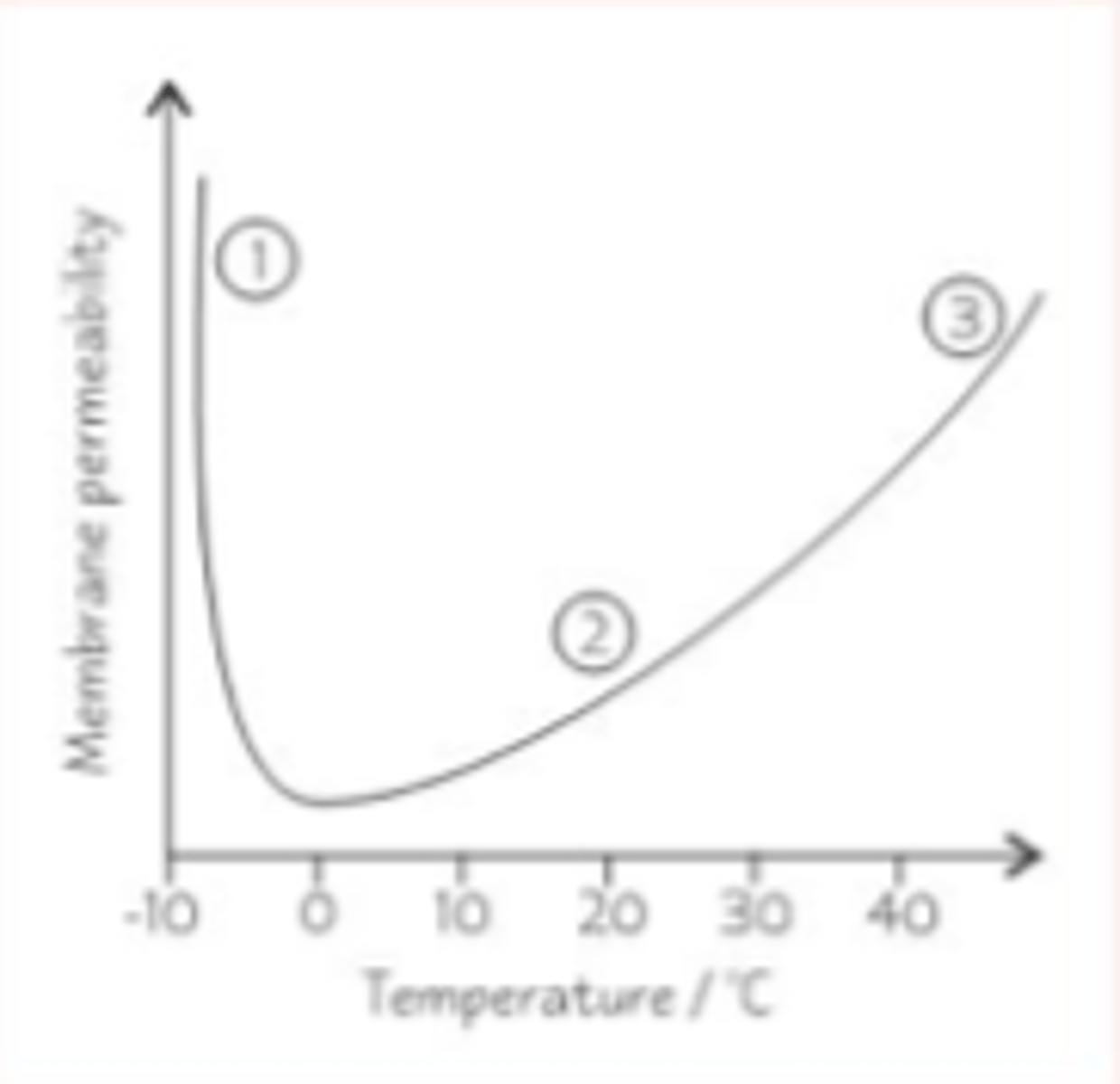
How does temperature affect membrane permeability
Permeability increases with temperature
- Below zero degrees, the phospholipids have little kinetic energy, so are rigid. However, transport proteins will be deformed, increasing permeability. Ice crystals may also form and pierce the membrane, further increasing permeability.
- Between 0 and 45 degrees, phospholipids gain more kinetic energy and pack less closely together, increasing permeability.
- Above 45 degrees, the phospholipid bilayer will begin to melt and the transport proteins will deform increasing permeability. Water molecules inside the cell will expand, putting pressure on the membrane
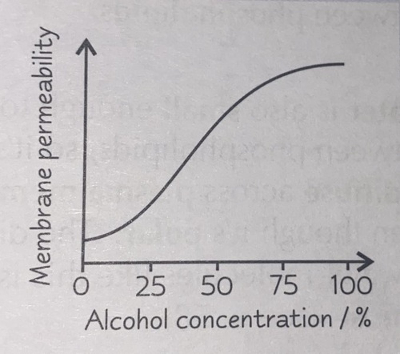
Affect of alcohol on membrane permeability
Alcohol disolves the lipids in the phospholipid bilayer, causing it to lose its structure, increasing permeability