IMSK/histo- nerve and bone visuals
 Call Kai
 Learn
Learn Practice Test
 Spaced Repetition
 Match
Match Flashcards
 Knowt Play
1/24

There's no tags or description
Looks like no tags are added yet.
Name | Mastery | Learn | Test | Matching | Spaced | Call with Kai | Chat |
|---|
No analytics yet
Send a link to your students to track their progress
25 Terms
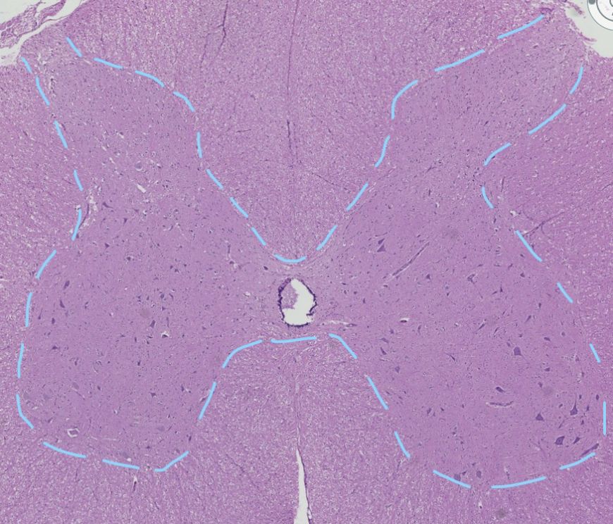
Is the outlines “butterfly shaped” region in this spinal cord grey or white matter?
grey matter

in this spinal cord grey matter, which region is outlined in yellow and which is outlined in green?
dorsal horn, ventral horn
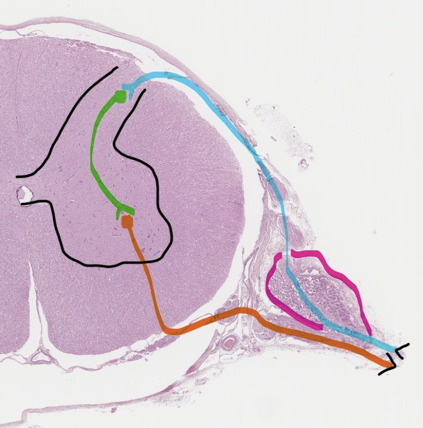
ID the aqua, green, and orange structures.
sensory affferent, interneruron, motor efferent

ID the pink outline and black outline
dorsal root ganglion, grey matter

What is outlined in blue in this image? (there are 3)
dorsal root of spinal nerve, dorsal root ganglion, dorsal rootlets

ID the two seperate portions of this spinal nerve located above & below the dashed blue line.
above- dorsal root, below-ventral root

which anatomical (shape) classification does the sensory neuron have, whose cell body is in the dorsal root ganglion?
unipolar
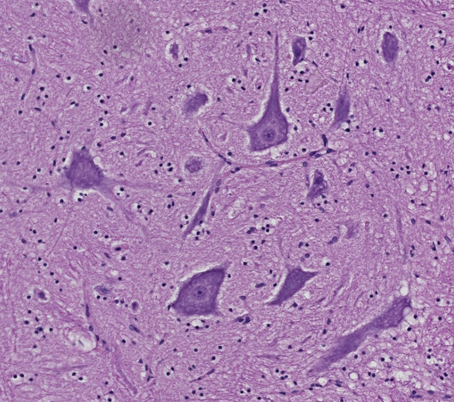
what is the anatomical classification of the motor neurons in the ventral horn? what do these somatic motor neurons innervate?
multipolar neurons, skeletal muscle

these are neurons in the dorsal horn of the spinal cord. what anatomical classification are these neurons? what functional classification?
multipolar, interneurons
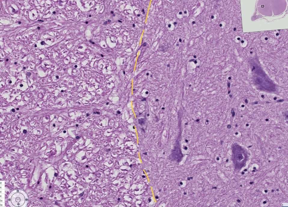
within this spinal cord, ID the types of matter (grey or white) to the left and right of the dashed yellow line
white, grey

this is white matter in the spinal cord, ID the shrunken structures indicated by the yellow arrows.
axons
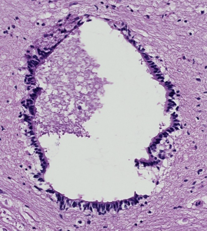
what do we call the fluid filled space within the spinal cord?
which glial cells line this space?
do these cells manufacture cerebrospinal fluid?
central canal, ependymal cells, in ventricle of brain (not central canal)
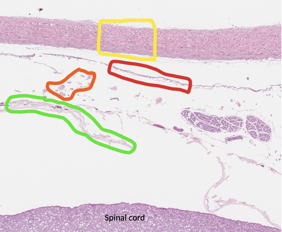
ID the indicated layers of meninges: yellow, red, orange, green
dura mater, arachnoid membrane, arachnoid trabeculae remnants within subarachnoid space, pia mater

what type of bone formation occurs within well vascularized CT without a cartilaninous precursor?
intramembranous
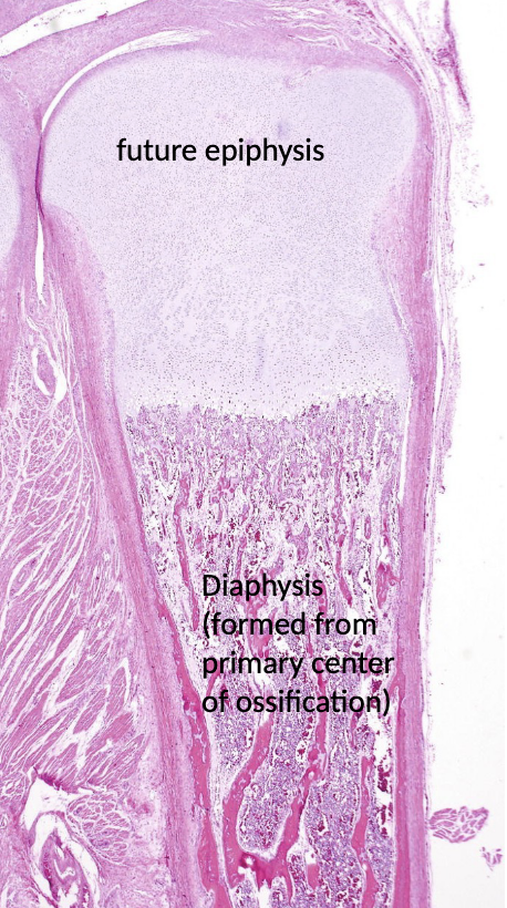
what tyoe of bone formation occurs within a cartilaginous precursor?
endochondral
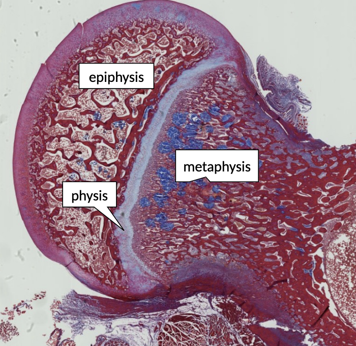
this is a portion of a growing long bone, illustrating the epiphysis, physis and metaphysis. what are synonyms for “physis”?
epiphyseal plate and growth plate
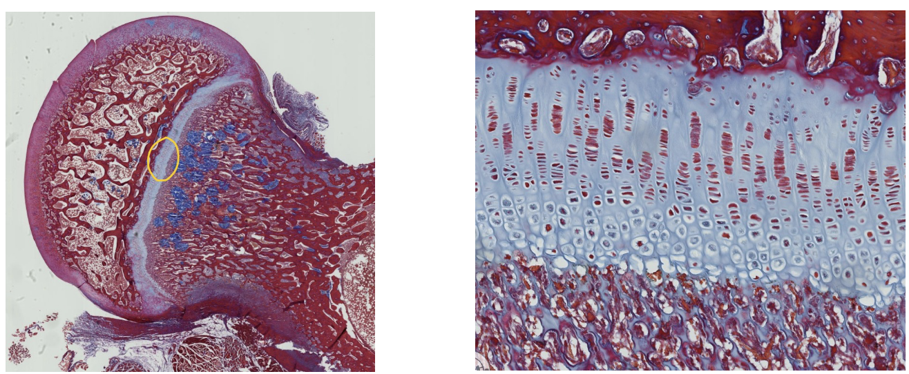
ID the specific type of CT that makes up the physis of the long bone shown in this region.
hyaline cartilage

Layers of the physis: which specific zone is encircled?
zone of hypertrophy
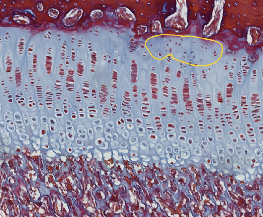
Layers of the physis: which specific zone is encircled?
zone of resting cartilage
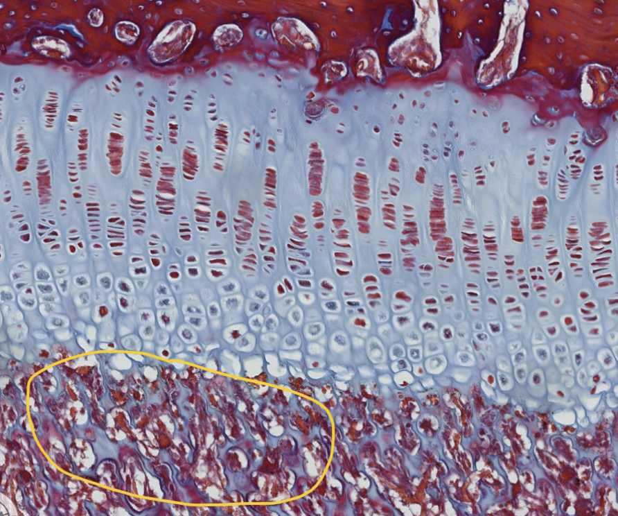
Layers of the physis: which specific zone is encircled?
zone of calcification/ossification
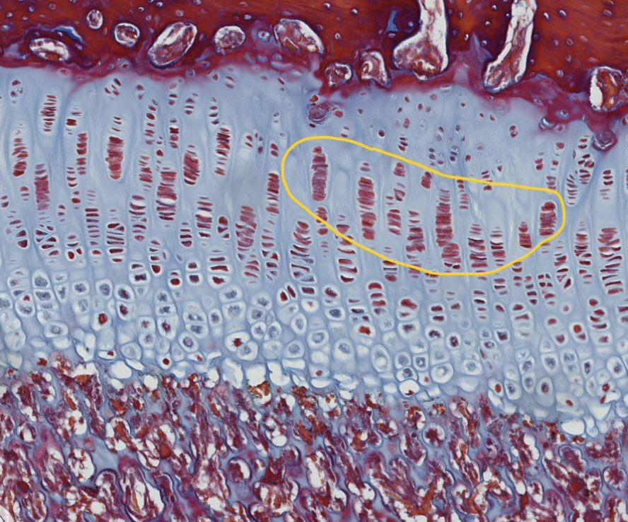
Layers of the physis: which specific zone is encircled?
zone of proliferation
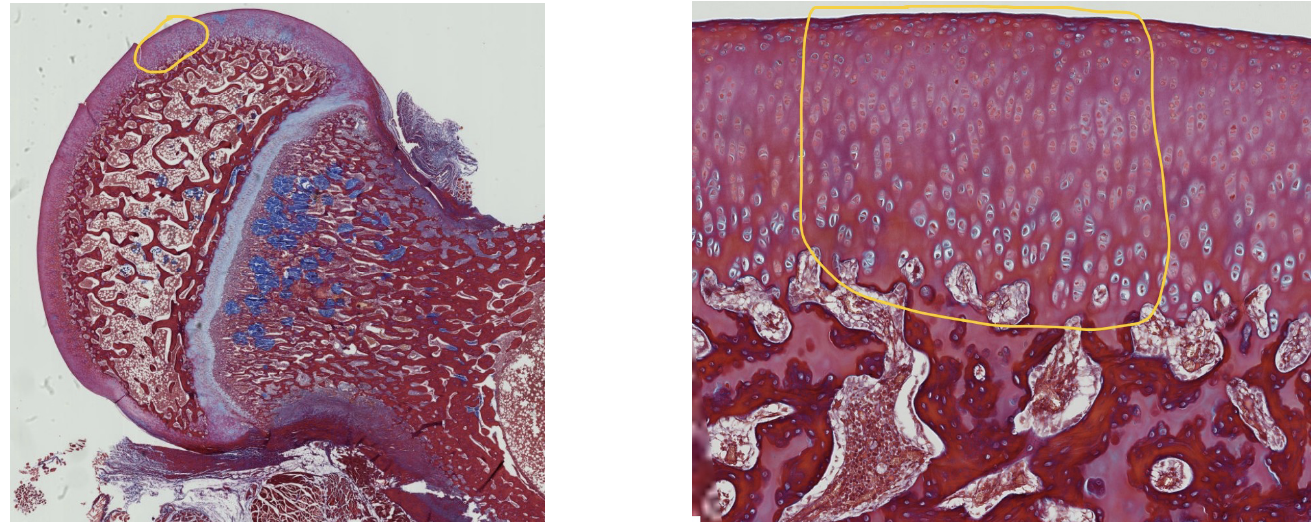
A portion of the articular surface of this long bone is encircled in the left image and is enlarged in the right image.
1. What do we call this tissue that covers the articulating surfaces of synovial joints?
2. ID the specific type of CT that makes up this region.
articular cartilage, hyaline cartilage
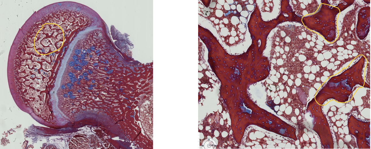
A portion of the epiphysis is encircled in the left image and is enlarged in the right image.
What are the encircled spicules of bone called? (include all synonyms)
spongy/cancellous/trabecular bone

FYI bone marrow: the region encircled in the upper image is enlarged in the lower image. the large cells with the multilobed nuclei are megakaryocyted, which produce platelets.
Which bone cell do these megakeryocytes resemble?
osteoclasts (multinucleated)
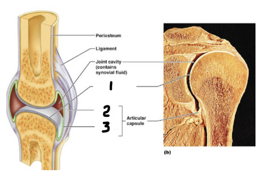
name the numbered layers of this synovial joint:
joint surface
outer layer of articular capsule
inner layer of articular capsule
articular cartilage, fibrous capsule, synovial membrane