Spinal Cord Pathologies
 Call Kai
 Learn
Learn Practice Test
 Spaced Repetition
 Match
Match Flashcards
 Knowt Play
1/23

There's no tags or description
Looks like no tags are added yet.
Name | Mastery | Learn | Test | Matching | Spaced | Call with Kai | Chat |
|---|
No analytics yet
Send a link to your students to track their progress
24 Terms
Types of Spinal Cord Pathologies
Congenital
Congenital pathologies are conditions that the patient is born with.
Tumors
Tumors are caused by abnormal tissue growth.
Trauma
Trauma occurs with injury.
Vascular
Vascular pathologies result from abnormal blood flow.
Infectious
Infectious pathologies are caused by viruses or bacteria.
Demyelinating/Inflammatory
Demyelinating/inflammatory pathologies occur as part of a bodily response or autoimmune function
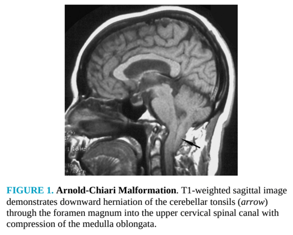
Arnold Chiari,
commonly shortened to Chiari malformation, is a structural defect that causes the cerebellar tonsils to slip below the foramen magnum into the spinal canal. This abnormal positioning can interfere with the normal flow of cerebrospinal fluid (CSF) and may cause compression of the brain stem or the spinal cord
Patients may be unaffected by this pathology, or the condition may be severe. Chiari malformation is classified as a congenital pathology. Chiari-like malformations, referred to as tonsillar ectopia (abnormal position), can be caused by brain swelling, tumors, or trauma to the head or spine.
Special CSF flow sequences, specifically phase contrast GRE, can be obtained at the level of the foramen magnum to determine flow obstruction.
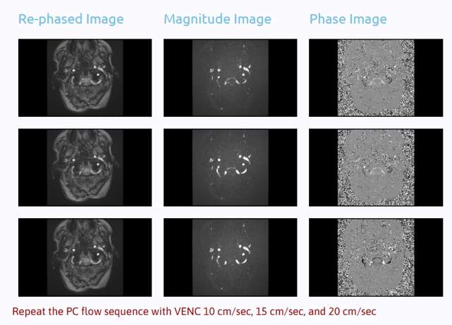
pulse gated sequence for chiari
Syringomyelia (syrinx) i
a cyst or fluid-filled area formed within the spinal cord. It can be congenital and is often seen with Chiari malformations. A syrinx can also develop due to tumors, trauma, or post-inflammatory changes. A syrinx can form at any level of the spinal cord.
MRI Appearance:
•
Best seen on T2-weighted imaging.
•
T1-weighted images: Hypointense (dark) compared to normal spinal cord tissue and may be difficult to distinguish unless large.
•
T2-weighted images: Hyperintense (bright) fluid signal, similar to CSF, showing a well-defined, elongated cavity within the central spinal cord.
•
STIR or FLAIR: Shows the syrinx as fluid intensity, though less commonly used for syrinx diagnosis.
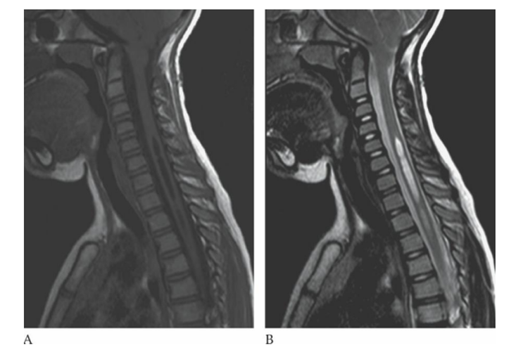
Syringomyelia (syrinx) i
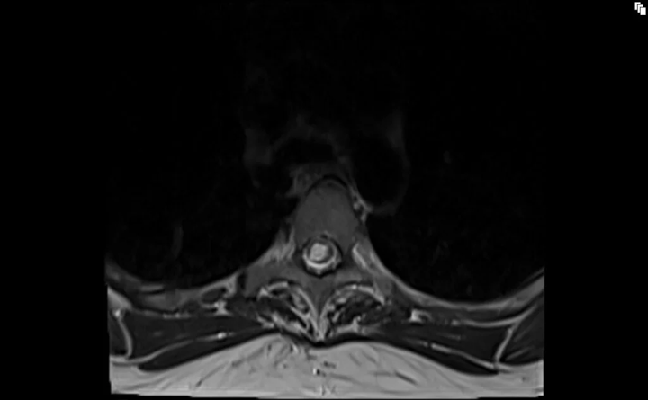
seroma
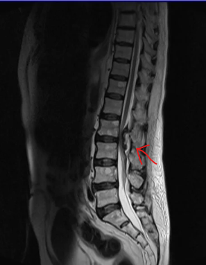
a collection of fluid that builds up after surgery.
MRI Appearance
•
T1-weighted images: Low signal intensity.
•
T2-weighted images: Classical fluid appearance.
•
Seromas do not enhance with contrast.
Tethered cord
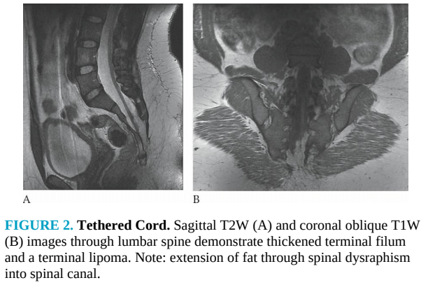
a condition where the spinal cord is abnormally attached (“tethered”) within the spinal canal, limiting movement and causing stretching, especially during growth. This prevents the conus medullaris from terminating in its normal position around L1.
MRI Appearance
Low-Lying Conus Medullaris
Below the normal L1–L2 level
Children: Conus should end at L1–L2
Adults: Conus below L2–L3 is abnormal
Tethered cord: Conus may terminate as low as L3–L5 or lower
Thickened Filum Terminale
Normal thickness: ≤ 2 mm
Thickened: > 2 mm, often fatty and hyperintense on T1
Fatty filum = lipoma of filum terminale
Demyelinating Inflammatory Disease
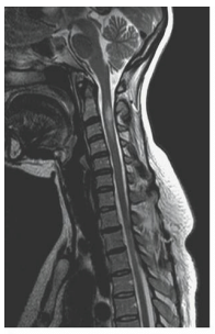
Multiple sclerosis (MS) is a demyelinating disease that can be found in the brain or spinal cord. It is represented on imaging by areas of inflammatory plaques
MRI Appearance
•
MS plaques will have high signal on T2-weighted images
•
Active plaques enhance on post-contrast (T1-weighted) images
•
T2 FLAIR imaging is the best sequence for detecting MS plaques
Ependymoma and Meningioma
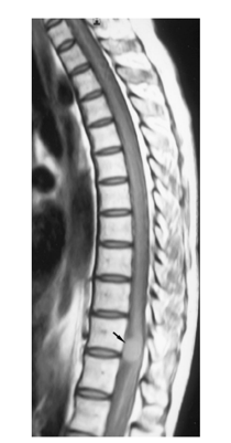
Spinal meningiomas are benign, hard, slow growing, and highly vascular. They may occur in the brain or spinal cord and are most commonly found in the thoracic spine.
MRI Appearance
•
T1-weighted images are typically isointense to the spinal cord.
•
T2-weighted images may present with either low-signal or high- signal intensity.
•
Contrast-enhanced T1-weighted images show a homogeneous enhancing high-signal intensity mass.
spinal meningioma
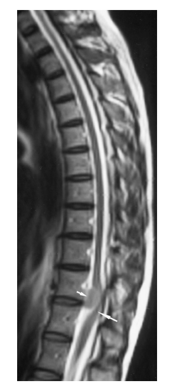
spinal meningioma
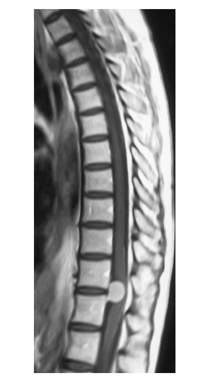
spinal meningioma
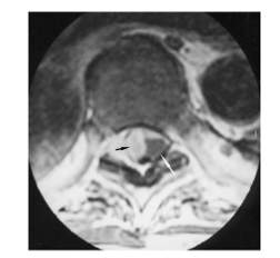
spinal meningioma
spinal cord hematoma
collection of blood in the spinal cord. The most common cause of a spinal cord hematoma is trauma. MRI appearance varies depending on the stage of the hematoma.
MRI Appearance by Stage
Acute Stage (Several Hours to 3 Days)
Slightly low signal on T1-weighted images.
Marked low signal with high signal on surrounding edema on T2-weighted images.
Subacute Stage (3 Days to 3 Weeks)
High signal on T1-weighted images.
High signal with low signal clot on T2-weighted images.
Chronic Stage (3 Weeks to Months)
High signal on T1-weighted images.
High signal with low-signal rim on T2-weighted images.
Gradient echo images appear with low-signal intensity.
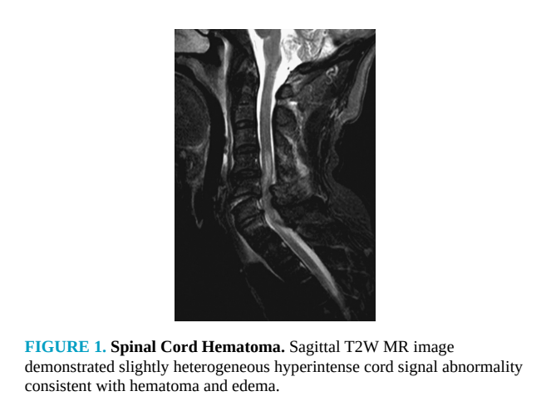
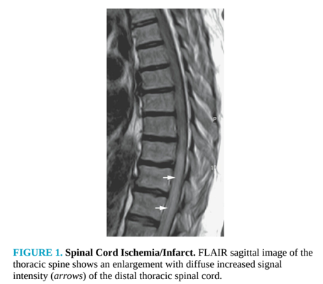
Spinal cord ischemia or infarct
occurs due to a decrease of blood supply to the spinal cord. While any area of the spinal cord may be affected, these findings are usually located in the lower thoracic or lumbar spine.
MRI Appearance
•
T1-weighted images: May show enlargement of the spinal cord.
•
T2-weighted images: Show high-signal intensity within the cord.
•
FLAIR images: Are very helpful due to suppression of the CSF signal.
cerebrospinal fluid (CSF) leak
occurs when fluid surrounding the brain or spinal cord leaks through a tear or hole in the dura mater. Common causes include trauma, surgery or procedure, or spontaneous occurrence.
MRI Appearance
•
T2-weighted images: CSF appears hyperintense.
•
T1-weighted images: CSF appears hypointense.
•
Surrounding areas: May demonstrate edema or hyperintensity due to the presence of CSF.
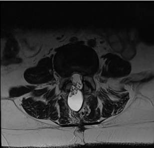
csf leak in lumbar t2
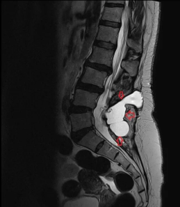
csf leak lumbar
Cauda equina syndrome
an emergent neurological condition that occurs when the nerve roots in the cauda equina are compressed. If this condition is not treated promptly, the resulting symptoms may become permanent.
Causes of cauda equina syndrome include disc herniation, trauma, tumor, infection, inflammation, or spinal stenosis.
MRI Appearance
The MRI appearance of cauda equina syndrome depends on the underlying cause, but it is notable for a void in the spinal canal near the area of the cauda equina. In an emergency room setting, there may be concern for cauda equina syndrome when a patient experiences urinary or fec— incontinence.
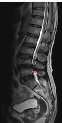
cauda equina