Neurologic System, Stroke, and Head Injury
 Call Kai
 Learn
Learn Practice Test
 Spaced Repetition
 Match
Match Flashcards
 Knowt Play
1/57

Earn XP
Description and Tags
Vocabulary practice cards covering focused neurologic assessment, diagnostic procedures, stroke types and emergency management, head injuries, intracranial pressure dynamics, and related nursing care.
Name | Mastery | Learn | Test | Matching | Spaced | Call with Kai | Chat |
|---|
No analytics yet
Send a link to your students to track their progress
58 Terms
Level of Consciousness (LOC)
A measurement reflecting the patient's ability to arouse, respond, and interact with their environment.
Glasgow Coma Scale (GCS)
A standardized scoring tool used to assess level of consciousness across eye opening, best verbal, and best motor responses, ranging from 3 (totally unresponsive) to 15 (best response), with a score of 8 or less indicating a comatose state.

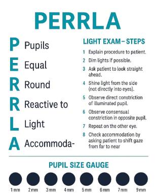
PERRLA
An acronym describing a normal pupillary assessment finding: Pupils Equal, Round, Reactive to Light, and Accommodation.
Pronator Drift
A physical assessment technique where the patient extends both arms forward with palms up and closes their eyes; downward drift or pronation of one arm indicates subtle motor weakness.
Expressive Aphasia
A language impairment where the patient understands what they want to say but experiences difficulty producing language.
Nursing Strategies:
ask simple questions
allow plenty of response time
use yes/no questions when helpful
provide communication boards/picutres
encourage gestures/writing if possible
do not finish every sentence for the patient
Receptive Aphasia
A language impairment characterized by difficulty understanding language, even though the patient's speech output may remain fluent.
Nursing Strategies:
use short, simple statements
give one instruction at a time
use gestures/visual cues
repeat/rephrase as needed
verify understanding
actively listen and be patient
Dysarthria
A motor speech disorder caused by impaired muscle control used for speech production, resulting in slurred or hard-to-understand speech while language comprehension remains intact.
Noncontrast Head CT
The essential initial imaging modality for suspected acute stroke used primarily to rapidly rule out intracranial hemorrhage before administering reperfusion therapies.
Magnetic Resonance Angiography (MRA)
A specialized non-invasive magnetic resonance study that provides detailed visualization of cerebral blood vessels to detect stenosis, occlusions, or aneurysms.
Cerebral Angiography
An invasive vascular procedure in which a catheter is threaded through an artery into cerebral circulation to visualize vessels, diagnose aneurysms or arteriovenous malformations, and guide endovascular therapy.
Key nursing responsibilities include checking contrast allergy history, reviewing kidney function (BUN/creatinine), verifying consent, keeping NPO as ordered, and assessing baseline neuro status and peripheral pulses before the procedure; after the scan, nurses must monitor the access site for bleeding/hematoma, evaluate distal pulses and neurovascular status, track
Lumbar Puncture (LP)
A diagnostic procedure where a needle is inserted into the subarachnoid space (typically while side-lying with knees flexed) to collect cerebrospinal fluid (CSF) for analysis.
This procedure is used to diagnose conditions such as infections, inflammatory/demyelinating disorders, bleeding, and inflammatory diseases of the central nervous system, and to measure intracranial pressure.
Key Nursing Responsibilities:
obtaining informed consent
ensuring the patient is in the correct position
monitoring vital signs during and after the procedure
assessing the puncture site for complications
Monitor for post-dural puncture headache (worse when upright, improves when lying down)
providing post-procedure care, such as ensuring adequate fluid intake to prevent headaches.
Unsafe in patients with increased intracranial pressure due to risk of herniation.
Electroencephalogram (EEG)
A diagnostic procedure that records the electrical activity of the brain using scalp electrodes to evaluate seizures, epilepsy, or unexplained altered mental status.
Key nursing responsibilities:
explain that the procedure is painless
hair should be clean and free of styling products
sleep deprivation may be ordered
medications are adjusted only if prescribed
patient may be exposed to flashing lights or hyperventilation during testing
External Ventricular Drain (EVD)
A device inserted into the lateral ventricles of the brain to monitor intracranial pressure and drain cerebrospinal fluid (CSF) to manage elevated ICP. This procedure is commonly used in cases of traumatic brain injury, hemorrhage, or hydrocephalus, providing both diagnostic and therapeutic benefits.
Nursing Priorities:
maintain prescribed drainage level
level system to ordered anatomical reference
repositioning/ambulation requires nursing assistance to maintain drainage pressures
monitor ICP
monitor amount/color of CSF drainage (to look for signs of infection)
assess neurologic status
maintain strict aseptic technique
monitor for infection
Craniotomy
A neurosurgical procedure where a portion of the skull (bone flap) is temporarily removed to access brain tissue and then replaced. Used to access the brain for tumors, hemorrhage, aneurysms, etc. Bone flap is replaced.
Craniectomy
A neurosurgical procedure where a portion of the skull is removed and not immediately replaced, providing space for severe cerebral swelling. This procedure is often indicated for conditions such as traumatic brain injury or large strokes, where removal of the skull can help relieve pressure on the brain.
Burr Holes
Small openings are drilled through the skull to drain hematomas or collections of fluid. Lower impact on the patient, but still a concern for infection.
Transient Ischemic Attack (TIA)
A temporary episode of focal neurological dysfunction caused by brain ischemia without acute tissue infarction, serving as an important warning sign for future stroke. Also known as a mini-stroke, it typically lasts less than 24 hours and may resolve quickly, indicating a need for urgent medical evaluation.
Thrombotic Ischemic Stroke
An ischemic stroke caused by a clot forming directly within a cerebral artery, frequently associated with underlying atherosclerotic disease. Ruptured plaque that cuts off blood supply.
Embolic Ischemic Stroke
An ischemic stroke caused by a clot or debris traveling from another part of the body (such as the heart or carotid arteries) and blocking a cerebral blood vessel. Atrial fibrillation is an important cause.
Lacunar Ischemic Stroke
An ischemic stroke resulting from the occlusion of small penetrating arteries deep within the brain is strongly linked to chronic hypertension and diabetes.
Ischemic Penumbra
The region of ischemic but structurally intact brain tissue surrounding an infarction core that remains viable if blood flow is rapidly restored.
BE FAST
A mnemonic identifying key signs of acute stroke: Balance loss, Eyes/vision changes, Face drooping, Arm weakness, Speech difficulty, and Time to call 911.

Last Known Well (LKW)
The precise time at which a patient was last observed or confirmed to be at their baseline normal neurological function, essential for establishing eligibility for reperfusion therapies, urgency and type of imaging, and treatment decision.
“When was the patient last known to be neurologically normal?” This information is critical in stroke care for determining treatment options.
NIH Stroke Scale (NIHSS)
A standardized 11-item clinical evaluation tool used to quantify acute stroke deficit severity, establish baseline impairment, and track neurological changes. Higher score = greater stroke. severity
Tenecteplase (TNK)
A weight-based IV thrombolytic agent administered as a single bolus for acute ischemic stroke reperfusion therapy. Must be given with 4.5 hours of stroke.
Alteplase (tPA)
A weight-based IV thrombolytic medication administered as an initial IV bolus followed by a 1-hour infusion for acute ischemic stroke reperfusion. Must be given with 4.5 hours of stroke.
Endovascular Thrombectomy (EVT)
An interventional procedure in which a catheter is advanced into cerebral vasculature to mechanically remove a blood clot causing a large-vessel occlusion.
Intracerebral Hemorrhage (ICH)
A subtype of hemorrhagic stroke involving direct blood vessel rupture and bleeding within brain tissue, frequently caused by chronic hypertension.
Presents as focal deficits, headache, decreased LOC, and nausea and vomiting.
Major concerns are hematoma, expansion, edema, and increased ICP
Treatment focus is control bleeding/ICP, surgery when indicated.
Subarachnoid Hemorrhage (SAH)
Bleeding into the subarachnoid space surrounding the brain, most often caused by a ruptured cerebral aneurysm, presenting with a sudden thunderclap headache.
Major concerns are rebleeding, vasospasm/delayed cerebral ischemia, and hydrocephalus
Treatment focus is secure aneurysm and prevent complications with surgical clipping or endovascular coiling, alongside supportive care.
Nimodipine
An enteral calcium channel blocker administered following aneurysmal subarachnoid hemorrhage to prevent or lessen delayed cerebral ischemia from vasospasm.
4-Factor Prothrombin Complex Concentrate (4-factor PCC)
An IV medication containing blood coagulation factors used for urgent reversal of warfarin-induced anticoagulation during acute bleeding emergencies.
Primary Brain Injury
Occurs at the moment of trauma.
Physical brain damage occurring at the direct moment of impact or trauma, such as contusions, direct tissue laceration, skull fractures, or axonal shearing.
CANNOT BE REVERSED
Secondary Brain Injury
Progressive brain damage unfolding after initial trauma caused by edema, hypoxia, hypotension, hypercapnia, elevated ICP, or impaired perfusion.
Concussion
A mild traumatic brain injury caused by mechanical forces that results in transient physical, cognitive, emotional, or sleep-related symptoms, occurring with or without loss of consciousness.
Seek immediate evaluation for:
worsening headache
repeated vomiting
increased confusion/agitation
increased drowsiness or inability to awaken
new weakness/numbness
slurred speech
seizure
one pupil larger than the other
new loss of coordination
loss of consciousness or worsening responsiveness
Epidural Hematoma
An accumulation of arterial blood between the skull and dura mater, often associated with skull fractures, classically marked by a brief loss of consciousness followed by a lucid interval and sudden deterioration.
NEUROSURGICAL EMERGENCY
May require rapid surgical evaluation, craniotomy/burr holes (for small bleeds), and ICP management.

Subdural Hematoma
A collection of venous blood between the dura mater and arachnoid mater caused by tearing of bridging veins, commonly seen in elderly individuals and patients with alcohol dependency.
Acute: symptoms within 24hrs of injury. Progress rapidly, high M&M due to lots of bleeding
Subacute: symptoms 2-10 days after injury. Slower bleed
Chronic: symptoms appear weeks to months after injury, often subtle and progressively worsening. Can get walled off and become highly osmotic, causing expanding mass.

Diffuse Axonal Injury (DAI)
Widespread damage to nerve axons throughout the brain resulting from severe acceleration, deceleration, or rotational forces, often leading to immediate, prolonged coma.
Monro-Kellie Hypothesis
The doctrine stating that the skull is a rigid container holding brain tissue, blood, and CSF; an increase in volume of any one compartment must be offset by a decrease in another or ICP will rise.
Cerebral Perfusion Pressure (CPP)
The net pressure gradient driving blood flow to the brain cells, calculated as CPP=MAP−ICP, with a normal target range of 60−80 mmHg.
Decorticate Posturing
An abnormal motor response to pain characterized by rigid leg extension and inward flexion of arms, wrists, and fingers toward the body's core, indicating damage above the brainstem. Arms toward the CORE
Decerebrate Posturing
An ominous motor posture characterized by rigid extension of legs, flexed wrists, and extended, pronated arms held straight at the sides, signaling brainstem involvement.
Generally indicates worse prognosis.

Cushing's Triad
A late sign of markedly increased intracranial pressure and impending brain herniation consisting of bradycardia, severe hypertension with a widening pulse pressure, and irregular respirations.
This is the body’s attempt to maintain blood flow to the brain by increasing the systemic arterial pressure.
Mannitol
An osmotic diuretic administered intravenously to pull fluid from hyper-edematous brain tissue into the intravascular space, lowering intracranial pressure.
CT with Iodinated Contrast
A diagnostic brain imaging study used to visualize vessels, tumors, and infections. Key nursing responsibilities include checking contrast allergy history, reviewing kidney function (BUN/creatinine), and ensuring IV access before the scan, followed by monitoring for allergic reactions, tracking renal function, and encouraging hydration afterwards.
MRI
A diagnostic imaging modality that provides detailed visualization of brain tissue without radiation, commonly used to evaluate ischemic injury, brain tumors, demyelinating disease, infection, and structural abnormalities.
Nursing Responsibilities:
Before
screen for pacemakers/ICDS
aneurysm clips
cochlear implants
Ensure no metal implants or devices that may contraindicate use.
claustrophobia
During
patient must remain still
provide hearing protection
maintain communication
Sedation may be needed for severe claustrophobia
CT vs MRI
A comparison of primary neuroimaging modalities: CT is fast, involves radiation, and is excellent for detecting acute hemorrhage in emergencies with minimal device concerns; MRI is slower, radiation-free, requires strict safety screening for metallic implants, and provides superior brain tissue detail with greater sensitivity for early ischemic stroke.
Intracranial Pressure Monitoring
A procedure to measure the pressure within the skull, used to assess conditions like traumatic brain injury or stroke. It helps guide treatment decisions and monitor cerebral perfusion.
Normal ICP: 5-15 mm Hg
Increased ICP: Sustained elevation requires evaluation and treatment to prevent brain damage and optimize outcomes. Continuous monitoring allows for timely interventions.
Postoperative Neurosurgical Care
The management and monitoring provided after neurosurgery to ensure recovery, prevent complications, and support neurological function. This includes assessing vital signs, neurological status, and managing pain and potential infections.
monitor BP closely
prevent hypotension
HOB generally elevated as prescribed
maintain neutral head/neck alignment
monitor LOC/GCS, pupils, motor strength, speech, and compare with preoperative baseline
Dysphagia
An impairment in swallowing function commonly caused by neurological injury, placing the patient at high risk for aspiration pneumonia.
Key Nursing & Safety Priorities:
Keep patient strictly NPO until swallowing safety is formally evaluated
Recognize that aspiration may be silent
Monitor for clinical signs including coughing or choking with food/fluids, a wet or gurgling voice, drooling, food pocketing, or difficulty initiating a swallow
Protecting Affected Extremities after Stroke
Involves measures taken to safeguard and maintain the function of limbs affected by a stroke. This may include repositioning, range of motion exercises, and the use of supportive devices to prevent complications such as contractures and pressure ulcers.
Upper Extremity
support affected arm during positioning/transfers
maintain appropriate alignment
avoid pulling on affected arm
monitor for shoulder pain/subluxation
encourage prescribed ROM
Lower Extremity
Maintain proper positioning
Monitor skin and pressure areas
Use appropriate transfer assistance
Prevent foot drop/contractures as directed by therapy
Sensory Loss
inspect skin frequently
protect from heat/cold
avoid pressure or injury
teach patient to visually inspect affected areas
Stroke Complications
Possible adverse effects that can occur after a stroke include physical, cognitive, and emotional challenges. Common complications include dysphagia, mobility issues, and risk of aspiration pneumonia. Other complications may involve aphasia, anosognosia, seizures, and changes in bladder or bowel control.
Nursing Prevention/Recognition:
Regularly assess swallowing ability
Implement mobility and rehabilitation strategies
Monitor for cognitive and emotional changes
Educate patient and family about potential complications and self-care strategies
High Yield Nursing Priorities for Stroke

Initial Assessment of Head Injury
First: Trauma priorities (Airway, Breathing, and Circulation) and Protect the cervical spine until injury is excluded.
Do a focused neurological assessment, look for evidence of head trauma, and record important history.
Cerebral Contusion
Bruising of the brain tissue. May cause local edema, hemorrhage, focal neurologic deficits, seizures, or increased ICP.
These may evolve/enlarge after the initial injury.
Increased ICP
Above 20 mmHg is considered pathologic and should be treated.
Initial symptoms: headache, projectile vomiting, papilledema
Late: Cushing’s Triad- bradycardia, hypertension (with widening pulse pressure), and irregular respiratory pattern.
Recognizing Increased ICP
Early findings:
change in level of consciousness
restlessness
confusion
increased drowsiness
difficulty following commands
headache
N/V
Progressive Findings:
Worsening LOC
Pupillary changes
New motor weakness
Abnormal posturing
Seizures
Changes in respiratory pattern
Managing Increased ICP
promote venous drainage
head of bed –30º
Maintain neutral head and neck alignment
Reduce cerebral edema
Maintain oxygenation and ventilation
Maintain cerebral perfusion
Decrease cerebral metabolic demand
Remove blood/CSF/mass when necessary
Avoid unnecessary spikes caused by
coughing, suctioning, straining, vomiting, agitation, pain, seizures, fever, hypoxia, etc
Nursing Strategies:
Avoid prolonged excessive stimulation
treat pain and agitation
Prevent constipation/straining
Control fever
Maintain oxygenation
Suction only when clinically necessary
Allow recovery between stimulating activities
head of bed