Echocardiography
1/48
There's no tags or description
Looks like no tags are added yet.
Name | Mastery | Learn | Test | Matching | Spaced | Call with Kai |
|---|
No study sessions yet.
49 Terms
Apical and Parasternal
What are the 2 cardiac windows?
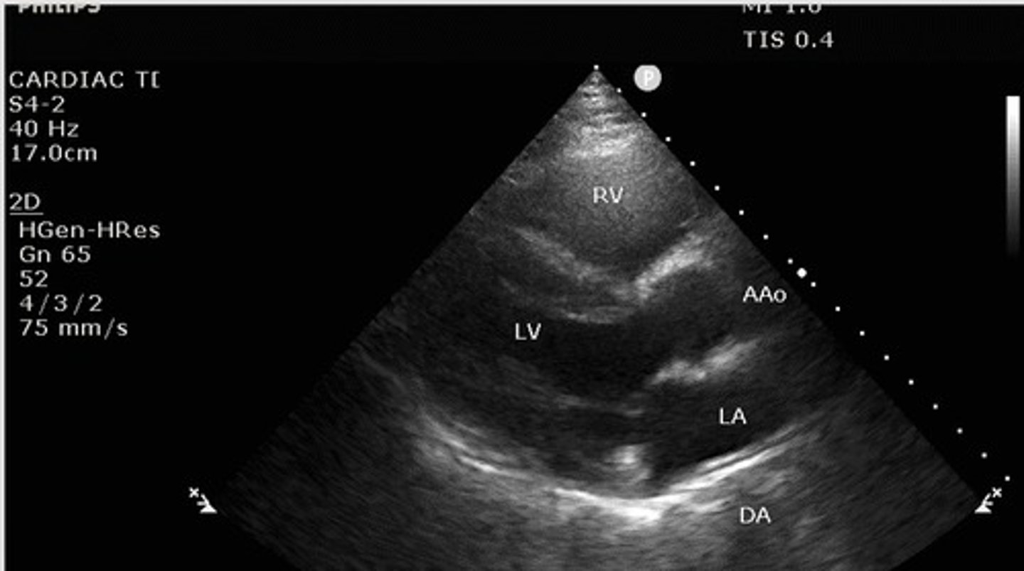
Parasternal long axis
What view of the heart is this?
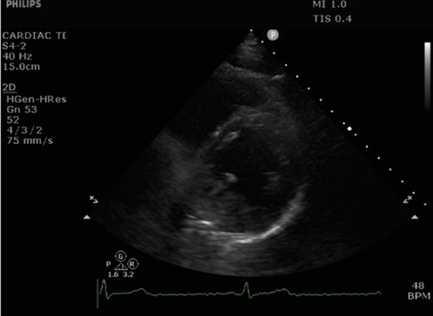
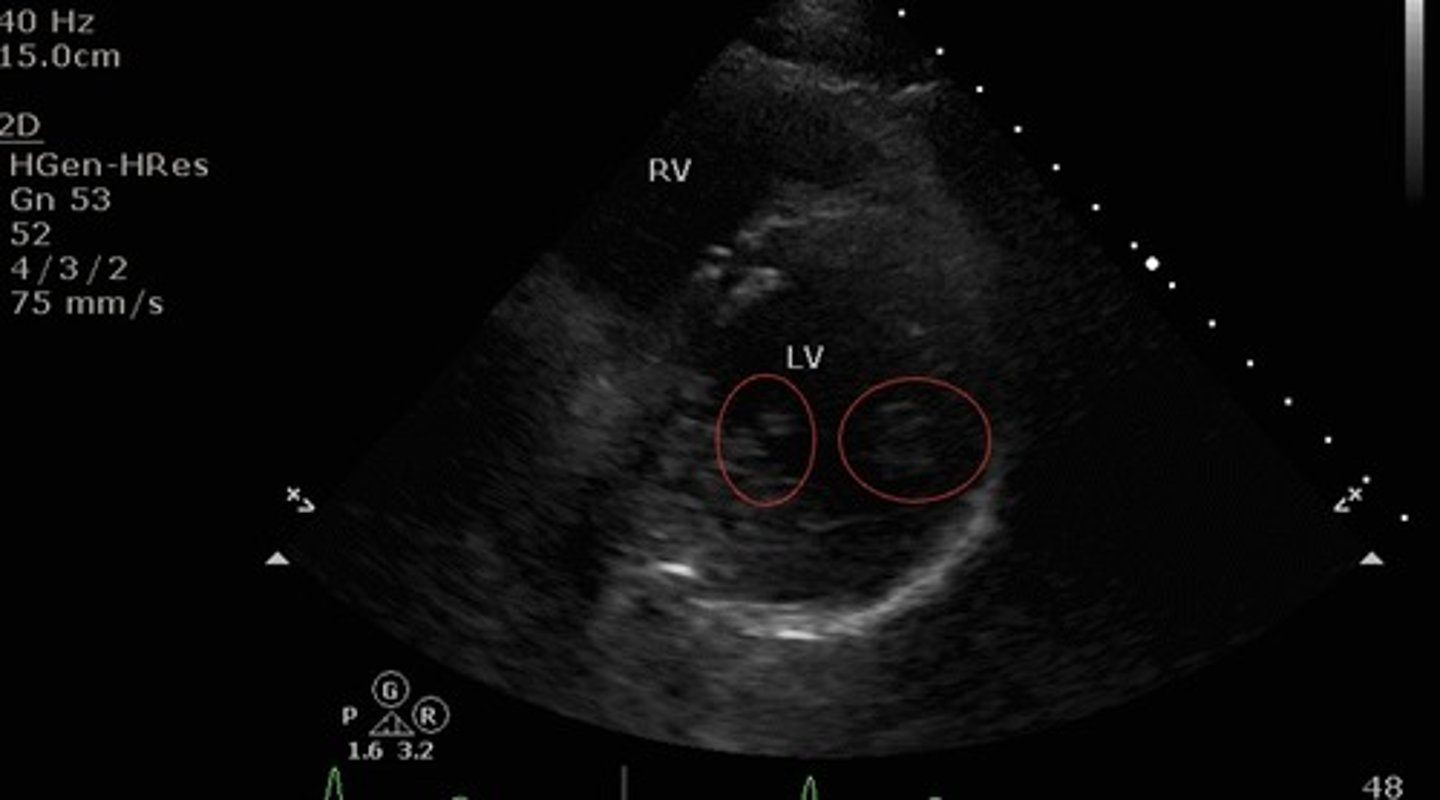
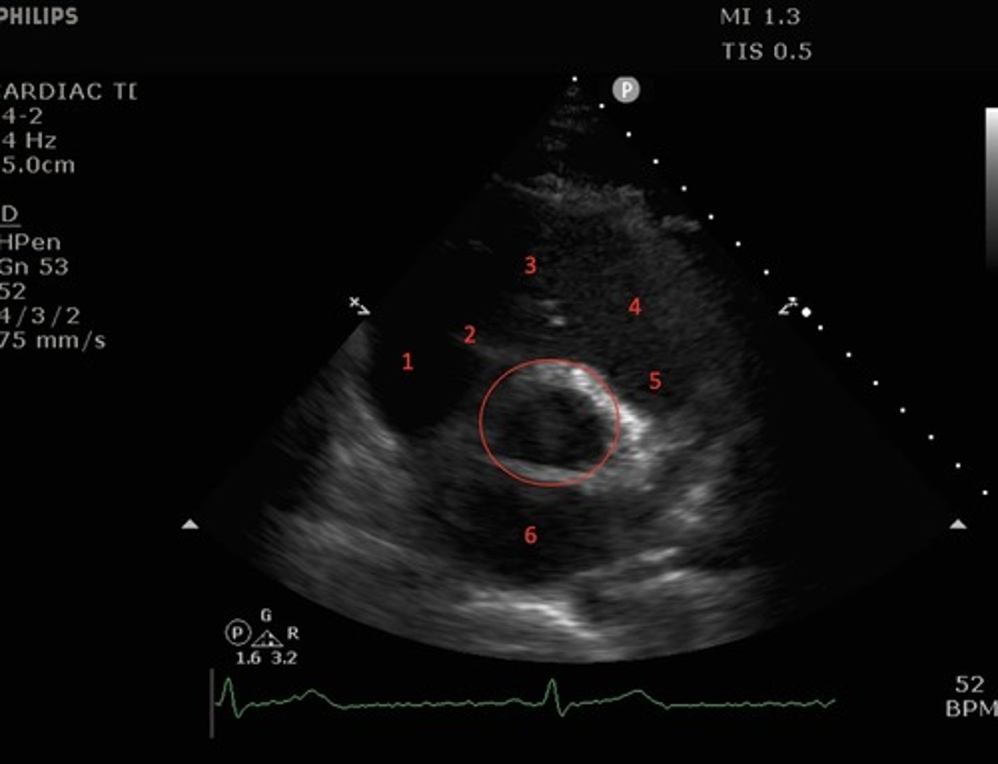
Parasternal short axis mid-ventricle
What view of the heart is this?
right ventricle, left ventricle, papillary muscles of the left ventricle
What structures are shown by the parasternal short axis mid ventricular view?
Papillary muscles
What structures are indicated by the red circles?
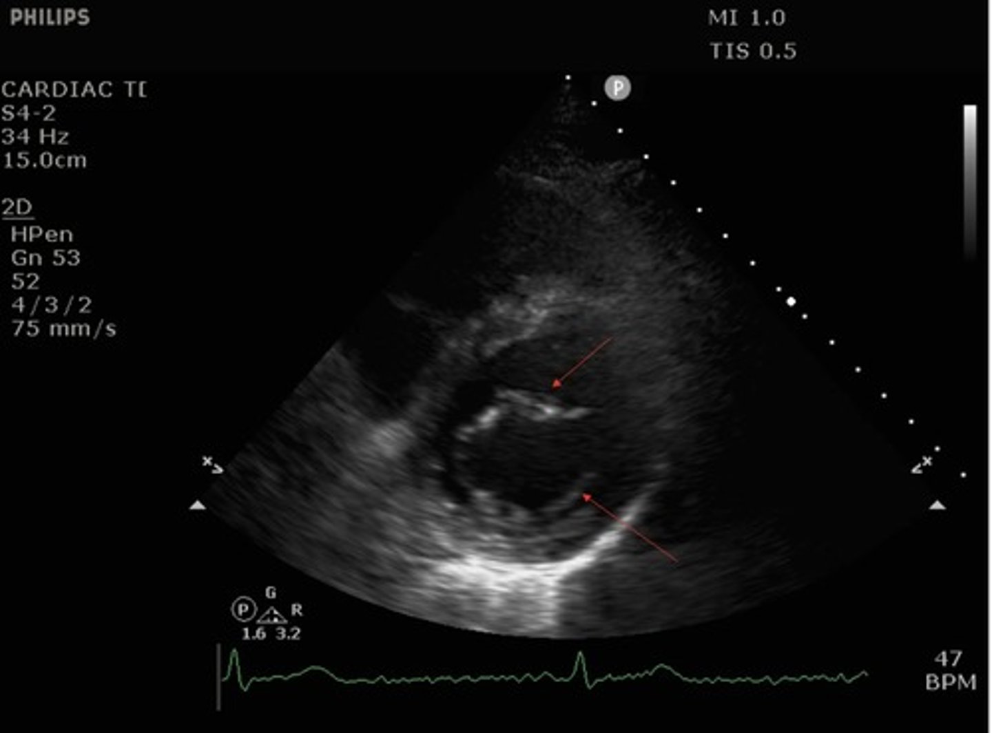
parasternal short axis mitral valve view
what view is this?

top arrow: anterior leaflet of mitral valve
bottom arrow: posterior leaflet of mitral valve
What structures are indicated by the red arrows?
Parasternal short axis aortic valve view
What view is this?
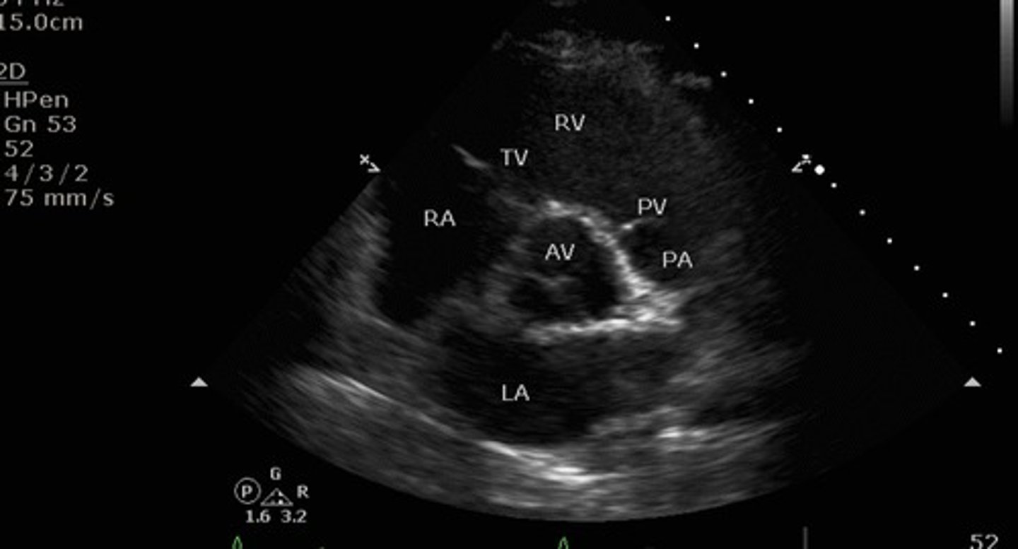
1. right atrium
2.tricuspid valve
3.right ventricle
4. pulmonic valve
5.pulmonary artery
6. left atrium
red circle: aortic valve
identify the numbered and circled structures
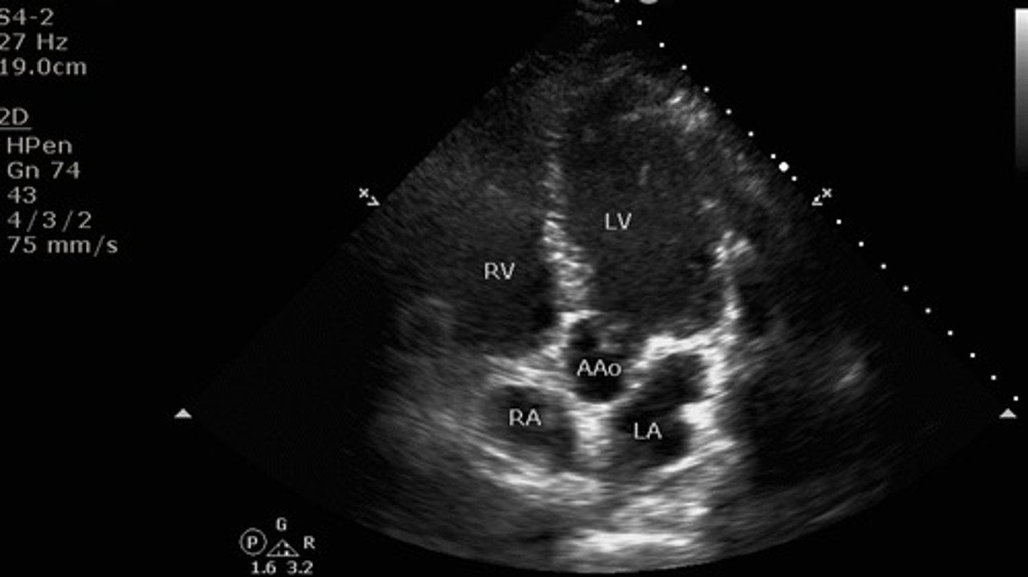
Apical 4 chamber view
what view is this?
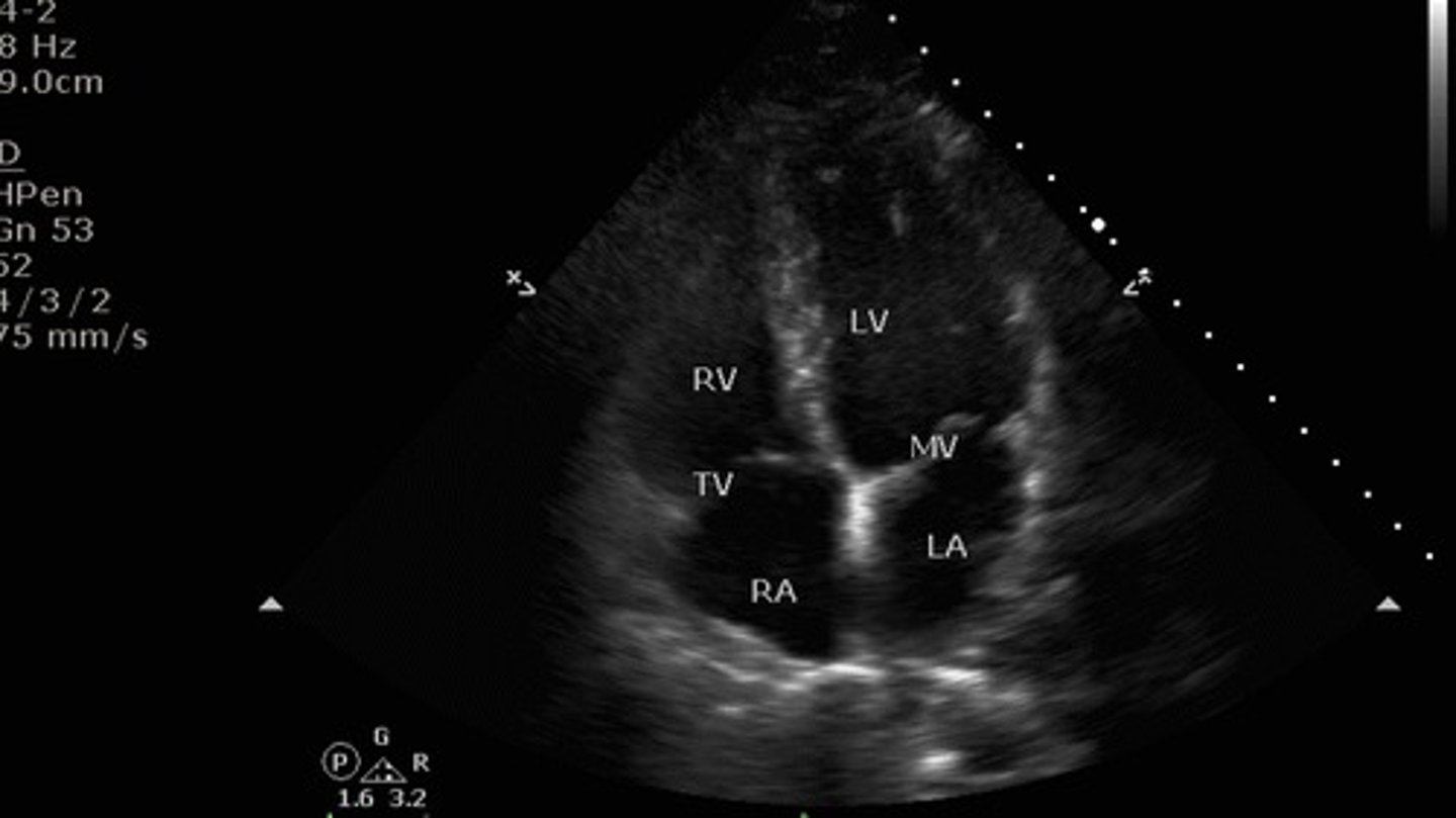
toward the bed with patient supine
where is the indicator mark pointed for the 4 chamber view?
1. right ventricle
2. tricuspid valve
3. right atrium
4. left ventricle
5. mitral valve
6. left atrium
identify the numbered structures
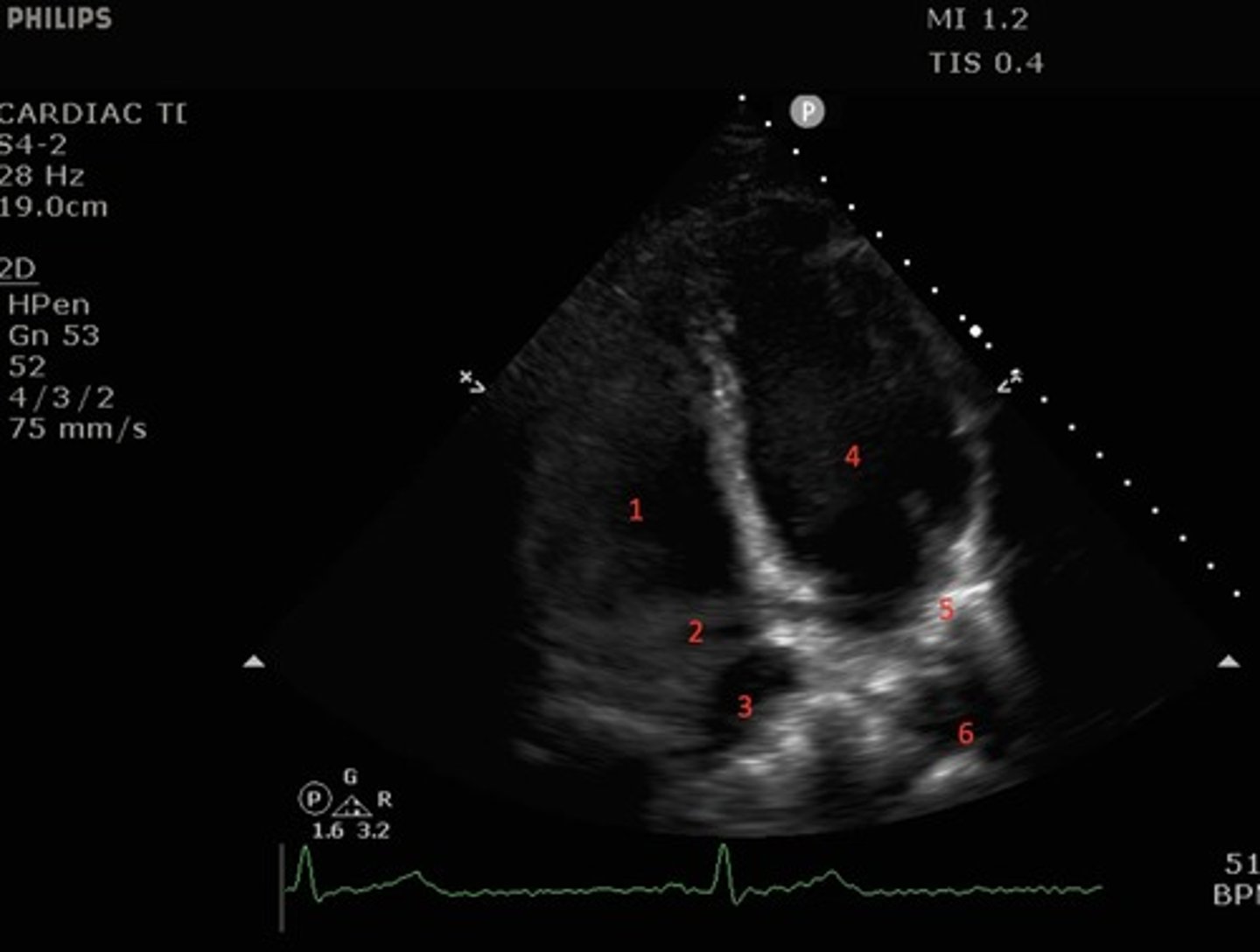
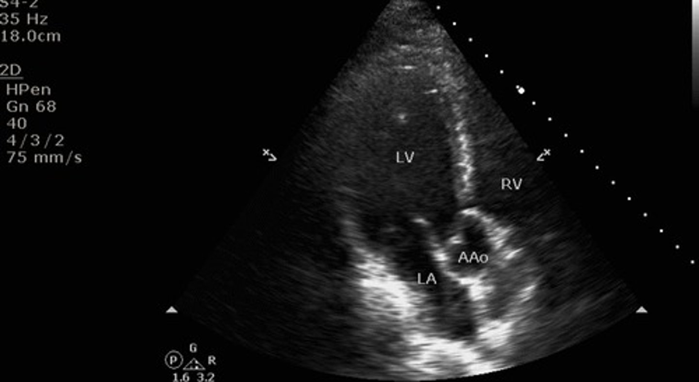
5 chamber apical view
what view is this?
the ascending aorta
what additional structure can you see when you achieve the 5 "chamber" view?
apical 2 chamber view
what view is this
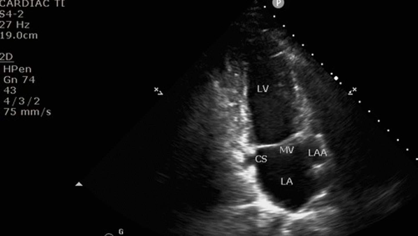
the right side
What side of the heart is omitted from the apical two chamber view?
apical 3 chamber view
what view is this?
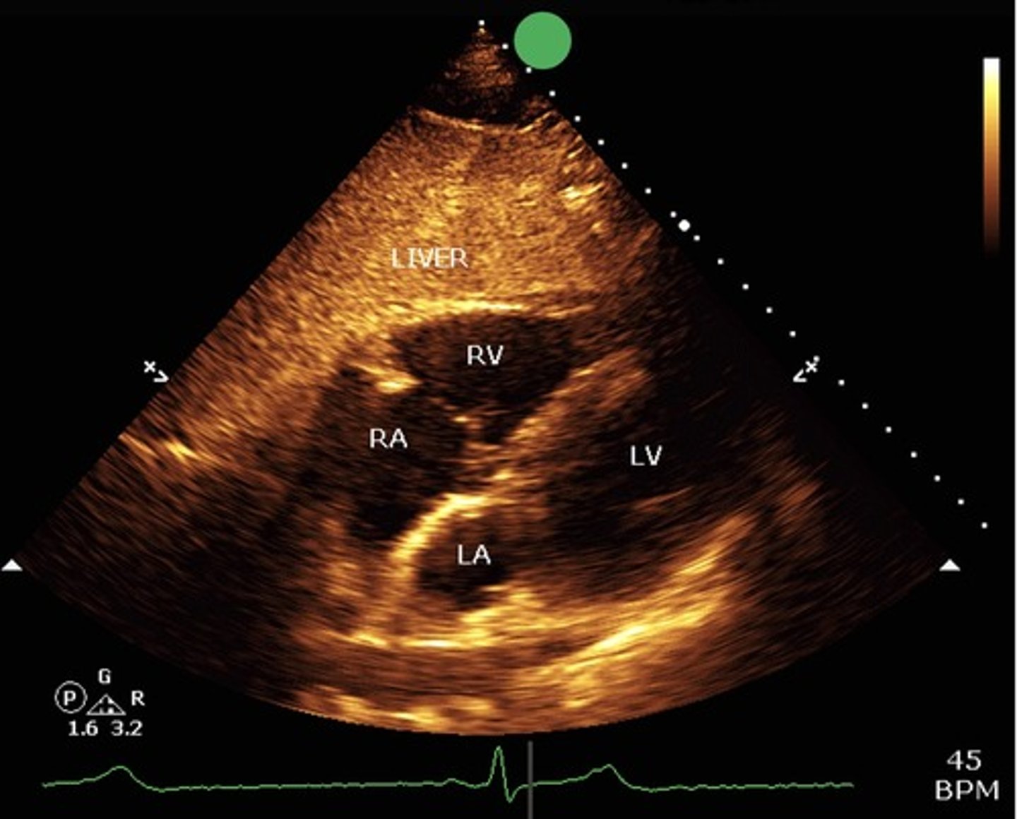
Subxiphoid view
what view is this?
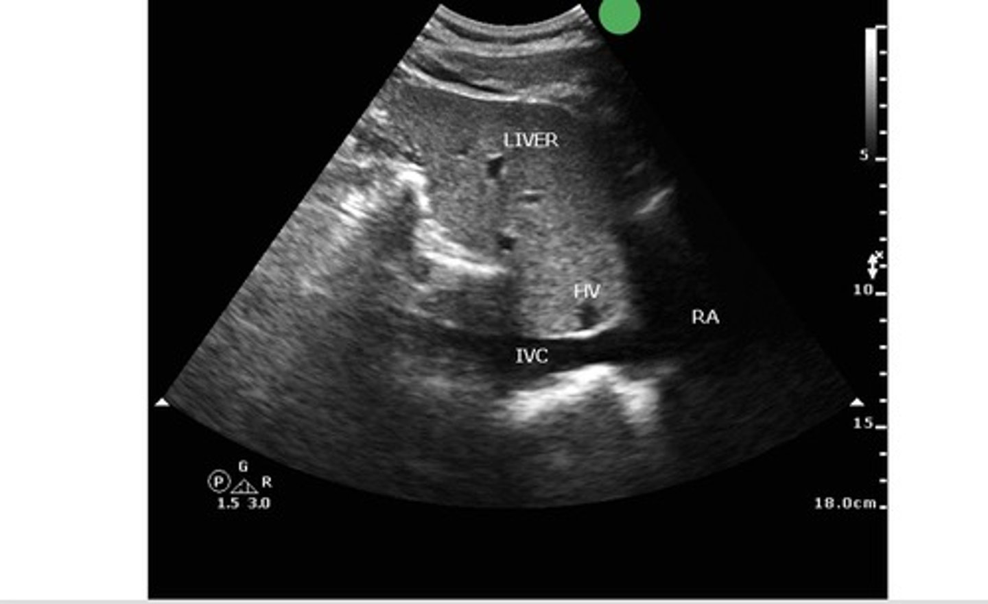
subxiphoid IVC view
what view is this?
left
which ventricle's function has the following features

right
which ventricle's function has the following features

hyperdynamic LV function
What does >70% EF indicate for LV function?
normal LV function
What does 55-70% EF indicate for LV function?
mildly reduced LV function
What does 45-54% EF indicate for LV function?
moderately reduced LV function
What does 30-44% EF indicate for LV function?
severely reduced LV function
What does <30% EF indicate for LV function?
Fractional Area Change and TAPSE
What are the two methods used to assess RV function?
the longitudinal displacement of the tricuspid valve annulus with systole (relying on M-mode echocardiography)
What does TAPSE measure?
parallel
the imaging plane should be chosen so that ultrasound beam is as ________ to the direction of blood flow as possible
determining pressure gradients across cardiac valves
what is the following equation used for

NO it's just qualitative: it indicated direction towards or away from the probe
Does color flow Doppler tell you the velocity or pressure of blood flow?
early diastolic filling, near the end of the T wave
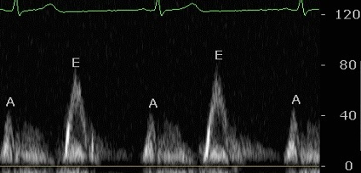
what does the E wave indicate on pulsed wave Doppler? What EKG feature does it correspond with?
flow from atrium to ventricle during atrial contraction (atrial kick), just after P wave
what does the A wave indicate on pulsed wave Doppler? What EKG feature does it correspond with?
velocity vs time: flow toward probe appears above graph baseline, flow away appears below baseline
What does the quantitive doppler graph indicate? What do the waves show?
pulsed wave doppler, the peaks are hollow
which type of quantitative doppler is this? How can you tell?
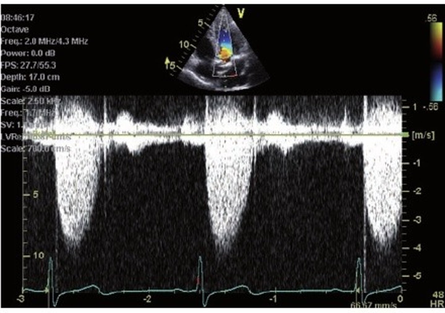
continuous wave doppler, the peaks are filled
which type of quantitative doppler is this? How can you tell?
LV, RV, outflow tract, mitral valve, aortic valve, ascending aorta, descending aorta
what structures are seen with parasternal long axis view?
LA, RA, RV, aortic valve, tricuspid valve, pulmonic valve, pulmonary artery
what structures are seen with parasternal short axis aortic valve view?
LV, RV, mitral valve
what structures are seen with parasternal short axis mitral valve view?
LV, RV, papillary muscles
what structures are seen with parasternal short axis mid-ventricular view?
LV, RV, LA, RA, mitral valve, tricuspid valve
what structures are seen with apical 4 chamber view?
LV, RV, LA, RA, mitral valve, tricuspid valve, aortic valve
what structures are seen with apical 5 chamber view?
LV, left atrium, mitral valve
what structures are seen with apical 2 chamber view?
LV, RV, outflow tract, mitral valve, aortic valve, ascending aorta, descending aorta
what structures are seen with apical 3 chamber view?
parasternal long axis and apical 3 chamber view
what two views have the same structures?
PSAX-aortic, A5C, A3C
in what views can you see the aortic valve
A4C, A5C, A2C, A3C
views where you can see the mitral valve
A4C, A5C, PSAX-AV view
views where you can see the tricuspid valve
PSAX-AV
optimal views to see the Pulmonic Valve