Lecture 2 (combined)
 Call Kai
 Learn
Learn Practice Test
 Spaced Repetition
 Match
Match Flashcards
 Knowt Play
1/33

There's no tags or description
Looks like no tags are added yet.
Name | Mastery | Learn | Test | Matching | Spaced | Call with Kai | Chat |
|---|
No analytics yet
Send a link to your students to track their progress
34 Terms
Memorize triad (on iPad)
done
n/a
n/a
n/a
v
n/a
n/a
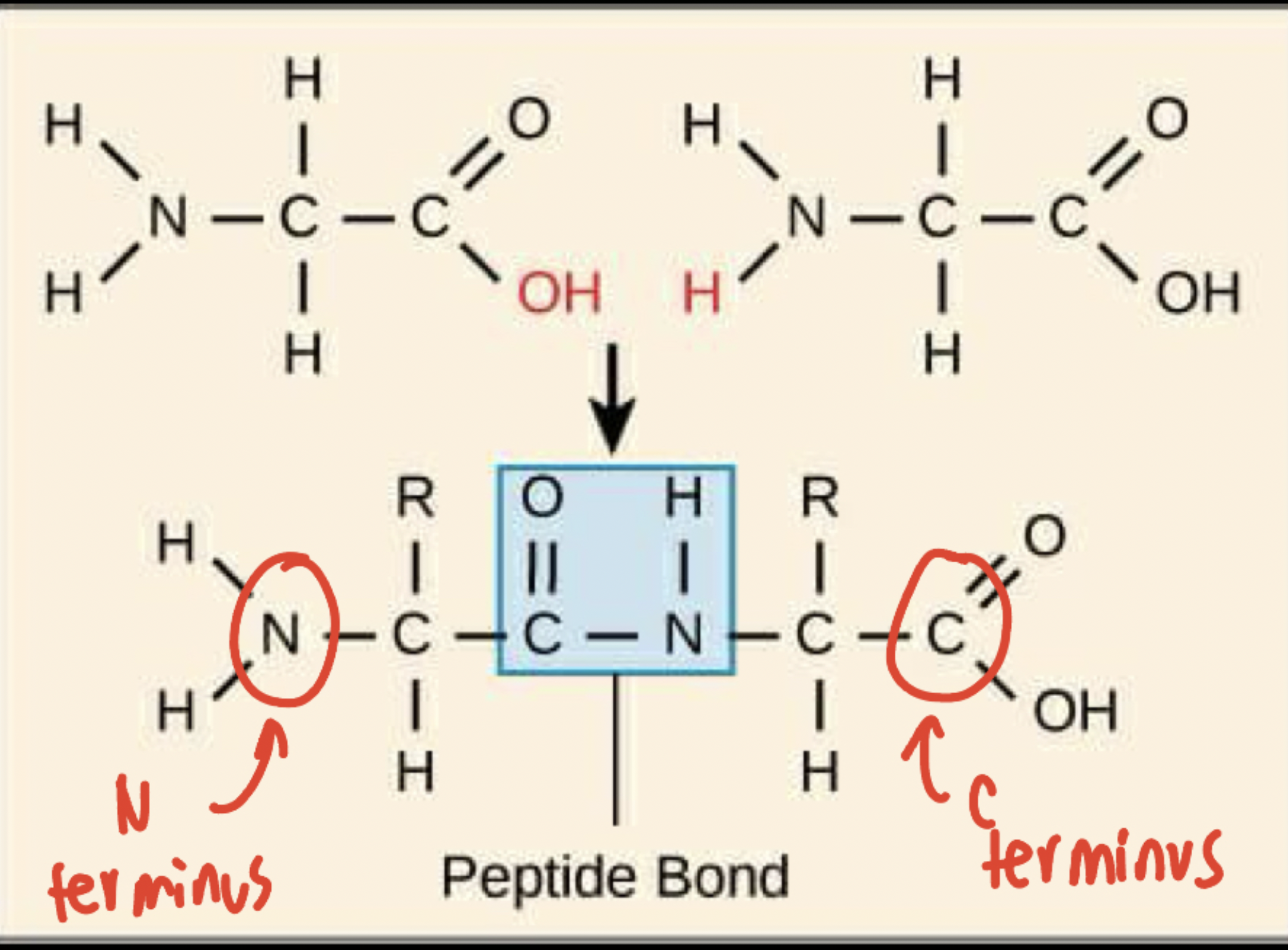
Primary structure + draw an example
An amino acid chain connected by a covalent peptide bond (C-N).
Nitrogen (from amino group) of the first amino acid connects with the carbon (carboxyl group) of the second amino acid
N-terminus: beginning of primary structure, refers to the nitrogen
C-terminus: end of primary structure, refers to the carbon
Residue
Amino acid (more specifically, each amino acid in the polypeptide chain)
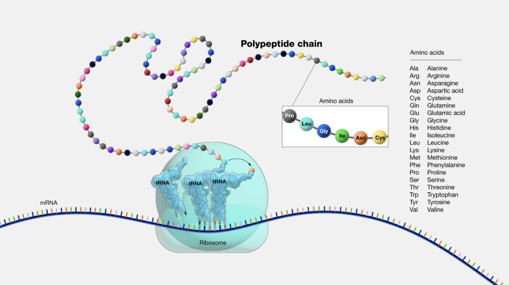
Peptide bond in relation to the ribosome and tRNAs + explain the process of translation
The ribosome is the “machine” that builds peptide bonds using tRNAs to bring in the right amino acids in the right order (aka translation)
The ribosome reads one mRNA codon at a time → each codon specifies which amino acid to bring
tRNA carries one specific amino acid and has an anticodon that matches the mRNA codon
Ribosome lines up the growing chain
Peptide bond forms — the ribosome catalyzes the reaction between the growing chain and the new amino acid
Ribosome shifts forward to the next codon, the used tRNA leaves, and a new tRNA comes in
Repeat until polypeptide chain is fully built
start codon
Met (AUG)
stop codons
UGA, UAG, UAA
Why is AUG special, and how does the ribosome know when it means "start" vs. just "insert methionine"?
AUG codes for methionine AND is the universal start codon
The ribosome can't tell which one from the codon alone → Context decides:
surrounding sequences and other factors at the genome dictate which one it is
What are the two ways to study peptides (broadly)?
separate them based on mass
Separate them based on the unique chemistry of their side chains
Class example: Insulin and peptide sequencing
alpha and beta chains come from two different genes, but they come together to form insulin
insulin is a conserved sequence (meaning that compared to a lot of other animals, our insulin sequence is very similar to there), meaning it is important/not very changed throughout evolution
Which enzymes chop up peptides? What do they chop up? Why?
Trypsin, Chymotrypsin → break peptide bonds
Trypsin: cuts C-terminal after Lysine or Arginine → This is because Trypsin’s active site has a negatively charged pocket (Asp residue at its bottom) → because Lysine and Arginine are positively charged and basic residues their side chains get pulled into that pocket
*only if there is not a Proline as the C terminus residue → this is because Proline forms a rigid ring, making it difficult for Trypsin to access the peptide bond
Chymotrypsin: cuts after large hydrophobic residues (The, Trp, Tyr)
Secondary structure
Polypeptide beginning to twist into four patterns, defined by weak and strong interactions
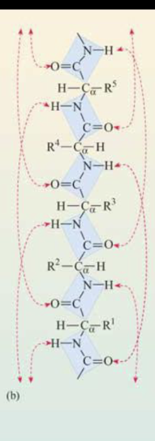
peptide bonds, H bonds between NH and C=O groups
Disulfide bridge
2 cysteines in proximity form a covalent bond (S-S) → either between regions on the same chain OR between chains
In secondary structure, what is proline, hydrophobic/hydrophillic interactions, H bonding, and cysteine used for?
“Tools” that help peptides start making structures
Proline: kinks
Hydrophobic/hydrophillic side chains: to attract like, to repel unlike
H bond: for weak interactions
Cysteine: for strong bonding
Alpha helix
polypeptide chain that twists into a rod-like structure
secondary structure, rod-like shape
inside: backbone
outside: R-chain groups (exposed for maximum reactivity)
some AAs are more likely to be in an alpha helix formation than others
Hydrogen bonds between carbonyl oxygens (C=O) and amine hydrogen 4 amino acids downstream
Beta pleated sheets
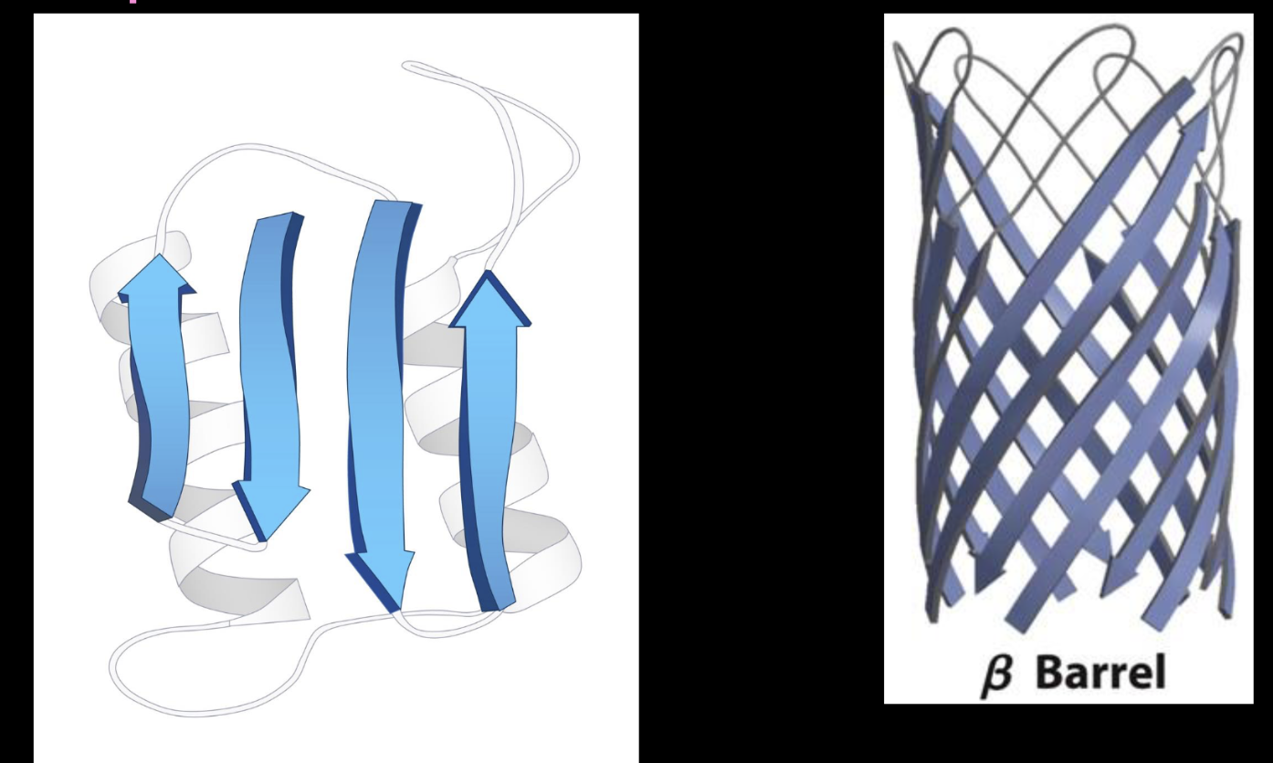
Polymers of amino acids are lined up either parallel or antiparallel to each other
secondary structure
antiparallel: line up perfectly
parallel: same direction, so they can’t line up perfectly
Tertiary structure
3D shape that the polypeptide chain forms
Secondary structures combine to form motifs (more complex structures)
Quaternary structure
2 or more polypeptide chains that bind together to form superstructures
Denaturation and renaturation
Denaturation: loss of protein structure and function
renaturation: When bonds that are broken are remade
Proteostasis
The life cycle of a protein
Transcription (DNA→RNA) → Translation (RNA→amino acid chain, made by ribosome)
Chaperones help the new chain fold
Protein can exist in 3 interconvertible states:
Native State = correctly folded, functional
Folding Intermediates = partially folded, in progress
Aggregates = misfolded clumps (bad — disease-associated)
These states can convert back and forth (folding ↔ unfolding, aggregation ↔ disaggregation
If a protein can't be fixed, it's degraded
How do chaperones help protein folding, and what happens without them?
Without a chaperone: unfolded protein folds on its own, but slowly → during this slow window it's exposed and can stick to other unfolded proteins, forming a nonfunctional aggregate.
With a chaperone: the unfolded protein is captured inside the chaperone's cavity, which shields/isolates it from other proteins. Protected from unwanted interactions, it folds correctly into its native shape, then is released ("removed for use") as a functional folded protein.
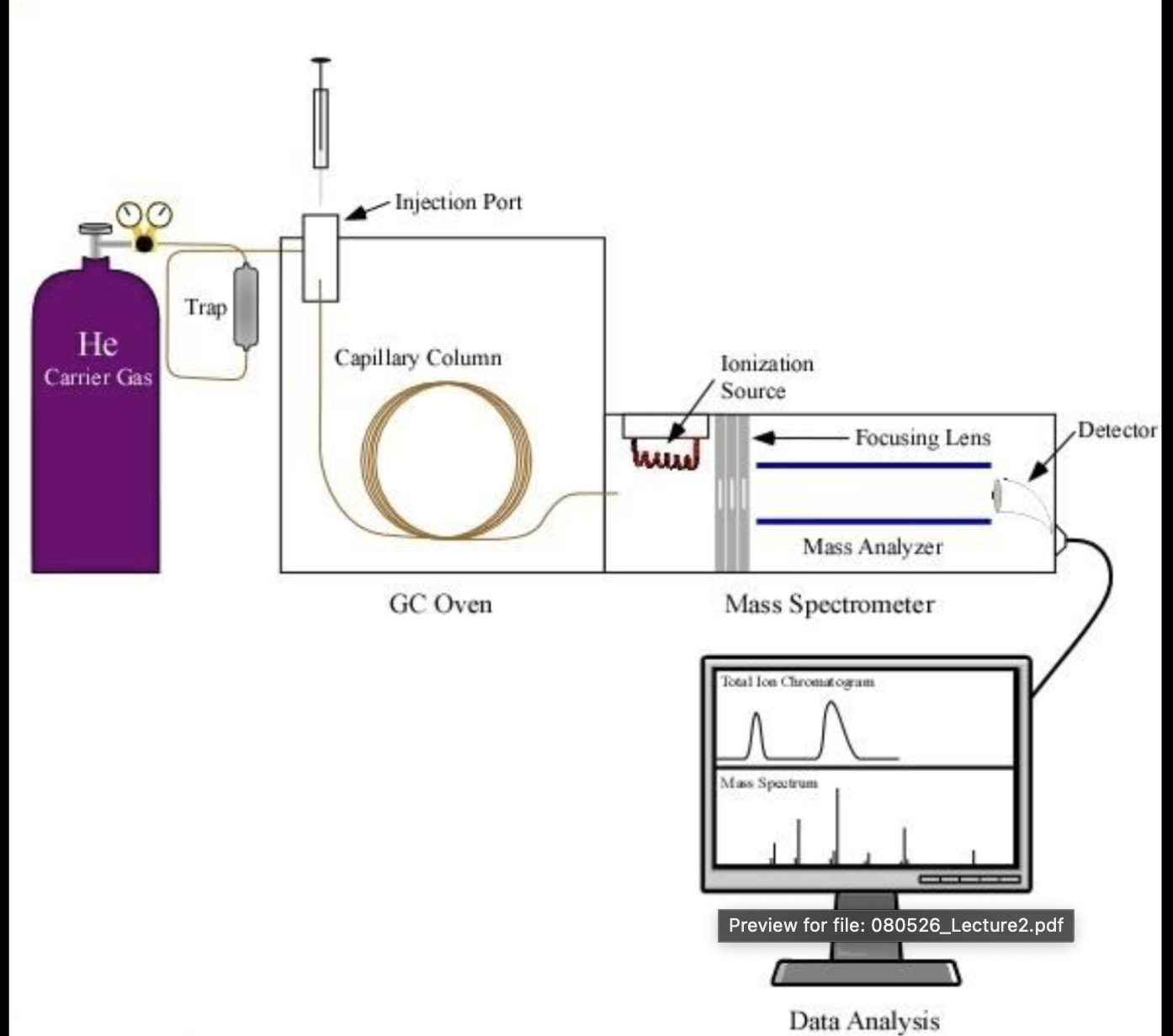
What is GC-MS and how does it work
used anytime you need to figure out what specific small molecules are in a sample (better for small stuff, not big stuff like DNA)
Inject liquid into port → turns into gas
Sample gas is pushed through a column using a different gas → its molecules stick differently.
Ones that stick better move through the column faster, the ones that don’t are delayed
MS blasts molecules with electrons → turns into fragments → fragments separated by mass → fragmentation pattern generated
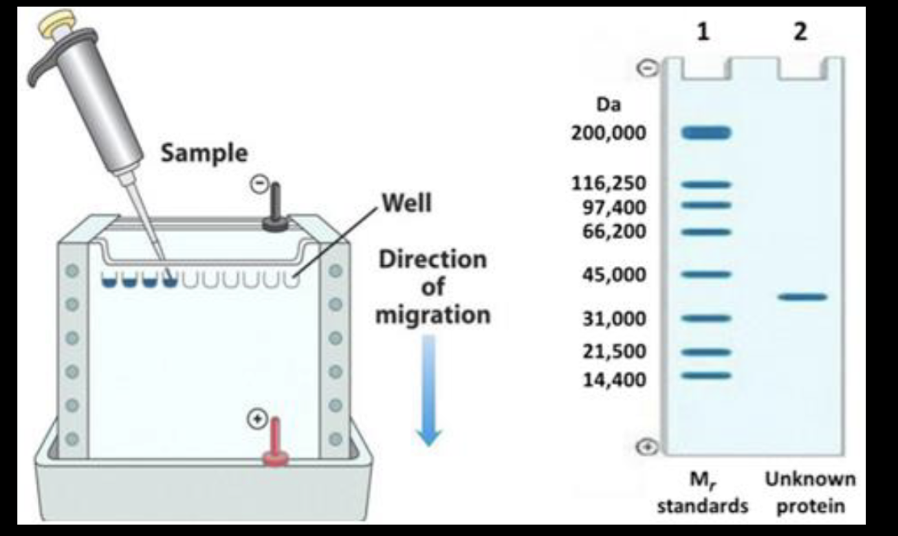
What is SDS PAGE and what is it used for?
used anytime you need to know what proteins are in a sample, how big they are, or how much of them there is
Get proteins and treat them with heat and SDS (a detergent that makes them all - charged)
Put proteins into wells on the top of the gel (- electrode on top, + electrode on bottom) → they all travel to the + electrode
small proteins → closer to bottom, large proteins → closer to top
Isoelectric point of a protein
pH at which the net charge is 0
average of the pKa of the carboxyl and amine group of the protein
What is 2D electrophoresis, and what is it used for?
Good if there are a lot of proteins you need to identify
Dimension 1: isoelectric focusing
lay proteins on gel strip with built in pH gradient
run electricity through it → each protein moves until its isoelectric point
Dimension 2: SDS page (as usual)
What is column chromatography, and what is it used for?
Separates proteins from a mixture
Fill tube with beads
pour protein mixture on top of the column
Elute (wash through) using solvent
Separate based on:
Size: big molecules come out first, small come out last
charge: + charge proteins stick to beads and don’t move, - charge proteins flow through and don’t stick (cus beads are - charge) and come out first, neutral proteins also flow through
Affinity: target protein sticks, everything else washes through
Collect fractions (liquid that pours through the bottom)
Check fractions for protein of interest
Peptide tags
A peptide sequence you attach onto a protein of interest, so you don’t have to use an antibody
Fluorescence
A glowing protein you can use to attach onto other proteins, so you don’t have to use an antibody (peptide tag)
6xHis tag
Peptide tag that gives your protein a metal binding property
6 His in a row, His is basically a metal magnet
Immunochemistry and antibodies in rabbits
antibodies are generated to target a protein of interest
ex) generating antibodies in rabbits: inject antigen in rabbit → antigen activates B-cells → plasma B cells provide antibodies → collect antibodies from rabbits
Problems with antibody generation
limited to what is immunogenic → some proteins don’t provoke a good immune response
generates antibodies specific for many different cell motifs → grabs lookalike proteins with similar motifs, not JUST your protein of interest
native vs denature protein antibodies → The immune system "sees" different things depending on the protein's shape at injection — a folded protein produces an antibody that recognizes 3D shape, while a denatured/unfolded protein produces an antibody that recognizes the linear amino acid sequence
What is immunohistochemistry? + immunofluorescence
Used to study in-situ observations
cut cells thin and attach to a microscope slide
primary antibody finds target protein in sample
secondary antibody finds primary antibody based on its constant (Fc) region
makes a brown color where antibody is bound (due to color changing enzyme)
immunofluorescence → secondary antibody is a fluorescent protein, not an enzyme