Skull anatomy
 Call Kai
 Learn
Learn Practice Test
 Spaced Repetition
 Match
Match Flashcards
 Knowt Play
1/192

There's no tags or description
Looks like no tags are added yet.
Name | Mastery | Learn | Test | Matching | Spaced | Call with Kai | Chat |
|---|
No analytics yet
Send a link to your students to track their progress
193 Terms
Frontal Bone
the large cranial bone forming the front part of the cranium: the forehead and the upper part of the orbits

Parietal Bone
either of two skull bones between the frontal and occipital bones and forming the top and sides of the cranium

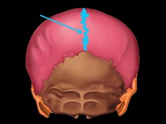
Sagital Suture
Located on the midline of the skull lies between both parietal bones (immovable joint)

Coronal Suture
the suture between the parietal and frontal bones of the skull

Lambdoidal Suture
suture that separates parietal bones from occipital bone

Squamosal Suture
between parietal bone and temporal bone on side of the skull, bordered in back by occipital bone

Optic Foramen
On either side ochiasmatic groove -transmits optic nerve (CN II) along with its meningeal coverings, and the ophthalmic artery

Sella turcica
depression in the sphenoid bone where the pituitary gland is located

Temporal Bone
bone that forms parts of the side of the skull and floor of the cranial activity.

Mastoid Process
projection of the temporal bone located behind the ear

Styloid
spike-like process extending downward from the temporal bone on each side of the skull
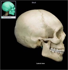
External Auditory Meatus
hole in the temporal bone extending medially to the inner ear

Jugular Foramen
hole at the back/base of the skull.

Carotid Foramen
opening for carotid artery

Mandibular fossa
depressions where manidble and cranium meet and join

Temporomandibular joint (TMJ)
the joint between the head of the lower jawbone and the temporal bone

Occipital Bone
a saucer-shaped membrane bone that forms the back of the skull

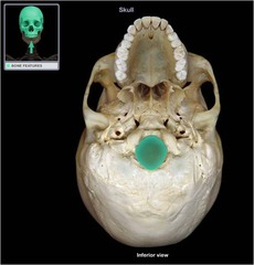
Foramen Magnum
the large opening at the base of the cranium through which the spinal cord passes
Occipital Condyle
ridges on left and right of foramen magnum

Middle nasal Concha
Curved bone inside the nasal cavity.

Crista Galli
vertical projection providing a point of attachment for the dura mater, helping to secure the brain within the skull

Cribiform plate
part of ethmoid bone, at the roof of the nasal cavity
Shape: Thin, flat plate with many tiny holes

Maxilla
Forms most of the upper jaw, and part of the hard palate, contains upper molars, premolars, and canines

Palatine Process
forms the anterior portion of the hard palate (roof) of the mouth also forms parts of the nasal cavity and eye orbits

Alveolar Process
a ridge that forms the borders of the upper and lower jaws and contains the sockets of the teeth

Lacrimal Bone
small fragile bone making up part of the front inner walls of each eye socket

Lacrimal Fossa
Anterior and lateral depression of roof of orbit which accommodates the lacrimal gland
Nasal Bone
an elongated rectangular bone that forms the bridge of the nose

Palatine Bone
either of two irregularly shaped bones that form the back of the hard palate and helps to form the nasal cavity and the floor of the orbits
Inferior nasal Concha
located on each side of the nasal septum, attached to the lateral wall of the nasal cavity; increase epithelial surface area and create turbulence in the inspired air

Vomer
thin trapezoidal bone of the skull forming the posterior and inferior parts of the nasal septum (the lower portion of the nasal septum)

Zygomatic Bone
the arch of bone beneath the eye that forms the prominence of the cheek

Mandible
the lower jawbone in vertebrates

Condylar Process
the condyle of the ramus of the mandible that articulates with the skull

Aveolar Process
bone surronding the teeth

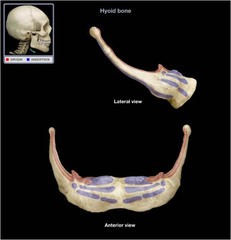
Hyoid Bone
bone that is suspended in the mid neck region and provides a moveable base for the tongue
Frontal

Maxilla

Mandible

Sphenoid
Butterfly-shaped bone in the middle of the skull.

Zygomatic

Temporal

Occipital

Sphenoid

Parietal

Nasal Bone

Coronal Suture

Squamous suture

Lambdoid suture

Sagittal suture
Mastoid Process
Bony bump behind the ear.

Styloid Process

External Acoustic Meatus

Mandibular Condyle
rounded projection at the top of the mandible that forms the TMJ with the temporal bone.

Mental Foramen

Ethmoid

Sphenoid

Lacrimal
tears → it’s the little bone by your eye associated with tear drainage.
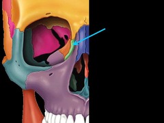
Vomer

Middle Nasal Conchae

Inferior Nasal Conchae

Foramen Magnum

Palatine

Sphenoid

Zygomatic Process
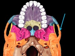
Temporal Process
Part of the zygomatic bone that extends back toward the temporal bone.

Frontal Bone
the large cranial bone forming the front part of the cranium: the forehead and the upper part of the orbits

Parietal Bone
either of two skull bones between the frontal and occipital bones and forming the top and sides of the cranium

Temporal Bone

Occipital Bone
a saucer-shaped membrane bone that forms the back of the skull

Maxilla
Forms most of the upper jaw, and part of the hard palate, contains upper molars, premolars, and canines

Lacrimal
small fragile bone making up part of the front inner walls of each eye socket

Nasal Bone
an elongated rectangular bone that forms the bridge of the nose

Palatine
Either of two irregularly shaped bones that form the back of the hard palate and helps to form the nasal cavity and the floor of the orbits
Zygomatic Bone
the arch of bone beneath the eye that forms the prominence of the cheek

Mandible
the lower jawbone in vertebrates
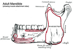
Sphenoid
Base and sides of skull; part of the eye orbits.

Ethmoid
Internal base of skull; structures in nasal cavity.
Frontal bone

Coronal suture

Infraorbital foramen

Mental foramen

Optic foramen

Parietal bone

Squamous suture

Temporal bone

Vomer

Nasal bone

Supraorbital foramen

Supraorbital foramen
holes above the eye socket

Inferior nasal conchae

Alveolar process
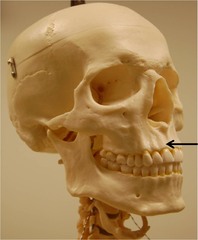
Maxilla

Condylar process
Rounded bump at the back/top of the mandible (jaw)

Mandibular angle

Mandibular notch

Zygomatic process of temporal bone

Zygomatic bone

Frontal Bone

Maxilla Bone
